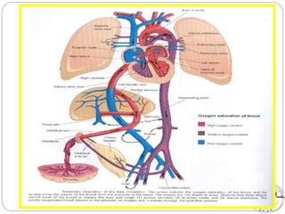
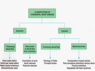
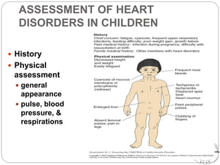
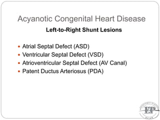
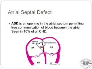
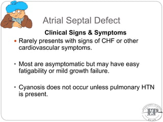
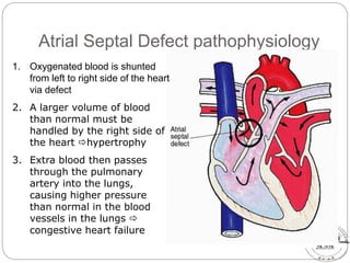
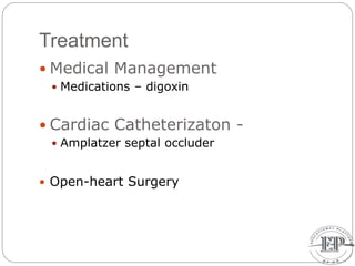
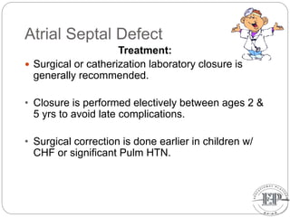
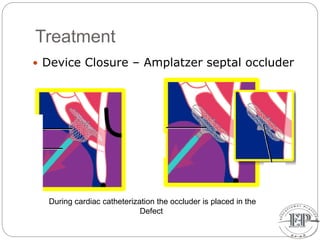
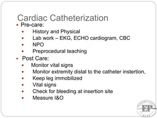
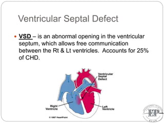
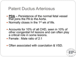
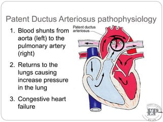
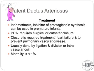
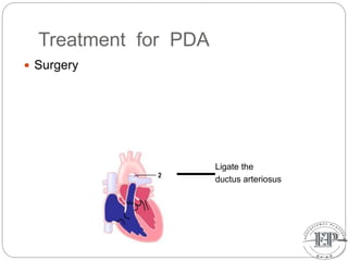
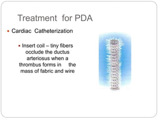
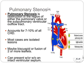
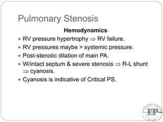
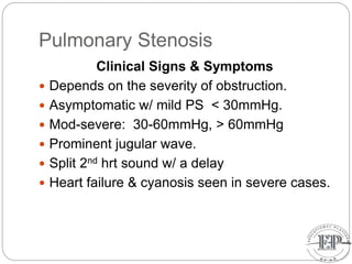
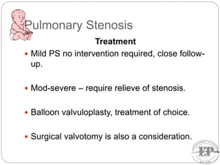
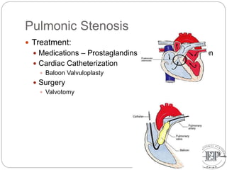
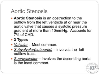
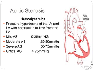
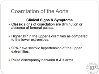
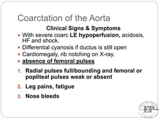
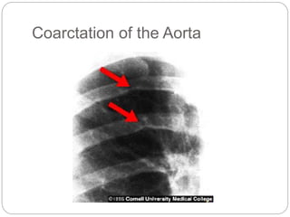
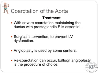
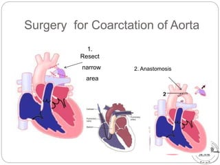
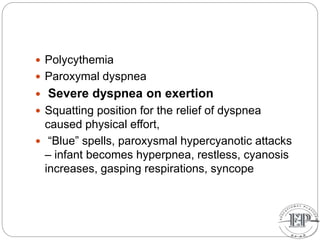
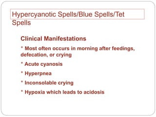
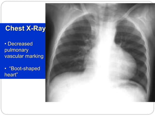
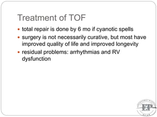
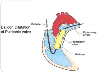
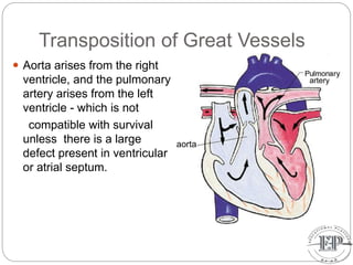
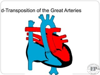
This document discusses fetal circulation and several congenital heart defects that can occur, including their pathophysiology, clinical presentation, and treatment. It begins by describing the normal fetal circulation, where oxygenated blood from the placenta travels through the umbilical vein, ductus venosus, and foramen ovale to supply the fetus. It then covers several defects: atrial septal defect, ventricular septal defect, patent ductus arteriosus, pulmonary stenosis, aortic stenosis, and coarctation of the aorta. For each, it provides an overview of how blood flow is disrupted, typical signs and symptoms, and treatment approaches like surgery, catheterization, or medication management.
Acyanotic Congenital Heart Diseases;
1. Left-to-right shunts
a. Ventricular Septal Defect(VSD)
b. Atrial Septal Defect(ASD)
c. Patent Ductus Arteriosus(PDA)
d. Atrioventricular Septal Defect(AVSD)
e. Aortopulmonary window
* Eisenmenger Syndrome – The shunt becomes right-to-left
2. Left-sided obstructive lesions
a. Coarctation of the Aorta(COA)
b. Congenital Aortic Stenosis
c. Mitral Stenosis
d. Interrupted Aortic Arch
Cyanotic Congenital Heart Diseases;
1. Right-to-left shunts
a. Tetralogy of Fallot
b. Pulmonary stenosis
c. Pulmonary atresia
d. Tricuspid atresia
e. Ebstein’s anomaly
2. Complete mixed lesions
a. Transposition of the great vessels
b. Double outlet right ventricle(DORV)
c. Total anomalous pulmonary venous return
d. Truncus arteriosus
e. Hypoplastic left heart syndrome
Congenital heart disease, by dr Shaymaa Fayad, El Nasr Hospital Port saidmohamed osama hussein
1. Blood from the placenta travels through the umbilical vein to the fetus's heart, bypassing the lungs via the foramen ovale. It then circulates through the aorta to the body.
2. The ductus arteriosus shunts blood away from the lungs, which are not used for gas exchange in utero.
3. At birth, closure of the ductus arteriosus and foramen ovale, along with an increase in pulmonary blood flow, directs blood through the lungs for the first time.
This document discusses various acyanotic congenital heart diseases. It begins by classifying congenital heart disease into cyanotic and acyanotic categories. It then further divides acyanotic congenital heart disease into those with a left-to-right shunt or increased pulmonary blood flow, such as atrial septal defect (ASD), ventricular septal defect (VSD), and patent ductus arteriosus (PDA), and those without a shunt such as coarctation of the aorta. The document then goes on to describe the pathophysiology, clinical features, investigations and management of ASD, VSD, and PDA in more detail. It also briefly discusses Eisenmenger syndrome.
This document discusses pediatric cardiac disorders, including:
1. Congenital heart defects (CHDs) are the most common birth defects and cause of infant mortality. CHDs can be acyanotic (left-to-right shunts) or cyanotic (right-to-left shunts). Common defects include atrial and ventricular septal defects, patent ductus arteriosus, tetralogy of Fallot, and transposition of the great arteries.
2. Assessment of suspected CHD involves history, physical exam including pulse oximetry, chest x-ray, EKG, and echocardiogram. Major signs are systolic murmurs, diastolic murmurs, cyan
Truncus arteriosus is a congenital heart defect where there is a single arterial trunk exiting the heart, giving rise to the pulmonary artery, aorta, and coronary arteries. This occurs when normal septation of the embryonic bulbar trunk fails to occur. Blood from the two ventricles mixes in the common trunk, resulting in decreased oxygen levels. Surgical repair is needed to separate pulmonary and systemic blood flow and close the ventricular septal defect. Without repair, complications like congestive heart failure and pulmonary hypertension can develop.
Transposition of the great arteries is a serious but rare heart defect present at birth (congenital), in which the two main arteries leaving the heart are reversed (transposed). The condition is also called dextro-transposition of the great arteries.
A 3-week-old male infant presented with cyanosis, tachypnea, fatigue with feeding, and sweating. On examination, he had tachycardia, low blood pressure, and a systolic murmur, consistent with a congenital heart defect such as tetralogy of Fallot or transposition of the great vessels. Further testing, including echocardiogram and chest x-ray, was needed to determine the specific condition and appropriate treatment.
The document discusses various types of congenital heart defects including cyanotic and acyanotic defects, summarizing key features such as symptoms, physical exam findings, treatments, and anesthetic considerations for septal defects, atrial septal defects, patent ductus arteriosus, interrupted aortic arch, tetralogy of Fallot, and single ventricle physiology. It provides an overview of important congenital heart conditions from an anesthesiology perspective.
The document discusses congenital heart diseases, which occur in approximately 1% of live births. It describes several types of congenital heart defects including atrial septal defect (ASD), ventricular septal defect (VSD), atrioventricular canal defect, and patent ductus arteriosus - all of which involve increased pulmonary blood flow. It also discusses obstructive defects like aortic stenosis and pulmonary stenosis. The document provides details on the pathophysiology, clinical manifestations, diagnosis, and treatment of these various congenital heart conditions.
This document provides an overview of congenital heart disease (CHD) and anesthesia considerations for CHD. It begins with definitions and classifications of CHD, including acyanotic and cyanotic defects. Specific conditions discussed include atrial septal defect, ventricular septal defect, patent ductus arteriosus, pulmonary stenosis, aortic stenosis, and coarctation of the aorta. For each condition, the document outlines etiology, pathophysiology, clinical presentation, diagnosis, and treatment considerations.
This document summarizes key aspects of acyanotic heart disease, including ventricular septal defects (VSDs), atrial septal defects (ASDs), patent ductus arteriosus (PDAs), aortic stenosis (AS), coarctation of the aorta, bicuspid aortic valve, and pulmonary stenosis. It covers clinical findings, management, natural history, complications, and treatment options for each condition. It also discusses ductal-dependent lesions that require prostaglandin E1 treatment, such as hypoplastic left heart syndrome.
This document discusses congenital heart diseases, beginning with a review of fetal circulation and how it transitions after birth. It then classifies common acyanotic and cyanotic congenital heart diseases. Ventricular septal defect, patent ductus arteriosus, and coarctation of the aorta are discussed in more detail, including their pathophysiology, clinical presentation, diagnosis, and management principles. The objective is to revise fetal circulation, classify congenital heart diseases, and discuss common types and their clinical approach.
Common heart conditions in children copy.pptxRamiHaris
This document discusses common congenital and acquired heart conditions in children. It begins by describing fetal circulation and how congenital heart diseases arise from defects present at birth. The conditions are classified as acyanotic or cyanotic depending on whether they allow mixing of oxygenated and deoxygenated blood. Common acyanotic conditions discussed include atrial septal defect, ventricular septal defect, and patent ductus arteriosus. Cyanotic conditions include tetralogy of Fallot and transposition of the great vessels. The document also covers acquired conditions like rheumatic fever and infective endocarditis, as well as their presentation, diagnosis, and management.
The document discusses various acyanotic heart diseases including atrial septal defect (ASD), ventricular septal defect (VSD), patent ductus arteriosus (PDA), coarctation of aorta, pulmonary stenosis, and aortic stenosis. It provides details on the pathophysiology, clinical features, investigations, and management of each condition. Key points include that ASD is the most common congenital heart disease, VSD has a pansystolic murmur at the lower left sternal edge, PDA causes a continuous murmur and loud P2 with pulmonary hypertension, coarctation causes hypertension and rib notching on CXR, and large defects can lead to pulmonary hypertension and
TAPVC is a congenital heart defect where the pulmonary veins do not connect normally to the left atrium, instead connecting to the right atrium, often via the superior vena cava. This causes oxygenated blood from the lungs to mix with deoxygenated blood. An ASD or PFO is always present to allow blood to reach the left side of the heart. Without treatment, TAPVC is fatal. Surgical repair is required to reconnect the pulmonary veins to the left atrium and close any defects. Post-surgery, patients require monitoring for complications, but long-term survival is generally good if repaired.
Congenital heart disease (CHD) refers to structural heart defects present at birth. Diagnosis involves history, physical exam, chest X-ray, ECG, and echocardiogram. Most CHDs can be corrected with surgery if done in a timely manner. Echocardiography can identify and determine severity of specific lesions. Pediatricians must also identify any associated conditions that could impact outcomes.
Congenital Heart disease or CHD refers to structural abnormalities in the heart present at birth. It is the most common type of birth defect, occurring in 5-8 per 1,000 live births. CHD encompasses several types of defects including atrial septal defect (ASD), ventricular septal defect (VSD), and patent ductus arteriosus (PDA). ASD involves an abnormal opening between the left and right atria, VSD is an opening in the ventricular septum between the ventricles, and PDA is the failure of the ductus arteriosus to close after birth, allowing blood to flow between the aorta and pulmonary artery. Symptoms vary depending on the size and
This document provides information about acyanotic heart defects, including definitions, causes, pathophysiology, clinical manifestations, diagnosis, management, and complications. It discusses several types of acyanotic heart defects such as atrial septal defect (ASD), ventricular septal defect (VSD), atrioventricular canal defect, patent ductus arteriosus (PDA), coarctation of the aorta, and aortic stenosis. For each condition, it outlines the definition, incidence, types if applicable, pathophysiology that occurs due to the structural abnormality, associated signs and symptoms, diagnostic evaluation, treatment options including medical management and surgery, and potential complications.
Atrial septal defect (ASD), ventricular septal defect (VSD), patent ductus arteriosus (PDA), and tetralogy of Fallot (TOF) are four common types of congenital heart disease. ASD is a hole in the atrial septum that allows blood to flow from the left to the right atrium. VSD is a hole in the ventricular septum that allows blood to flow between the ventricles. PDA is a persistent opening between the aorta and pulmonary artery that normally closes after birth. TOF involves four abnormalities that reduce pulmonary blood flow.
1. The document discusses various types of acyanotic congenital heart disease including ventricular septal defect (VSD), atrial septal defect (ASD), patent ductus arteriosus (PDA), and coarctation of aorta.
2. It provides details on the classification, symptoms, diagnosis, and treatment options for each condition.
3. The conditions are characterized by shunting of blood between the left and right sides of the heart without cyanosis, and can cause heart failure if left untreated.
This document provides information on the approach to heart failure in the intensive care unit. It begins with statistics on the incidence and prevalence of heart failure. It then discusses the morbidity, mortality, definitions, types, etiology, pathophysiology, signs and symptoms, diagnostic tests, compensatory mechanisms, and prognosis of heart failure. Key points include that heart failure is a leading cause of hospitalization, the median age of presentation is 76, and the 5-year mortality rate is around 50-60%. Common causes include hypertension, coronary artery disease, and alcohol abuse. The pathophysiology involves an imbalance in preload and afterload. Diagnostic tests include BNP/NT-proBNP levels and echocardiogram. Comp
Seminar congenital cardiac disorders (pda,TA and AP Window)Uma Binoy
Patent ductus arteriosus is a congenital heart disorder where the ductus arteriosus, a blood vessel connecting the pulmonary artery and aorta, fails to close after birth as it normally would. This allows blood to shunt from the aorta to the pulmonary artery, potentially causing heart failure. It can be diagnosed via echocardiogram, electrocardiogram, or chest x-ray and may be treated with medications like indomethacin or surgery.
1. An atrial septal defect is an opening in the septum between the left and right atria, allowing blood to shunt from the left to the right side of the heart.
2. It is one of the most common congenital heart defects found in adults.
3. Symptoms range from none in small defects to fatigue and shortness of breath from right heart strain in large defects that cause significant shunting of blood from the left to the right atrium.
Complete transposition of the great arteries (D-TGA.pptxDrPNatarajan2
Complete transposition of the great arteries (D-TGA) is a congenital heart defect where the aorta arises from the right ventricle and the pulmonary artery arises from the left ventricle. This results in deoxygenated blood circulating to the body and oxygenated blood recirculating to the lungs. Without intervention, D-TGA is fatal in infancy due to hypoxia and heart failure. Treatment involves atrial septostomy to improve mixing, followed by surgical repair such as the arterial switch operation or atrial baffle procedures to redirect blood flow. The arterial switch operation has better long-term outcomes.
There are over 100 different types of congenital heart disease that can be categorized into 9 major groups. The most common are ventricular septal defects (VSD), atrial septal defects (ASD), patent ductus arteriosus (PDA), and tetralogy of Fallot. For any congenital heart disease, the anesthesiologist must understand the pathophysiology of the defect and how changes in systemic and pulmonary vascular resistance may impact blood flow and oxygen saturation. The goal is to avoid maneuvers that could increase intracardiac shunting and hypoxemia based on whether it is a left-to-right or right-to-left shunt. Thorough preoperative evaluation including echocardi
This document provides an overview of congenital heart disease, including prevalence, circulatory adjustments at birth, hemodynamic classifications, and descriptions of specific conditions like atrial septal defect (ASD) and ventricular septal defect (VSD). It notes that congenital heart defects affect 6-8 per 1000 live births and can range widely in severity. Diagnosis typically occurs by 1 week or 1 month of age. After birth, clamping of the umbilical cord and expansion of the lungs cause pressure changes and closure of passages between circulations. Conditions are classified as acyanotic or cyanotic depending on oxygen saturation levels. ASD and VSD are both described in detail including typical clinical features, imaging findings, and management
Similar to Unit 9; Peadiatric Cardiology, Educational Platform.pptx (20)
Physiology and chemistry of skin and pigmentation, hairs, scalp, lips and nail, Cleansing cream, Lotions, Face powders, Face packs, Lipsticks, Bath products, soaps and baby product,
Preparation and standardization of the following : Tonic, Bleaches, Dentifrices and Mouth washes & Tooth Pastes, Cosmetics for Nails.
ISO/IEC 27001, ISO/IEC 42001, and GDPR: Best Practices for Implementation and...PECB
Denis is a dynamic and results-driven Chief Information Officer (CIO) with a distinguished career spanning information systems analysis and technical project management. With a proven track record of spearheading the design and delivery of cutting-edge Information Management solutions, he has consistently elevated business operations, streamlined reporting functions, and maximized process efficiency.
Certified as an ISO/IEC 27001: Information Security Management Systems (ISMS) Lead Implementer, Data Protection Officer, and Cyber Risks Analyst, Denis brings a heightened focus on data security, privacy, and cyber resilience to every endeavor.
His expertise extends across a diverse spectrum of reporting, database, and web development applications, underpinned by an exceptional grasp of data storage and virtualization technologies. His proficiency in application testing, database administration, and data cleansing ensures seamless execution of complex projects.
What sets Denis apart is his comprehensive understanding of Business and Systems Analysis technologies, honed through involvement in all phases of the Software Development Lifecycle (SDLC). From meticulous requirements gathering to precise analysis, innovative design, rigorous development, thorough testing, and successful implementation, he has consistently delivered exceptional results.
Throughout his career, he has taken on multifaceted roles, from leading technical project management teams to owning solutions that drive operational excellence. His conscientious and proactive approach is unwavering, whether he is working independently or collaboratively within a team. His ability to connect with colleagues on a personal level underscores his commitment to fostering a harmonious and productive workplace environment.
Date: May 29, 2024
Tags: Information Security, ISO/IEC 27001, ISO/IEC 42001, Artificial Intelligence, GDPR
-------------------------------------------------------------------------------
Find out more about ISO training and certification services
Training: ISO/IEC 27001 Information Security Management System - EN | PECB
ISO/IEC 42001 Artificial Intelligence Management System - EN | PECB
General Data Protection Regulation (GDPR) - Training Courses - EN | PECB
Webinars: https://pecb.com/webinars
Article: https://pecb.com/article
-------------------------------------------------------------------------------
For more information about PECB:
Website: https://pecb.com/
LinkedIn: https://www.linkedin.com/company/pecb/
Facebook: https://www.facebook.com/PECBInternational/
Slideshare: http://www.slideshare.net/PECBCERTIFICATION
This presentation was provided by Steph Pollock of The American Psychological Association’s Journals Program, and Damita Snow, of The American Society of Civil Engineers (ASCE), for the initial session of NISO's 2024 Training Series "DEIA in the Scholarly Landscape." Session One: 'Setting Expectations: a DEIA Primer,' was held June 6, 2024.
Executive Directors Chat Leveraging AI for Diversity, Equity, and InclusionTechSoup
Let’s explore the intersection of technology and equity in the final session of our DEI series. Discover how AI tools, like ChatGPT, can be used to support and enhance your nonprofit's DEI initiatives. Participants will gain insights into practical AI applications and get tips for leveraging technology to advance their DEI goals.
A workshop hosted by the South African Journal of Science aimed at postgraduate students and early career researchers with little or no experience in writing and publishing journal articles.
Strategies for Effective Upskilling is a presentation by Chinwendu Peace in a Your Skill Boost Masterclass organisation by the Excellence Foundation for South Sudan on 08th and 09th June 2024 from 1 PM to 3 PM on each day.
This presentation includes basic of PCOS their pathology and treatment and also Ayurveda correlation of PCOS and Ayurvedic line of treatment mentioned in classics.
Assessment and Planning in Educational technology.pptxKavitha Krishnan
In an education system, it is understood that assessment is only for the students, but on the other hand, the Assessment of teachers is also an important aspect of the education system that ensures teachers are providing high-quality instruction to students. The assessment process can be used to provide feedback and support for professional development, to inform decisions about teacher retention or promotion, or to evaluate teacher effectiveness for accountability purposes.
2. Fetal Circulation
Before birth, blood from the placenta,
about 80% saturated with oxygen, returns
to the fetus by way of the umbilical vein.
On approaching the liver, most of this
blood flows through the ductus venosus
directly into the inferior vena cava,
bypassing the liver.
After a short course in the inferior vena
cava, where placental blood mixes with
deoxygenated blood returning from the
lower limbs, it enters the right atrium.
3. Fetal Circulation
Here it is guided toward the oval foramen by
the valve of the inferior vena cava, and most
of the blood passes directly into the left
atrium.
From the left atrium, where it mixes with a
small amount of desaturated blood returning
from the lungs, blood enters the left ventricle
and ascending aorta.
Since the coronary and carotid arteries are
the first branches of the ascending aorta, the
heart musculature and the brain are supplied
with well-oxygenated blood.
4. Fetal Circulation
A small amount from the IVC is prevented
from entering the left atrium and remains
in the right atrium.
It mixes with desaturated blood returning
from the head and arms by way of the
superior vena cava.
Desaturated blood from the superior vena
cava flows by way of the right ventricle
into the pulmonary trunk.
5. Fetal Circulation
During fetal life, resistance in the
pulmonary vessels is high, such that most
of this blood passes directly through the
ductus arteriosus into the descending
aorta, where it mixes with blood from the
proximal aorta.
After coursing through the descending
aorta, blood flows toward the placenta by
way of the two umbilical arteries.
The oxygen saturation in the umbilical
arteries is approximately 58%.
6. Fetal Circulation
N.B.
A. A small amount of blood enters the liver sinusoids
and mixes with blood from the portal circulation.
B. A sphincter mechanism in the ductus venosus,
close to the entrance of the umbilical vein, regulates
flow of umbilical blood through the liver sinusoids.
This sphincter closes when a uterine contraction
renders the venous return too high, preventing a
sudden overloading of the heart.
During its course from the placenta to the organs of
the fetus, blood in the umbilical vein gradually loses
its high oxygen content as it mixes with desaturated
blood.
7. Fetal Circulation
Theoretically, mixing may occur in the following
places :
1) In the liver by mixture with a small amount of
blood returning from the portal system.
2) In the inferior vena cava which carries
deoxygenated blood returning from the lower
extremities, pelvis, and kidneys.
3) In the right atrium by mixture with blood returning
from the head and limbs.
4) In the left atrium by mixture with blood returning
from the lungs.
5) At the entrance of the ductus arteriosus into the
descending aorta.
8.
9. Congenital heart disease (CHD)
Congenital heart disease is a defect in the
structure of the heart and
great vessels which is present at birth
• CHD are the main cause of defect-related deaths
• Incidence is 8-9/1000 live births
• More common in premature infants
• May be associated with a significant musculoskeletal
defect
(e.g. diaphragmatic hernia, tracheo-oesophageal fistula,
imperforate anus)
10. Congenital heart disease (CHD)
• Causes are multifactorial and
include maternal illness
(diabetes mellitus,
phenylketonuria, and systemic
lupus erythematosus),
maternal infections (Rubella),
drugs (lithium, thalidomide),
known teratogens, harmful
habits (alcohol, hydantoin)
and associations with
chromosomal abnormality or
other recognized patterns of
malformation or syndrome; Trisomy 18
100% have CHD
11. Central cyanosis
• noted in the trunk, tongue, mucous membranes
• due to reduced oxygen saturation
Peripheral cyanosis
• noted in the hands and feet, around mouth
• due to reduced local blood flow
Recognition of Cyanosis
12.
13. ASSESSMENT OF HEART
DISORDERS IN CHILDREN
History
Physical
assessment
general
appearance
pulse, blood
pressure, &
respirations
15. Atrial Septal Defect
ASD is an opening in the atrial septum permitting
free communication of blood between the atria.
Seen in 10% of all CHD.
16. Atrial Septal Defect
Clinical Signs & Symptoms
Rarely presents with signs of CHF or other
cardiovascular symptoms.
• Most are asymptomatic but may have easy
fatigability or mild growth failure.
• Cyanosis does not occur unless pulmonary HTN
is present.
17. Atrial Septal Defect pathophysiology
1. Oxygenated blood is shunted
from left to right side of the heart
via defect
2. A larger volume of blood
than normal must be
handled by the right side of
the heart hypertrophy
3. Extra blood then passes
through the pulmonary
artery into the lungs,
causing higher pressure
than normal in the blood
vessels in the lungs
congestive heart failure
18. Treatment
Medical Management
Medications – digoxin
Cardiac Catheterizaton -
Amplatzer septal occluder
Open-heart Surgery
19. Atrial Septal Defect
Treatment:
Surgical or catherization laboratory closure is
generally recommended.
• Closure is performed electively between ages 2 &
5 yrs to avoid late complications.
• Surgical correction is done earlier in children w/
CHF or significant Pulm HTN.
20. Treatment
Device Closure – Amplatzer septal occluder
During cardiac catheterization the occluder is placed in the
Defect
21. Cardiac Catheterization
Pre-care:
History and Physical
Lab work – EKG, ECHO cardiogram, CBC
NPO
Preprocedural teaching
Post Care:
Monitor vital signs
Monitor extremity distal to the catheter instertion,
Keep leg immobilized
Vital signs
Check for bleeding at insertion site
Measure I&O
22. Ventricular Septal Defect
VSD – is an abnormal opening in the ventricular
septum, which allows free communication
between the Rt & Lt ventricles. Accounts for 25%
of CHD.
23. Ventricular Septal Defect
Hemodynamics
The left to right shunt occurs secondary to
PVR being < SVR, not the higher pressure in
the LV.
This leads to elevated RV & pulmonary
pressures & volume hypertrophy of the LA &
LV.
24. Ventricular Septal Defect
Clinical Signs & Symptoms
• Small - moderate VSD, 3-6mm, are usually
asymptomatic and 50% will close
spontaneously
by age 2yrs.
• Moderate – large VSD, almost always have
symptoms and will require surgical repair.
26. Ventricle Septal Defect
pathophysiology
1. Oxygenated blood is shunted
from left to right side of the
heart via defect
2. A larger volume of blood
than normal must be
handled by the right side of
the heart hypertrophy
3. Extra blood then passes
through the pulmonary
artery into the lungs,
causing higher pressure
than normal in the blood
vessels in the lungs
congestive heart failure
27. Ventricular Septal Defect
Treatment
• Small VSD - no surgical intervention, no
physical restrictions, just reassurance and
periodic follow-up and endocarditis prophylaxis.
• Symptomatic VSD - Medical treatment
initially with afterload reducers & diuretics.
28. Ventricular Septal Defect
Treatment
Indications for Surgical Closure:
Large VSD with medically uncontrolled
symptomatology & continued FTT.
Ages 6-12 mo with large VSD & Pulm. HTN
30. Patent Ductus Arteriosus
PDA – Persistence of the normal fetal vessel
that joins the PA to the Aorta.
Normally closes in the 1st wk of life.
Accounts for 10% of all CHD, seen in 10% of
other congenital hrt lesions and can often play
a critical role in some lesions.
Female : Male ratio of 2:1
Often associated with coarctation & VSD.
31. Patent Ductus Arteriosus
Hemodynamics
As a result of higher aortic pressure, blood
shunts L to R through the ductus from Aorta
to PA.
Extent of the shunt depends on size of the
ductus & PVR:SVR.
Small PDA, pressures in PA, RV, RA are
normal.
32. Patent Ductus Arteriosus
Clinical Signs & Symptoms
Small PDA’s are usually asymptomatic
Large PDA’s can result in symptoms of CHF,
growth restriction, FTT.
Bounding arterial pulses
Widened pulse pressure
Enlarged heart, prominent apical impulse
Classic continuous machinary systolic murmur
Mid-diastolic murmur at the apex
33. Patent Ductus Arteriosus pathophysiology
1. Blood shunts from
aorta (left) to the
pulmonary artery
(right)
2. Returns to the
lungs causing
increase pressure
in the lung
3. Congestive heart
failure
34. Patent Ductus Arteriosus
Treatment
Indomethacin, inhibitor of prostaglandin synthesis
can be used in premature infants.
PDA requires surgical or catheter closure.
Closure is required treatment heart failure & to
prevent pulmonary vascular disease.
Usually done by ligation & division or intra
vascular coil.
Mortality is < 1%
36. Treatment for PDA
Cardiac Catheterization
Insert coil – tiny fibers
occlude the ductus
arteriosus when a
thrombus forms in the
mass of fabric and wire
38. Pulmonary Stenosis
Pulmonary Stenosis is
obstruction in the region of
either the pulmonary valve or
the subpulmonary ventricular
outflow tract.
Accounts for 7-10% of all
CHD.
Most cases are isolated
lesions
Maybe biscuspid or fusion of
2 or more leaflets.
Can present w/or w/o an
intact ventricular septum.
39. Pulmonary Stenosis
Hemodynamics
RV pressure hypertrophy RV failure.
RV pressures maybe > systemic pressure.
Post-stenotic dilation of main PA.
W/intact septum & severe stenosis R-L shunt
cyanosis.
Cyanosis is indicative of Critical PS.
40. Pulmonary Stenosis
Clinical Signs & Symptoms
Depends on the severity of obstruction.
Asymptomatic w/ mild PS < 30mmHg.
Mod-severe: 30-60mmHg, > 60mmHg
Prominent jugular wave.
Split 2nd hrt sound w/ a delay
Heart failure & cyanosis seen in severe cases.
41. Pulmonary Stenosis
Treatment
Mild PS no intervention required, close follow-
up.
Mod-severe – require relieve of stenosis.
Balloon valvuloplasty, treatment of choice.
Surgical valvotomy is also a consideration.
42. Pulmonic Stenosis
Treatment:
Medications – Prostaglandins to keep the PDA open
Cardiac Catheterization
Baloon Valvuloplasty
Surgery
Valvotomy
43. Aortic Stenosis
Aortic Stenosis is an obstruction to the
outflow from the left ventricle at or near the
aortic valve that causes a systolic pressure
gradient of more than 10mmHg. Accounts for
7% of CHD.
3 Types
Valvular – Most common.
Subvalvular(subaortic) – involves the left
outflow tract.
Supravalvular – involves the ascending aorta
is the least common.
44. Aortic Stenosis
Hemodynamics
Pressure hypertrophy of the LV and
LA with obstruction to flow from the
LV.
Mild AS 0-25mmHG
Moderate AS 25-50mmHg
Severe AS 50-75mmHg
Critical AS > 75mmHg
45. Aortic Stenosis
Clinical Signs & Symptoms
Mild AS may present with exercise
intolerance, easy fatigabiltity, but usually
asymptomatic.
Moderate AS – Chest pain, dypsnea on
exertion, dizziness & syncope.
Severe AS – Weak pulses, left sided heart
failure, Sudden Death.
LV thrust at the Apex.
Systolic thrill @ rt base/suprasternal notch
46. Aortic Stenosis
Treatment
Balloon valvuloplasty is the standard of
treatment. Aortic insufficiency & re-stenosis is
likely after surgery and may require valve
replacement.
Activity should not be restricted in Mild AS.
Mod-severe AS, no competitive sports.
47. Coarctation of the Aorta
Coarctation- is narrowing of the aorta at
varying points anywhere from the transverse
arch to the iliac bifurcation.
98% of coarctations are juxtaductal
Male: Female ratio 3:1.
Accounts for 7 % of all CHD.
48. Coarctation of the Aorta
Hemodynamics
Obstruction of left ventricular outflow pressure
hypertrophy of the LV.
49. Coarctation of the Aorta
Clinical Signs & Symptoms
Classic signs of coarctation are diminution or
absence of femoral pulses.
Higher BP in the upper extremities as compared
to the lower extremities.
90% have systolic hypertension of the upper
extremities.
Pulse discrepancy between rt & lt arms.
50. Coarctation of the Aorta
Clinical Signs & Symptoms
With severe coarc LE hypoperfusion, acidosis,
HF and shock.
Differential cyanosis if ductus is still open
Cardiomegaly, rib notching on X-ray.
absence of femoral pulses
1. Radial pulses full/bounding and femoral or
popliteal pulses weak or absent
2. Leg pains, fatigue
3. Nose bleeds
52. Coarctation of the Aorta
Treatment
With severe coarctation maintaining the
ductus with prostaglandin E is essential.
Surgical intervention, to prevent LV
dysfunction.
Angioplasty is used by some centers.
Re-coarctation can occur, balloon angioplasty
is the procedure of choice.
57. Cyanotic Spell
most signif prob to develop in infants and toddlers
with cyanotic heart disease
brought on by crying, feeding, exercise, warm
bath, or straining to defecate
during a hypoxic spell, child will often squat knee
to chest to decrease venous return (by incr
systemic vascular resistance) from LE which decr
CO and relieves the cyanotic spell.
58. Tetralogy of Fallot
combination of four defects
pulmonary stenosis: degree determines severity
VSD
over-riding of the aorta
RVH
accounts for 10% of CHD
elevated R sided pressures: R to L shunt
xray: boot shaped heart d/t RVH
risk for metabolic acidosis and syncope.
59. Tetralogy of Fallot’s
Tetralogy of Fallot
Four anomalies
Pulmonary stenosis
VSD
Dextroposition of the
aorta
Hypertrophy of right
ventricle
60. Clinical manifestation of TOF
Symptoms are variable depending of
degree of obstruction
Cyanosis
Digital clubbing and hyperpnea at rest are
directly related to the degree of cyanosis
Tachycardia
Mental retardation
Retarded growth and development
RV heave
Systolic ejection murmur is heard along the left
sternal border
61. Polycythemia
Paroxymal dyspnea
Severe dyspnea on exertion
Squatting position for the relief of dyspnea
caused physical effort,
“Blue” spells, paroxysmal hypercyanotic attacks
– infant becomes hyperpnea, restless, cyanosis
increases, gasping respirations, syncope
62. Hypercyanotic Spells/Blue Spells/Tet
Spells
Clinical Manifestations
٭ Most often occurs in morning after feedings,
defecation, or crying
٭ Acute cyanosis
٭ Hyperpnea
٭ Inconsolable crying
٭ Hypoxia which leads to acidosis
64. Treatment of TOF
total repair is done by 6 mo if cyanotic spells
surgery is not necessarily curative, but most have
improved quality of life and improved longevity
residual problems: arrhythmias and RV
dysfunction
67. Surgical treatment
cardiac catheterization, which may include
procedural treatment in the cath lab
valve replacement
conduit placement
cardiac transplant
69. Treatment of the Child with TOF
Decrease cardiac workload
Prevention of intercurrent infection
Prevention of hemoconcentration
Surgical repair – palliative or corrective surgery
71. Nursing Care:
Monitor VS
I&O
Medications
Position
Metabolic rest
Assess and document child/family
interactions
Parent teaching
72. Transposition of Great Vessels
Aorta arises from the right
ventricle, and the pulmonary
artery arises from the left
ventricle - which is not
compatible with survival
unless there is a large
defect present in ventricular
or atrial septum.
artery
aorta
74. Transposition of the Great Arteries
Pathophysiology
Cyanosis due to failure of delivery of pulmonary venous
blood to the systemic circulation
Two parallel circulations with no mixing
Open atrial septum (fossa ovalis) allows some left-to-right
shunt, enhanced by a left-to-right ductus arteriosus shunt
Presence of ventricular septal defect facilitates mixing
75. Transposition of the Great
Arteries
Aorta from right ventricle, pulmonary artery from
left ventricle.
Cyanosis from birth, hypoxic spells sometimes
present.
Heart failure often present.
Cardiac enlargement and diminished pulmonary
artery segment on x-ray.
76. Transposition of the Great
Arteries
Anatomic communication must exist between
pulmonary and systemic circulation, VSD, ASD,
or PDA.
Untreated, the vast majority of these infants
would not survive the neonatal period.
77. Transposition of the Great Arteries
Clinical Manifestations
Cyanosis, tachypnea are most often recognized
within the 1st hrs or days of life.
Hypoxemia is usually moderate to severe,
depending on the degree of atrial level shunting and
whether the ductus is partially open or totally closed.
Physical findings, other than cyanosis, may be
remarkably nonspecific.
Murmurs may be absent, or a soft systolic ejection
murmur may be noted at the midleft sternal border.
78. Transposition of the Great Arteries
Chest film
Oval-shaped heart
Narrow mediastinum
Normal or increased pulmonary vascular markings
79. D-Transposition of the Great Arteries
This condition is a medical emergency,
and only early diagnosis and appropriate intervention can
avert the development of prolonged severe hypoxemia and
acidosis,
80. Treatment
When transposition is suspected, an infusion of prostaglandin
E1 should be initiated immediately to maintain patency of the
ductus arteriosus and improve oxygenation.
Endotracheal intubation
Infants who remain severely hypoxic or acidotic despite
prostaglandin infusion should undergo Rashkind balloon atrial
septostomy
A Rashkind atrial septostomy is also usually performed in all
patients in whom any significant delay in surgery is necessary.
81. Preventing Birth
Defects
Stop smoking
Avoid drinking alcohol while pregnant
Take a daily vitamin containing folic acid
Antenatal to make sure any medication (over-the-
counter or prescription) is safe to take during
pregnancy
Stop use of any illegal or "street" drugs
82. Nursing interventions pre and post
cardiac catheterization
Assessment pre-op for baselines
Assessment post-op:
Vital signs (which ones are priority?)
Extremities
Activity
Hydration
Medications
Comfort measures
83. Teaching after cardiac catheterization
Parental teaching
Watch for s/s of bleeding, bruising at site
Loss of sensation in foot on side of cath
When to call the physician
If any of above s/s noted within 1st 24 hrs
84. Rheumatic Fever
inflammatory connective tissue disorder that
follows initial infection by group A beta-hemolytic
streptococci
may lead to permanent mitral or aortic valve
damage
migratory polyarthritis, subcutaneous nodules,
fevers, chorea movements.
85. Rheumatic fever
S/S
Systolic murmur
Chorea (sudden involuntary movement of the limbs)
Macular rash on the trunk
Swollen and tender joints, Subcutaneous nodules
Positive ASO titer and increased ESR and C-reactive protein
Fever
Lethargy/general malaise
Anorexia
Splenomegaly
Retinal hemorrhages
86. Treatment for Rheumatic Fever
antibiotics to treat the strept infection: pcn,
erythromycin
Analgasic for joint pain and fever
monitored by cardiac echo (serial)
steroids for severe carditis with CHF
long term antibiotics until adulthood
1x/mo IM (Pen G)
Bedrest
Prognosis depends on how much heart involvement
87. Principles that apply to all cardiac
conditions:
Encourage normal growth and
development
Counsel parents to avoid overprotection
Address parents’ concerns and anxieties
Educate parents about conditions, tests,
planned treatments, medications
Assist parents in developing ability to
assess child’s physical status
88. Rheumatic heart disease
Rheumatic heart disease is a condition
in which the heart valves have been
permanently damaged by rheumatic
fever. The heart valve damage may start
shortly after untreated or under-treated
streptococcal infection such as strep
throat or scarlet fever. An immune
response causes an inflammatory
condition in the body which can result in
on-going valve damage
89. Sign and symptoms
THEORITICAL
These are the most common
sign and symptoms of
Rheumatic fever:
• Fever
• Swollen, tender, red and
extremely painful joints —
particularly the knees and
ankles
• Nodules (lumps under the skin)
• Red, raised, lattice-like rash,
usually on the chest, back, and
abdomen
• Shortness of breath and chest
discomfort
CLINICAL
Carditis
Arthritis
Chorea
Erythema marginatum
Arthralgia
90. CONTI
THEORITICAL
• Uncontrolled movements of
arms, legs, or facial
muscles
• Weakness
Symptoms of Rheumatic
heart disease depend on
the degree of valve
damage and may include:
• Shortness of breath
(especially with activity or
when lying down)
• Chest pain
• Swelling
CLINICAL
Subcutaneous
nodules
91. DIAGNOSIS
THEORITICAL
History
Physical examination
Tests.
chest x ray
ECG
ECHO
Cardiac MRI
Blood test
CLINICAL
Elevated ESR ,
Elevated C reactive
protein and leukocytosis
ECG P-R interval
prolong
Chest X rays shows
enlarged heart
92. TREATMENT
THEORITICAL
Treatment depends in
large part on how
much damage has
been done to the
heart valves. In
severe cases,
treatment may include
surgery to replace or
repair a badly
damaged valve.
CLINICAL
Benzyle penicillin
Aspirin
Erythromycin
94. COMPLICATIONS
THEORY
Some complications of rheumatic heart
disease include:
• Heart failure. This can occur from either
a severely narrowed or leaking heart
valve.
• Bacterial endocarditis. This is an
infection of the inner lining of the heart,
and may occur when rheumatic fever has
damaged the heart valves.
• Complications of pregnancy and
delivery due to heart damage. Women
with rheumatic heart disease should
discuss their condition with their
healthcare provider before getting
pregnant.
• Ruptured heart valve. This is a medical
emergency that must be treated with
surgery to replace or repair the heart
valve.
CLINICAL
Atrial hypertension
Heart failure
Atrial fibrillation
Recurrence of acute
rheumatic fever
Endocarditis
95. Nursing management
Assess the child’s pain perception using an
appropriate scale every 2 to 3 hours
. Provides information about the pain level
of the child.
Assess changes in behavior, such as high-
pitched cry, irritability , restlessness, refusal to
move, facial grimace, aggressive or
dependent behavior.
96. Nonverbal pain descriptions that are age-related
as child or infant may be unable to describe pain;
fear and anxiety associated with pain cause
changes in behavioral responses.
Examine affected joints, degree of joint pain, level
of joint movement.