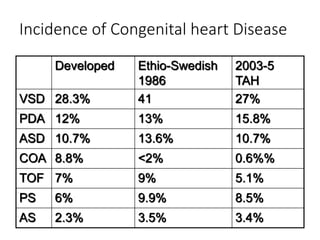
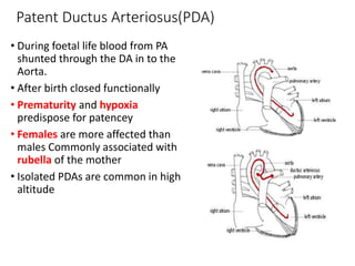
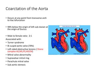
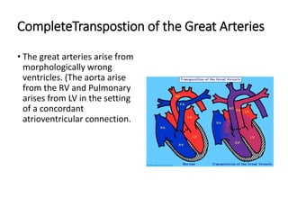
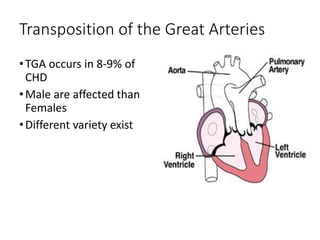
CONGENITAL HEART DISEASES
ETSEGEGENET GEDLU
Department of Pediatrics and Child Health
AAU Medical Faculty
2009
Objective
Revise the fetal circulation in short
Classification of congenital heart disease
Discuses the common acyanotic and cyanotic congenital heart diseases and their
clinical presentation, Investigation and principles of management.
Fetal circulation
The right and left ventricles exist in a parallel circuit.
In the fetus gas and metabolite exchange provided by the placenta
Parallel circuit maintained
 Foramen ovale
 Ductus arteriosus
 Ductus venosus
Fetal circulation
placenta
 largest amount of combined ventricular output
 Has lowest vascular resistance in the fetus
 Placenta empties into IVC therefore IVC oxygen saturation
greater than SVC saturation
 Highest PO2 (32 mmHg )is found in the umbilical vein
Ductus Venosus
 Oxygenated blood running from the placenta flow to the fetus
through umbilical vein and 50 % bypass the liver enters IVC
via DV
Fetal circulation
Foramen Ovale
About one third of IVC blood directed in to LA through
the foramen ovale and to LV ejected to the ascending
aorta
The result is higher saturated blood being delivered to
the brain and coronary arteries
Most SVC blood drains into RV through tricuspid valve
From the RV to the pulmonary artery and because of
pulmonary arterial circulation constricted only 10% of the
RV out put enters the lungs .
Fetal circulation
Ductus arteriosus
Less oxygenated blood shunted through the ductus
arteriosus away from the lungs and into the descending
aorta to perfuse the lower part of the body.
The blood return to the placenta via the to umbilical
arteries
This low oxygen content increases the efficiency of
oxygen exchange in the placenta
Transitional circulation
1. Interruption of Umbilical cord
 Removal of the low resistance placental circulation result in
an Increased systemic vascular resistance
 lack of blood flow through the placenta leads to closure of
Ductus venosus
2. Expansion of lungs
 Mechanical expansion of the lungs and increased arterial P02
result in a rapid decrease in pulmonary vascular resistance
 The output from RV flows entirely to the pulmonary
circulation
 The increased blood volume from the pulmonary circulation
increased the LA volume and pressure sufficiently to close
the foramen ovale
Transitional circulation
3. PDA closure
The ductus flow become left to right and later the ductus will
obliterated and become ligamentum arteriosus
Functional closure at 10 to 15 hours after birth
Increased systemic O2 saturation is strongest stimulus or
constriction of ductal smooth muscle
Gestational age: decreased responsiveness of ductal smooth
muscle to oxygen in premature infant
Decreasing prostaglandin levels leads to closure
Transitional circulation
The right ventricle is coupled with low
resistance pulmonary circulation and its wall
thickness and mass decreased
The left ventricle coupled to high resistance
systemic circulation and its wall thickness and
mass increased and deliver the entire systemic
cardiac out put.
Congenital Heart disease
Introduction:
Def: Structural or functional heart disease that present at birth.
It is not static ,there is always a continuous anatomical or physiological
change
• The incidence is higher in abortus and still births
• Estimate in live birth range from 4-10.2 per 1000live birth.
Cont….
• The incidence of specific type of CHD varies from one country to
another
• Specific aetiology only known 10%
• 8% genetic
• 2% environmental (rubella, foetal-alcohol syndrome
• 90% Multifactorial inheritance
Incidence of Congenital heart Disease
Developed Ethio-Swedish
1986
2003-5
TAH
VSD 28.3% 41 27%
PDA 12% 13% 15.8%
ASD 10.7% 13.6% 10.7%
COA 8.8% <2% 0.6%%
TOF 7% 9% 5.1%
PS 6% 9.9% 8.5%
AS 2.3% 3.5% 3.4%
cont.….
• The risk of recurrence in siblings varies from 1-4%
• Third Pregnancy 20-30%
• Parents with CHD 4-6%
• Varies with type of inheritance
• Except PDA and ASD males are more affected than females.
Classification of Congenital Heart
Diseases:
Common Acyanotic Heart Diseases:
• Ventricular septal defect
• Atrial septal defect
• Patent ductus arteriosus
• Coarctation of the aorta
• Pulmonary stenosis
• Aortic stenosis
Classification of CHD
Common cyanotic Heart Diseases:
• Tetralogy of Fallot
• Transposition of the Great Arteries
• Truncus Arteriosus
• Severe Pulmonary stenosis
• Pulmonary Atresia
• Tricuspid Atresia
• Hypoplastic heart diseases
Clinical manifestation of CHD
CHD suspected in any child with:
• Feeding difficulty
•Recurrent attack of respiratory tract infection
•Growth failure
•Cyanosis unresponsive to 100% oxygen-failed
hyperoxia test(pO2<100)
•Tachycardia
•Respiratory distress
•Rhythm disturbance
•Murmur
Managementt of CHD
General principle
• 1) Correction of underlying defect (timing depend on the type and severity).
• 2) Treatment of Congestive Heart Failure
Diuretics
Inotropic support
After load reduction
• 3)Prevention and treatment of complication
Pulmonary hpt : early surgical correction
Infective endocarditis: Administration of antibiotic chemoprophylaxis as indicated.
Non infective thromboembolism : prevent polycythemia
 polycythemia :partial exchange transfusion
• Counselling of parents on the risk of recurrence
Ventricular Septal Defect
•Most common CHD
•Both sexes are equally
affected
•Incidence of 1/3000
•Can be single or multiple
•Can be associated with
other congenital heart
diseases
VSD cont.…
Types of VSD
• 70% membranous close to pulmonary valve and Pulmonary artery
• 20% muscular
• 5% Aortic valve (sub aortic)
• 5% near junction of mitral and tricuspid valve (A-V canal defect)
Clinical manifestations
I) Asymptomatic:
• Small VSD , trivial shunt ,the pulmonary pressure is normal
• Loud harsh Holosystolic murmur at LLSB, with thrill
• X-ray is normal
• EKG normal
VSD CONT….
II ) Large defects:
• Excessive pulmonary blood flow lead to pulmonary hypertension
• Dyspnea, feeding difficulty
• Poor growth
• Profuse perspiration
• Recurrent pulmonary infection
Physical Examination:
• Prominence of the precordium
• Palpable parasternal lift
• Apical trust with systolic thrill
• Holosystolic murmur at LLSB less harsh and more blowing
• Diastolic murmur at the apex
• Increased P2 indicate pulmonary hpt.
VSD cont….
Severity depend on :
•The size of the defect
•Level of pulmonary resistance to systemic
resistance.
•Defects < 0.5 restrictive
> 1cm sq non restrictive
•Majority closes spontaneously
•Large defects lead to CHF at early age
Chest X-Ray (VSD)
•Cardiomegally
(LA, LV,RV,PA)
• Increased vascular
marking
Diagnostics cont…
ECG:
Bi-ventricular
hypertrophy
P wave peaked and
notched
Echocardiography (VSD)
•Position and size of VSD
• Chamber size
• Pressure gradient
across the defect
• Direction of shunt
Cardiac catheterization
Angiography :
CLINICAL COURSE:
Small sized defects
• are closed spontaneously in the first year of life
• The risk of endocarditis is independent of the size
Moderate to large defects:
• decreased in size but not closed
• Heart failure and growth failure is common at the early age
• Risk of pulmonary hypertension lead to pulmonary vascular
diseases.
• Eisenmengers syndrome due to reversal of shunt which
presented with absence of thrill and cyanosis, decreased heart
size.
Treatment
Small size
• Reassurance
• No surgical treatment
• Maintain integrity of primary and permanent teeth
• Give anti- infective endocarditis prophylaxis
 Antibiotic prophylaxis before
 dental visit
 Tonsillectomy
 instrumentation of GUT,GIT
Treatment cont….
Large VSD:
• Control CHF
•Prevent development of Pulmonary vascular
disease.
 Surgical closure in the first year of life (6M-12M).
Palliative : pulmonary banding if surgery is not
possible for the time being.
Device closure of the VSD with Amplatzer device
Umbrella)
Patent Ductus Arteriosus(PDA)
• During foetal life blood from PA
shunted through the DA in to the
Aorta.
• After birth closed functionally
• Prematurity and hypoxia
predispose for patencey
• Females are more affected than
males Commonly associated with
rubella of the mother
• Isolated PDAs are common in high
altitude
Pathophysiology
Blood flow from the aorta to the pulmonary.
Extent of the shunt depend on
• size of the ductus
• ratio of pulmonary and systemic vascular resistance
Clinical manifestation
Depend on the :
•Size of the defect and direction of flow
•Small defects no symptom
•Large defect result in Large left to right shunt
CHF
Growth Failure
Repeated ARI
Reversal of shunt ,(Eisenmengers ) result in dyspnea and
cyanosis
Physical EXAMINATION
•Bounding pulse
•Wide pulse pressure
•Heave , thrill in the 2nd ics
•Continuous machinery murmur 2nd ics
•EKG: bi-ventricular hyperthrophy
•X-ray: prominent PA, increases PA marking enlarged
chambers(LA,LV),
•ECHO: size of the PDA, direction of flow, chamber size
•Catheterization: a step up oxygen saturation, PDA
anatomy in angiography
Clinical course:
•Small defects : few or no cardiac symptoms
•Large defects:
•CHF
•Infective endocarditis
•Systemic emboli
•Calcification of the ductus
•Non infective thrombosis with embolization
•Paradoxical emboli
•Eisenmengers syndrome if left untreated.
Treatment
Medical therapy:
• Congestive heart failure treatment
• infective endocarditis prophylaxis
• Surgical closure of the PDA (banding)
• Closure of the PDA coil embolization
Coarctation of the Aorta
• Occurs at any point from transverse arch
to iliac bifurcation
• 98% below the origin of left sub clavian at
the origin of Ductus
• Male to female ratio: 2:1
Associated with:
• Turner syndrome
• Bi cuspid aortic valve (70%)
• Left sided obstructive lesions ( Shone
complex-AS,MS,PS,HOCM)
• Mitral valve abnormality
• Supravalvar mitral ring
• Parachute mitral valve
• Sub aortic stenosis
Pathophysiology
• Collaterals develop to by pass the obstruction.
• Hypertension of the aortic branch proximal to coarctation
• In Pre ductal type the RV blood ejected through the ductus to supply the
descending aorta lead to differential cyanosis.
Clinical Manifestations
History
• Usually asymptomatic
• Older children:
Headaches
Epistaxis
Claudication, cold feet
Physical Examination:
• Weak or absent femoral pulses
• Increased B/P in the upper extremities
• B/P difference between upper and lower extremities
• Radio Femoral pulse delay ( Collaterals)
• A2 is loud , systolic murmur 3rd and 4th ULSB
Cont….
Chest x_ray:
•Dilated descending
Aorta, enlarged LV.
•Rib notching-due to
erosion of ribs by
collaterals
ECG: normal in childhood,
later LV hypertrophy.
ECHO
: Measure the stenotic
area
gradient
Treatment
•Neonatal: closure of the ductus lead to hypo perfussion
and acidosis, thus give infusion of prostaglandin(PGE1-
Alprostadil) to reopen the ductus, after stabilization
surgical treatment.
•Older children with CHF and no hypertension medical
treatment followed by surgery or angioplasty.
•Re -stenosis balloon angioplasty is safe.
Incidence of CHD
Developed Ethio-Swedish
1986
2003-5
TAH
VSD 28.3% 41 27%
PDA 12% 13% 15.8%
ASD 10.7% 13.6% 10.7%
COA 8.8% <2% 0.6%%
TOF 7% 9% 5.1%
PS 6% 9.9% 8.5%
AS 2.3% 3.5% 3.4%
Tetralogy of Fallot
•Common cyanotic
congenital cardiac anomaly
•Four anatomical
components of TOF:
•VSD
•Overriding Aorta
•Right ventricular outflow
obstruction
•Right ventricular
hypertrophy
Pathophysiology:
•Severity directly proportional to the degree of RVOT obstruction.
•Change in pulmonary and systemic vascular resistance and the degree of
RVOT obstruction affect degree of R-L shunt.
•Infundibular stenosis is progressive.
Clinical Manifestations:
Variable depend on RVOT obstruction
 pink to cyanosis
CHF is not a usual manifestation of TOF
Squatting
Dyspnoea on exertion
Hypoxic spells
Growth failure
Physical Examination:
Cyanosis variable
Clubbing
Usually S2 is single,
Quite precordium
Thrill at the pulmonary area(-+)
Systolic ejection murmur at the pulmonary area(LUSB)
Clinical pictures cont….
Electrocardiography (TOF)
•Right axis deviation
•Right atrial and right
ventricular enlargement
Chest x ray (TOF)
• Normal sized, boot
shaped heart.
•Reduced pulmonary
vascular marking
(oligemic depend on the
degree of RVOT
obstruction.
Echocardiography (TOF)
• Location and size of the VSD
• The aortic override
• The degree of RVOT obstruction
• The size of pulmonary valve
annulus
• Look for additional pulmonary
artery branch stenosis.
• Look for other associated
anomalies
Right aortic arch
Coronary arteries anatomic
variations
Cardiac catheterization and
Angiography
Hemodynanmic
 Anatomical information
Therapeutic
Complications:
Cardiovascular accidents : occurs in 4-5% of
cases is due to cerebral embolism.
Brain abscess: rare in the first two years of life
may be due to small cerebral infract which is
super infected due to bacteraemia.
Infective endocarditis
Polycythemia
Management
Medical management:
A) Neonatal period if pulmonary flow is dependent of DA, give
prostaglandin to prevent ductal closure and followed by palliative
shunt (B-T Shunt)
B)Recognition and treatment of hyper cyanotic spell.
Knee chest positioning of the patient
Administration of oxygen
Volume expansion
Correction of acidosis
Sedation with morphine
Propranolol
C) Treat infective endocarditis
D) Anaemia should be corrected
E) Severe polycythemia correction with phlebotomy should be
done.
Management
Surgical Management:
A) Palliative: Modified Blalock-Taussig shunt between pulmonary
artery branch and subclavian artery
B) Corrective surgery : closing of the VSD and reliving all possible
source of RVOT obstruction.
CompleteTranspostion of the Great Arteries
• The great arteries arise from
morphologically wrong
ventricles. (The aorta arise
from the RV and Pulmonary
arises from LV in the setting
of a concordant
atrioventricular connection.
Transposition of the Great Arteries
•TGA occurs in 8-9% of
CHD
•Male are affected than
Females
•Different variety exist
Hemodynamics
The RV pressure is systemic
The blood returning from the lung pass to the lung via PA
Systemic venous return passes back to the systemic
circulation via the aorta.
Both ventricles are volume overloaded.
The right ventricle also pressure overloads and result in CHF
Survival depend on associated Large ASD, VSD,PDA.
Clinical features:
Cyanosis detected 87% of the neonates
immediately ; rest later at the age of 1 to 3
month and is progressive.
Clubbing
Squatting only 20% of patients
Dyspnoea
Cardiac failure
Cough
Physical findings
1)TGA with no VSD
Cyanosis intense
Precordial lift
Loud first sound
Splitted S2
Murmur may not be heard
2)TGA with large VSD
Cyanosis less intense
Cardio -Respiratory distress, retraction
Oedema
Hepatomegally
Left and right ventricular pulsations are prominent
Systolic murmur at the LLSB
Diastolic rumble at the apex.
Radiology (TGA)
•Increased pulmonary
vascular marking
•Large heart with egg - on
side appearance.
Electrocardiography (TGA)
TGA with intact ventricular septum:
•P wave tall
•P upright T wave in lead V1 and V3R
•Right ventricular hypertrophy
•Right axis deviation
TGA with VSD:
•Bi atrial enlargement
•Bi Ventricular hypertrophy
•Right axis deviation
Echocardiography(TGA)
Shows the origion of :
pulmonary artery from
the LV
Aorta from RV
•Associated lesions like
VSD and ASD be
identified.
•Coronary artery anatomy
Prognosis
• Patient die of anoxia and or CHF in the first 6 month of life if there is
little communication.
• TGA with VSD and PS favourable
Management
Medical management:
•Prostaglandin to keep the DA open until palliation
done
•Give oxygen
•Treat metabolic acidosis with bicarbonate.
Surgical:
•Palliative balloon septostomy creating /increasing
ASD Rashkind procedure.
•Large VSD do pulmonary banding
•Intra atrial switch (Mustard procedure)
•Arterial Switch (Jaten Procedure)
1)Congenital HD 2009.ppt

1)Congenital HD 2009.ppt

  • 1.
    CONGENITAL HEART DISEASES ETSEGEGENETGEDLU Department of Pediatrics and Child Health AAU Medical Faculty 2009
  • 2.
    Objective Revise the fetalcirculation in short Classification of congenital heart disease Discuses the common acyanotic and cyanotic congenital heart diseases and their clinical presentation, Investigation and principles of management.
  • 3.
    Fetal circulation The rightand left ventricles exist in a parallel circuit. In the fetus gas and metabolite exchange provided by the placenta Parallel circuit maintained  Foramen ovale  Ductus arteriosus  Ductus venosus
  • 4.
    Fetal circulation placenta  largestamount of combined ventricular output  Has lowest vascular resistance in the fetus  Placenta empties into IVC therefore IVC oxygen saturation greater than SVC saturation  Highest PO2 (32 mmHg )is found in the umbilical vein Ductus Venosus  Oxygenated blood running from the placenta flow to the fetus through umbilical vein and 50 % bypass the liver enters IVC via DV
  • 5.
    Fetal circulation Foramen Ovale Aboutone third of IVC blood directed in to LA through the foramen ovale and to LV ejected to the ascending aorta The result is higher saturated blood being delivered to the brain and coronary arteries Most SVC blood drains into RV through tricuspid valve From the RV to the pulmonary artery and because of pulmonary arterial circulation constricted only 10% of the RV out put enters the lungs .
  • 6.
    Fetal circulation Ductus arteriosus Lessoxygenated blood shunted through the ductus arteriosus away from the lungs and into the descending aorta to perfuse the lower part of the body. The blood return to the placenta via the to umbilical arteries This low oxygen content increases the efficiency of oxygen exchange in the placenta
  • 7.
    Transitional circulation 1. Interruptionof Umbilical cord  Removal of the low resistance placental circulation result in an Increased systemic vascular resistance  lack of blood flow through the placenta leads to closure of Ductus venosus 2. Expansion of lungs  Mechanical expansion of the lungs and increased arterial P02 result in a rapid decrease in pulmonary vascular resistance  The output from RV flows entirely to the pulmonary circulation  The increased blood volume from the pulmonary circulation increased the LA volume and pressure sufficiently to close the foramen ovale
  • 8.
    Transitional circulation 3. PDAclosure The ductus flow become left to right and later the ductus will obliterated and become ligamentum arteriosus Functional closure at 10 to 15 hours after birth Increased systemic O2 saturation is strongest stimulus or constriction of ductal smooth muscle Gestational age: decreased responsiveness of ductal smooth muscle to oxygen in premature infant Decreasing prostaglandin levels leads to closure
  • 9.
    Transitional circulation The rightventricle is coupled with low resistance pulmonary circulation and its wall thickness and mass decreased The left ventricle coupled to high resistance systemic circulation and its wall thickness and mass increased and deliver the entire systemic cardiac out put.
  • 10.
    Congenital Heart disease Introduction: Def:Structural or functional heart disease that present at birth. It is not static ,there is always a continuous anatomical or physiological change • The incidence is higher in abortus and still births • Estimate in live birth range from 4-10.2 per 1000live birth.
  • 11.
    Cont…. • The incidenceof specific type of CHD varies from one country to another • Specific aetiology only known 10% • 8% genetic • 2% environmental (rubella, foetal-alcohol syndrome • 90% Multifactorial inheritance
  • 12.
    Incidence of Congenitalheart Disease Developed Ethio-Swedish 1986 2003-5 TAH VSD 28.3% 41 27% PDA 12% 13% 15.8% ASD 10.7% 13.6% 10.7% COA 8.8% <2% 0.6%% TOF 7% 9% 5.1% PS 6% 9.9% 8.5% AS 2.3% 3.5% 3.4%
  • 13.
    cont.…. • The riskof recurrence in siblings varies from 1-4% • Third Pregnancy 20-30% • Parents with CHD 4-6% • Varies with type of inheritance • Except PDA and ASD males are more affected than females.
  • 14.
    Classification of CongenitalHeart Diseases: Common Acyanotic Heart Diseases: • Ventricular septal defect • Atrial septal defect • Patent ductus arteriosus • Coarctation of the aorta • Pulmonary stenosis • Aortic stenosis
  • 15.
    Classification of CHD Commoncyanotic Heart Diseases: • Tetralogy of Fallot • Transposition of the Great Arteries • Truncus Arteriosus • Severe Pulmonary stenosis • Pulmonary Atresia • Tricuspid Atresia • Hypoplastic heart diseases
  • 16.
    Clinical manifestation ofCHD CHD suspected in any child with: • Feeding difficulty •Recurrent attack of respiratory tract infection •Growth failure •Cyanosis unresponsive to 100% oxygen-failed hyperoxia test(pO2<100) •Tachycardia •Respiratory distress •Rhythm disturbance •Murmur
  • 17.
    Managementt of CHD Generalprinciple • 1) Correction of underlying defect (timing depend on the type and severity). • 2) Treatment of Congestive Heart Failure Diuretics Inotropic support After load reduction • 3)Prevention and treatment of complication Pulmonary hpt : early surgical correction Infective endocarditis: Administration of antibiotic chemoprophylaxis as indicated. Non infective thromboembolism : prevent polycythemia  polycythemia :partial exchange transfusion • Counselling of parents on the risk of recurrence
  • 18.
    Ventricular Septal Defect •Mostcommon CHD •Both sexes are equally affected •Incidence of 1/3000 •Can be single or multiple •Can be associated with other congenital heart diseases
  • 19.
    VSD cont.… Types ofVSD • 70% membranous close to pulmonary valve and Pulmonary artery • 20% muscular • 5% Aortic valve (sub aortic) • 5% near junction of mitral and tricuspid valve (A-V canal defect)
  • 20.
    Clinical manifestations I) Asymptomatic: •Small VSD , trivial shunt ,the pulmonary pressure is normal • Loud harsh Holosystolic murmur at LLSB, with thrill • X-ray is normal • EKG normal
  • 21.
    VSD CONT…. II )Large defects: • Excessive pulmonary blood flow lead to pulmonary hypertension • Dyspnea, feeding difficulty • Poor growth • Profuse perspiration • Recurrent pulmonary infection
  • 22.
    Physical Examination: • Prominenceof the precordium • Palpable parasternal lift • Apical trust with systolic thrill • Holosystolic murmur at LLSB less harsh and more blowing • Diastolic murmur at the apex • Increased P2 indicate pulmonary hpt.
  • 23.
    VSD cont…. Severity dependon : •The size of the defect •Level of pulmonary resistance to systemic resistance. •Defects < 0.5 restrictive > 1cm sq non restrictive •Majority closes spontaneously •Large defects lead to CHF at early age
  • 24.
    Chest X-Ray (VSD) •Cardiomegally (LA,LV,RV,PA) • Increased vascular marking
  • 25.
  • 26.
    Echocardiography (VSD) •Position andsize of VSD • Chamber size • Pressure gradient across the defect • Direction of shunt
  • 27.
  • 28.
    CLINICAL COURSE: Small sizeddefects • are closed spontaneously in the first year of life • The risk of endocarditis is independent of the size Moderate to large defects: • decreased in size but not closed • Heart failure and growth failure is common at the early age • Risk of pulmonary hypertension lead to pulmonary vascular diseases. • Eisenmengers syndrome due to reversal of shunt which presented with absence of thrill and cyanosis, decreased heart size.
  • 29.
    Treatment Small size • Reassurance •No surgical treatment • Maintain integrity of primary and permanent teeth • Give anti- infective endocarditis prophylaxis  Antibiotic prophylaxis before  dental visit  Tonsillectomy  instrumentation of GUT,GIT
  • 30.
    Treatment cont…. Large VSD: •Control CHF •Prevent development of Pulmonary vascular disease.  Surgical closure in the first year of life (6M-12M). Palliative : pulmonary banding if surgery is not possible for the time being. Device closure of the VSD with Amplatzer device Umbrella)
  • 31.
    Patent Ductus Arteriosus(PDA) •During foetal life blood from PA shunted through the DA in to the Aorta. • After birth closed functionally • Prematurity and hypoxia predispose for patencey • Females are more affected than males Commonly associated with rubella of the mother • Isolated PDAs are common in high altitude
  • 32.
    Pathophysiology Blood flow fromthe aorta to the pulmonary. Extent of the shunt depend on • size of the ductus • ratio of pulmonary and systemic vascular resistance
  • 33.
    Clinical manifestation Depend onthe : •Size of the defect and direction of flow •Small defects no symptom •Large defect result in Large left to right shunt CHF Growth Failure Repeated ARI Reversal of shunt ,(Eisenmengers ) result in dyspnea and cyanosis
  • 34.
    Physical EXAMINATION •Bounding pulse •Widepulse pressure •Heave , thrill in the 2nd ics •Continuous machinery murmur 2nd ics •EKG: bi-ventricular hyperthrophy •X-ray: prominent PA, increases PA marking enlarged chambers(LA,LV), •ECHO: size of the PDA, direction of flow, chamber size •Catheterization: a step up oxygen saturation, PDA anatomy in angiography
  • 35.
    Clinical course: •Small defects: few or no cardiac symptoms •Large defects: •CHF •Infective endocarditis •Systemic emboli •Calcification of the ductus •Non infective thrombosis with embolization •Paradoxical emboli •Eisenmengers syndrome if left untreated.
  • 36.
    Treatment Medical therapy: • Congestiveheart failure treatment • infective endocarditis prophylaxis • Surgical closure of the PDA (banding) • Closure of the PDA coil embolization
  • 37.
    Coarctation of theAorta • Occurs at any point from transverse arch to iliac bifurcation • 98% below the origin of left sub clavian at the origin of Ductus • Male to female ratio: 2:1 Associated with: • Turner syndrome • Bi cuspid aortic valve (70%) • Left sided obstructive lesions ( Shone complex-AS,MS,PS,HOCM) • Mitral valve abnormality • Supravalvar mitral ring • Parachute mitral valve • Sub aortic stenosis
  • 38.
    Pathophysiology • Collaterals developto by pass the obstruction. • Hypertension of the aortic branch proximal to coarctation • In Pre ductal type the RV blood ejected through the ductus to supply the descending aorta lead to differential cyanosis.
  • 39.
    Clinical Manifestations History • Usuallyasymptomatic • Older children: Headaches Epistaxis Claudication, cold feet
  • 40.
    Physical Examination: • Weakor absent femoral pulses • Increased B/P in the upper extremities • B/P difference between upper and lower extremities • Radio Femoral pulse delay ( Collaterals) • A2 is loud , systolic murmur 3rd and 4th ULSB
  • 41.
    Cont…. Chest x_ray: •Dilated descending Aorta,enlarged LV. •Rib notching-due to erosion of ribs by collaterals ECG: normal in childhood, later LV hypertrophy.
  • 42.
    ECHO : Measure thestenotic area gradient
  • 43.
    Treatment •Neonatal: closure ofthe ductus lead to hypo perfussion and acidosis, thus give infusion of prostaglandin(PGE1- Alprostadil) to reopen the ductus, after stabilization surgical treatment. •Older children with CHF and no hypertension medical treatment followed by surgery or angioplasty. •Re -stenosis balloon angioplasty is safe.
  • 45.
    Incidence of CHD DevelopedEthio-Swedish 1986 2003-5 TAH VSD 28.3% 41 27% PDA 12% 13% 15.8% ASD 10.7% 13.6% 10.7% COA 8.8% <2% 0.6%% TOF 7% 9% 5.1% PS 6% 9.9% 8.5% AS 2.3% 3.5% 3.4%
  • 46.
    Tetralogy of Fallot •Commoncyanotic congenital cardiac anomaly •Four anatomical components of TOF: •VSD •Overriding Aorta •Right ventricular outflow obstruction •Right ventricular hypertrophy
  • 47.
    Pathophysiology: •Severity directly proportionalto the degree of RVOT obstruction. •Change in pulmonary and systemic vascular resistance and the degree of RVOT obstruction affect degree of R-L shunt. •Infundibular stenosis is progressive.
  • 48.
    Clinical Manifestations: Variable dependon RVOT obstruction  pink to cyanosis CHF is not a usual manifestation of TOF Squatting Dyspnoea on exertion Hypoxic spells Growth failure
  • 49.
    Physical Examination: Cyanosis variable Clubbing UsuallyS2 is single, Quite precordium Thrill at the pulmonary area(-+) Systolic ejection murmur at the pulmonary area(LUSB)
  • 50.
  • 51.
    Electrocardiography (TOF) •Right axisdeviation •Right atrial and right ventricular enlargement
  • 52.
    Chest x ray(TOF) • Normal sized, boot shaped heart. •Reduced pulmonary vascular marking (oligemic depend on the degree of RVOT obstruction.
  • 53.
    Echocardiography (TOF) • Locationand size of the VSD • The aortic override • The degree of RVOT obstruction • The size of pulmonary valve annulus • Look for additional pulmonary artery branch stenosis. • Look for other associated anomalies Right aortic arch Coronary arteries anatomic variations
  • 54.
  • 55.
    Complications: Cardiovascular accidents :occurs in 4-5% of cases is due to cerebral embolism. Brain abscess: rare in the first two years of life may be due to small cerebral infract which is super infected due to bacteraemia. Infective endocarditis Polycythemia
  • 56.
    Management Medical management: A) Neonatalperiod if pulmonary flow is dependent of DA, give prostaglandin to prevent ductal closure and followed by palliative shunt (B-T Shunt) B)Recognition and treatment of hyper cyanotic spell. Knee chest positioning of the patient Administration of oxygen Volume expansion Correction of acidosis Sedation with morphine Propranolol C) Treat infective endocarditis D) Anaemia should be corrected E) Severe polycythemia correction with phlebotomy should be done.
  • 57.
    Management Surgical Management: A) Palliative:Modified Blalock-Taussig shunt between pulmonary artery branch and subclavian artery B) Corrective surgery : closing of the VSD and reliving all possible source of RVOT obstruction.
  • 59.
    CompleteTranspostion of theGreat Arteries • The great arteries arise from morphologically wrong ventricles. (The aorta arise from the RV and Pulmonary arises from LV in the setting of a concordant atrioventricular connection.
  • 60.
    Transposition of theGreat Arteries •TGA occurs in 8-9% of CHD •Male are affected than Females •Different variety exist
  • 61.
    Hemodynamics The RV pressureis systemic The blood returning from the lung pass to the lung via PA Systemic venous return passes back to the systemic circulation via the aorta. Both ventricles are volume overloaded. The right ventricle also pressure overloads and result in CHF Survival depend on associated Large ASD, VSD,PDA.
  • 62.
    Clinical features: Cyanosis detected87% of the neonates immediately ; rest later at the age of 1 to 3 month and is progressive. Clubbing Squatting only 20% of patients Dyspnoea Cardiac failure Cough
  • 63.
    Physical findings 1)TGA withno VSD Cyanosis intense Precordial lift Loud first sound Splitted S2 Murmur may not be heard 2)TGA with large VSD Cyanosis less intense Cardio -Respiratory distress, retraction Oedema Hepatomegally Left and right ventricular pulsations are prominent Systolic murmur at the LLSB Diastolic rumble at the apex.
  • 64.
    Radiology (TGA) •Increased pulmonary vascularmarking •Large heart with egg - on side appearance.
  • 65.
    Electrocardiography (TGA) TGA withintact ventricular septum: •P wave tall •P upright T wave in lead V1 and V3R •Right ventricular hypertrophy •Right axis deviation TGA with VSD: •Bi atrial enlargement •Bi Ventricular hypertrophy •Right axis deviation
  • 66.
    Echocardiography(TGA) Shows the origionof : pulmonary artery from the LV Aorta from RV •Associated lesions like VSD and ASD be identified. •Coronary artery anatomy
  • 68.
    Prognosis • Patient dieof anoxia and or CHF in the first 6 month of life if there is little communication. • TGA with VSD and PS favourable
  • 69.
    Management Medical management: •Prostaglandin tokeep the DA open until palliation done •Give oxygen •Treat metabolic acidosis with bicarbonate. Surgical: •Palliative balloon septostomy creating /increasing ASD Rashkind procedure. •Large VSD do pulmonary banding •Intra atrial switch (Mustard procedure) •Arterial Switch (Jaten Procedure)