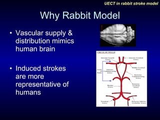
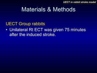
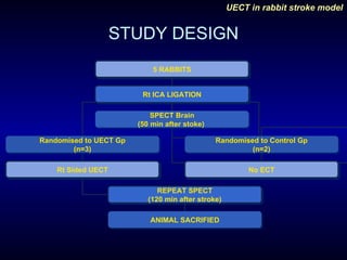
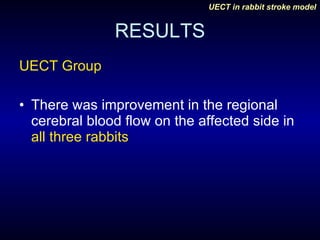
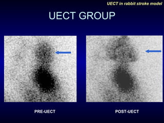
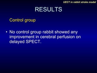
Unilateral Electroconvulsive Therapy in Rabbit Stroke Model: Evaluation Using SPECT  Deepak Agrawal, NK Gowda*, R Sagar**, PK Yadav***, Departments of Neurosurgery, Nuclear medicine*, Psychiatry** & Experimental Animal Facility*** All India Institute of Medical Sciences, New Delhi, India-110029
Background rCBF Increases following induced seizure Sestoft D, Meden P, Hemmingsen R, et al: Disparity in regional cerebral blood flow during electrically induced seizure. Acta Psychiatr Scand 1993;88:140-143.
Hypothesis This increase in rCBF may have a positive effect on the infarct size following stroke
AIMS & OBJECTIVES To evaluate the efficacy of unilateral electroconvulsive therapy (UECT) for cerebral protection following induced stroke in rabbits
Why Rabbit Model Vascular supply & distribution mimics human brain Induced strokes are more representative of humans
Why UECT? ECT has been previously shown to improve rCBF UECT causes marked improvement in CBF in the ipsilateral hemisphere B/L ECT improves perfusion in striatum Devanand DP, Dwork AJ, Hutchinson ER, Bolwig TG, Sackeim HA: Does ECT alter brain structure? Am J Psychiatry 1994;151:957-970 .
Materials & Methods Prospective randomized controlled study  Randomization: computer generated random number method Approval was taken from the institutional ethics committee for animal experiments.
Materials & Methods RABBITS New Zealand strain Weight- 2-3 kg Both Male and female animals were used
Materials & Methods Anesthesia Pre-anesthetized by 10% ketamine (20 mg/kg) given IM Continuous infusion of midazolam 0.2mg/kg and ketamine (0.2 ml/ kg/hour). 2ml of 0.5% lignocaine was infiltrated S/C
Materials & methods Operative Procedure Femoral cut down for IV access 2cm long incision along ant border of SCM Using blunt dissection CCA  exposed ICA identified & ligated with silk. Wound closed
Materials & Methods 2 mCi of Tc-99m HMPAO (Amersham, England) was then injected into the femoral vein, 10 minutes after induced stroke (carotid ligature).
Materials & Methods Animal  transported to nuclear medicine on a modified trolley, on ventimask and IV. Planar and SPECT images of the brain were taken 50 minutes after stroke.
Materials & Methods UECT Group rabbits Unilateral Rt ECT was given 75 minutes after the induced stroke.
Materials & Methods ECT Procedure  Modified Thymatron (TM) DGx, (Somatics LLC, Lake Bluff, IL, USA) ECT machine A brief-pulse square wave (0.5ms) & constant current of 0.9 A. Stimulus kept at 10% with impedence (resistance) of 800 ohms  Induced seizure was considered adequate when its duration > 20 sec.
Materials & Methods CONTROL RABBITS NO UECT
Materials & Methods Both groups  received 6 mCi of Tc-99m HMPAO after 90 minutes of stroke. A second set of planar and SPECT images were taken 150 minutes after stroke.
Materials & Methods Relative uptake of the radiotracer in both groups of rabbits was displayed on computer monitor and was analyzed using a graded grey scale.  Cerebellum was used as reference site (100% maximum value) for all comparisons
Materials & Methods Rabbits sacrificed using high dose of pancuronium and thiopentone given IV.
STUDY DESIGN 5 RABBITS Rt ICA LIGATION SPECT Brain (50 min after stoke)  Randomised to UECT Gp (n=3) Randomised to Control Gp (n=2) Rt Sided UECT No ECT REPEAT SPECT (120 min after stroke) ANIMAL SACRIFIED
RESULTS Study terminated prematurely as Ethics committee withdrew approval after 5 rabbits. UECT Group – 3 Rabbits Control Group- 2 Rabbits
RESULTS The right cerebrum showed decrease uptake of radiotracer compared to left cerebrum in  all five rabbits  confirming induction of ischemia.
RESULTS UECT Group There was improvement in the regional cerebral blood flow on the affected side in  all three rabbits
UECT GROUP PRE-UECT POST-UECT
RESULTS Control group No control group rabbit showed any improvement in cerebral perfusion on delayed SPECT.
LIMITATIONS No of rabbits studied was small Embolic stroke model would have been preferred to carotid ligation PET would have been better in imaging the induced stroke
CONCLUSIONS All rabbits receiving UECT had improvement in cerebral perfusion following induced stroke, vis-à-vis the control group.
CONCLUSIONS This study shows that UECT decreases the infarct size and increases rCBF in rabbit stroke model. Further studies using a larger sample size will be required for confirmation.
Thank You

Unilateral electroconvulsive therapy_in_rabbit_stroke_model1

  • 1.
    Unilateral Electroconvulsive Therapyin Rabbit Stroke Model: Evaluation Using SPECT Deepak Agrawal, NK Gowda*, R Sagar**, PK Yadav***, Departments of Neurosurgery, Nuclear medicine*, Psychiatry** & Experimental Animal Facility*** All India Institute of Medical Sciences, New Delhi, India-110029
  • 2.
    Background rCBF Increasesfollowing induced seizure Sestoft D, Meden P, Hemmingsen R, et al: Disparity in regional cerebral blood flow during electrically induced seizure. Acta Psychiatr Scand 1993;88:140-143.
  • 3.
    Hypothesis This increasein rCBF may have a positive effect on the infarct size following stroke
  • 4.
    AIMS & OBJECTIVESTo evaluate the efficacy of unilateral electroconvulsive therapy (UECT) for cerebral protection following induced stroke in rabbits
  • 5.
    Why Rabbit ModelVascular supply & distribution mimics human brain Induced strokes are more representative of humans
  • 6.
    Why UECT? ECThas been previously shown to improve rCBF UECT causes marked improvement in CBF in the ipsilateral hemisphere B/L ECT improves perfusion in striatum Devanand DP, Dwork AJ, Hutchinson ER, Bolwig TG, Sackeim HA: Does ECT alter brain structure? Am J Psychiatry 1994;151:957-970 .
  • 7.
    Materials & MethodsProspective randomized controlled study Randomization: computer generated random number method Approval was taken from the institutional ethics committee for animal experiments.
  • 8.
    Materials & MethodsRABBITS New Zealand strain Weight- 2-3 kg Both Male and female animals were used
  • 9.
    Materials & MethodsAnesthesia Pre-anesthetized by 10% ketamine (20 mg/kg) given IM Continuous infusion of midazolam 0.2mg/kg and ketamine (0.2 ml/ kg/hour). 2ml of 0.5% lignocaine was infiltrated S/C
  • 10.
    Materials & methodsOperative Procedure Femoral cut down for IV access 2cm long incision along ant border of SCM Using blunt dissection CCA exposed ICA identified & ligated with silk. Wound closed
  • 11.
    Materials & Methods2 mCi of Tc-99m HMPAO (Amersham, England) was then injected into the femoral vein, 10 minutes after induced stroke (carotid ligature).
  • 12.
    Materials & MethodsAnimal transported to nuclear medicine on a modified trolley, on ventimask and IV. Planar and SPECT images of the brain were taken 50 minutes after stroke.
  • 13.
    Materials & MethodsUECT Group rabbits Unilateral Rt ECT was given 75 minutes after the induced stroke.
  • 14.
    Materials & MethodsECT Procedure Modified Thymatron (TM) DGx, (Somatics LLC, Lake Bluff, IL, USA) ECT machine A brief-pulse square wave (0.5ms) & constant current of 0.9 A. Stimulus kept at 10% with impedence (resistance) of 800 ohms Induced seizure was considered adequate when its duration > 20 sec.
  • 15.
    Materials & MethodsCONTROL RABBITS NO UECT
  • 16.
    Materials & MethodsBoth groups received 6 mCi of Tc-99m HMPAO after 90 minutes of stroke. A second set of planar and SPECT images were taken 150 minutes after stroke.
  • 17.
    Materials & MethodsRelative uptake of the radiotracer in both groups of rabbits was displayed on computer monitor and was analyzed using a graded grey scale. Cerebellum was used as reference site (100% maximum value) for all comparisons
  • 18.
    Materials & MethodsRabbits sacrificed using high dose of pancuronium and thiopentone given IV.
  • 19.
    STUDY DESIGN 5RABBITS Rt ICA LIGATION SPECT Brain (50 min after stoke) Randomised to UECT Gp (n=3) Randomised to Control Gp (n=2) Rt Sided UECT No ECT REPEAT SPECT (120 min after stroke) ANIMAL SACRIFIED
  • 20.
    RESULTS Study terminatedprematurely as Ethics committee withdrew approval after 5 rabbits. UECT Group – 3 Rabbits Control Group- 2 Rabbits
  • 21.
    RESULTS The rightcerebrum showed decrease uptake of radiotracer compared to left cerebrum in all five rabbits confirming induction of ischemia.
  • 22.
    RESULTS UECT GroupThere was improvement in the regional cerebral blood flow on the affected side in all three rabbits
  • 23.
  • 24.
    RESULTS Control groupNo control group rabbit showed any improvement in cerebral perfusion on delayed SPECT.
  • 25.
    LIMITATIONS No ofrabbits studied was small Embolic stroke model would have been preferred to carotid ligation PET would have been better in imaging the induced stroke
  • 26.
    CONCLUSIONS All rabbitsreceiving UECT had improvement in cerebral perfusion following induced stroke, vis-à-vis the control group.
  • 27.
    CONCLUSIONS This studyshows that UECT decreases the infarct size and increases rCBF in rabbit stroke model. Further studies using a larger sample size will be required for confirmation.
  • 28.