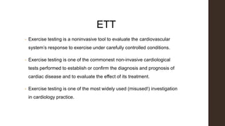
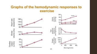
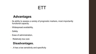
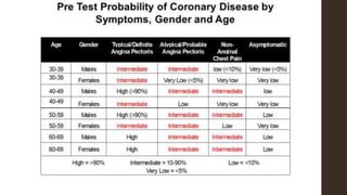
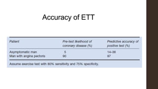
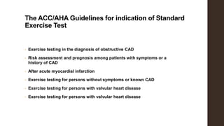
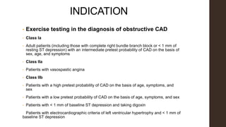
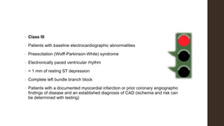
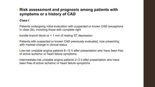
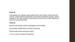
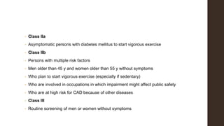
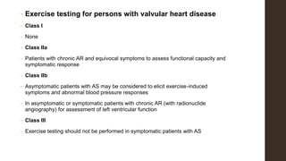
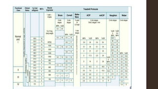
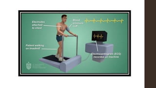
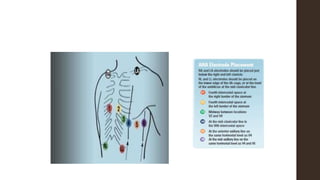
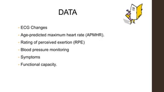
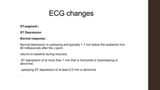
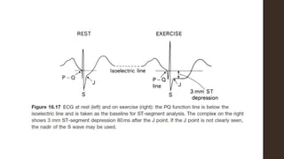
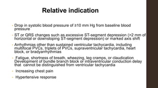
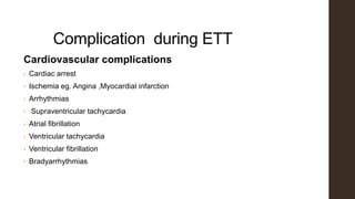
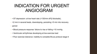
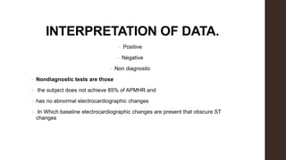
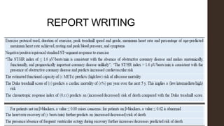
Exercise Tolerlance Test
Dr.Chinmoy Saha
Phase B Resident
M.D (Cardiology)
Dhaka Medical College
ETT
• Exercise testing is a noninvasive tool to evaluate the cardiovascular
system’s response to exercise under carefully controlled conditions.
• Exercise testing is one of the commonest non-invasive cardiological
tests performed to establish or confirm the diagnosis and prognosis of
cardiac disease and to evaluate the effect of its treatment.
• Exercise testing is one of the most widely used (misused!) investigation
in cardiology practice.
Graphs of the hemodynamic responses to
exercise
ETT
Advantages
• Its ability to assess a variety of prognostic markers, most importantly
functional capacity.
• Widespread availability,
• Safety
• Ease of administration,
• Relatively low cost.
Disadvantages.
• . It has a low sensitivity and specificity
Accuracy of ETT
The ACC/AHA Guidelines for indication of Standard
Exercise Test
• Exercise testing in the diagnosis of obstructive CAD
• Risk assessment and prognosis among patients with symptoms or a
history of CAD
• After acute myocardial infarction
• Exercise testing for persons without symptoms or known CAD
• Exercise testing for persons with valvular heart disease
INDICATION
• Exercise testing in the diagnosis of obstructive CAD
• Class Ia
• Adult patients (including those with complete right bundle branch block or < 1 mm of
resting ST depression) with an intermediate pretest probability of CAD on the basis of
sex, age, and symptoms
• Class IIa
• Patients with vasospastic angina
• Class IIb
• Patients with a high pretest probability of CAD on the basis of age, symptoms, and
sex
• Patients with a low pretest probability of CAD on the basis of age, symptoms, and sex
• Patients with < 1 mm of baseline ST depression and taking digoxin
Patients with electrocardiographic criteria of left ventricular hypertrophy and < 1 mm of
baseline ST depression
• Class III
• Patients with baseline electrocardiographic abnormalities
• Preexcitation (Wolff-Parkinson-White) syndrome
• Electronically paced ventricular rhythm
• > 1 mm of resting ST depression
• Complete left bundle branch block
• Patients with a documented myocardial infarction or prior coronary angiographic
findings of disease and an established diagnosis of CAD (ischemia and risk can
be determined with testing)
• Risk assessment and prognosis among patients with
symptoms or a history of CAD
• Class I
• Patients undergoing initial evaluation with suspected or known CAD (exceptions
in class 2b), including those with complete right
• bundle branch block or < 1 mm of resting ST depression
• Patients with suspected or known CAD previously evaluated, now presenting
with marked change in clinical status
• Low-risk unstable angina patients 8–12 h after presentation who have been free
of active ischemic or heart failure symptoms
• Intermediate-risk unstable angina patients 2–3 d after presentation who have
been free of active ischemic or heart failure symptoms
• Class IIa
• Intermediate-risk unstable angina patients with initial cardiac markers that are
normal, a repeat electrocardiographic study without significant change, cardiac
markers 6–12 h after symptom onset that are normal, and no other evidence of
ischemia during observation
• Class IIb
• Patients with baseline electrocardiographic abnormalities
• Preexcitation (Wolff-Parkinson-White) syndrome
• Electronically paced ventricular rhythm
• 1 mm or more of resting ST depression
• Complete left bundle branch block or any interventricular conduction defect with
QRS duration > 120 milliseconds
• Patients with a stable clinical course who undergo periodic monitoring to guide
treatment
• Class III
• Patients with severe comorbidity likely to limit life expectancy and/or candidacy
for revascularization
• High-risk unstable angina patients
• After acute myocardial infarction
• Class I
• Before discharge for prognostic assessment, activity prescription, or evaluation
of medical therapy (submaximal at about 4–6 d)
• Early after discharge for prognostic assessment and cardiac rehabilitation if the
predischarge exercise test was not performed
• (symptom limited, about 14–21 d)
• Late after discharge for prognostic assessment, activity prescription, evaluation
of medical therapy, and cardiac rehabilitation if the
• early exercise test was submaximal (symptom limited, about 3–6 wk)
• Class IIb
• Patients with electrocardiographic abnormalities
• Complete left bundle branch block
• Preexcitation (Wolff-Parkinson-White) syndrome
• Left ventricular hypertrophy
• Digoxin therapy
• Electronically paced ventricular rhythm
• > 1 mm of resting ST depression
• Periodic monitoring for patients who continue to participate in exercise
training or cardiac rehabilitation
• Class III
• Severe comorbidity likely to limit life expectancy or candidacy for revascularization
• Patients with acute myocardial infarction and uncompensated congestive heart
failure, cardiac arrhythmia, or noncardiac conditions
• that severely limit exercise ability
• Before discharge, patients who have been selected for or have undergone cardiac
catheterization (stress imaging tests are
• recommended)
• Exercise testing for persons without symptoms or known
CAD
• Class I
• None
• Class IIa
• Asymptomatic persons with diabetes mellitus to start vigorous exercise
• Class IIb
• Persons with multiple risk factors
• Men older than 45 y and women older than 55 y without symptoms
• Who plan to start vigorous exercise (especially if sedentary)
• Who are involved in occupations in which impairment might affect public safety
• Who are at high risk for CAD because of other diseases
• Class III
• Routine screening of men or women without symptoms
• Exercise testing for persons with valvular heart disease
• Class I
• None
• Class IIa
• Patients with chronic AR and equivocal symptoms to assess functional capacity and
symptomatic response
• Class IIb
• Asymptomatic patients with AS may be considered to elicit exercise-induced
symptoms and abnormal blood pressure responses
• In asymptomatic or symptomatic patients with chronic AR (with radionuclide
angiography) for assessment of left ventricular function
• Class III
• Exercise testing should not be performed in symptomatic patients with AS
• Exercise testing for persons with congenital heart
disease
• Class I
• None
• Class IIa
• Asymptomatic young adults < 30 y of age to determine exercise capability,
symptoms, and blood pressure response
• Adolescent or young adult patient with AS who has a Doppler mean gradient > 30
mm Hg or a peak velocity > 50 mm Hg if the patient is interested in athletic
participation or if the clinical findings and Doppler findings are disparate
• Asymptomatic young adult with a mean Doppler gradient > 40 mm Hg or a peak
Doppler gradient > 64 mm Hg or when the patient anticipates athletic participation or
pregnancy
• In patients with atrial septal defect with symptoms that are discrepant with clinical
findings or to document changes in oxygensaturation in patients with mild or
moderate PAH
• In patients with subvalvular AS testing to determine exercise capability,
symptoms, ECG changes or arrhythmias, or increase in LVOT gradient is
reasonable in the presence of otherwise equivocal indications for intervention
• In patients with supravalvular AS (along with other imaging modalities) testing
can be useful to evaluate the adequacy of myocardial perfusion
• Class IIb
• In patients with aortic coarctation, testing may be performed at intervals
determined in consultation with the regional ACHD center
• Class III
• Patients with atrial septal defect or patent ductus arteriosus with severe PAH
• Symptomatic patients with AS or those with repolarization abnormality on ECG
or systolic dysfunction on echocardiography
Absolute contraindications
• Acute myocardial infarction (within 2 d)
• High-risk unstable angina
• Uncontrolled cardiac arrhythmias causing symptoms or hemodynamic compromise
• Symptomatic, severe aortic stenosis
• Uncontrolled symptomatic heart failure
• Acute pulmonary embolus or pulmonary infarction
• Suspected or known dissecting aneurysm
• Active or suspected myocarditis, pericarditis, or endocarditis
• Acute noncardiac disorder that may affect exercise performance or be aggravated by exercise
(e.g., infection, renal failure, or thyrotoxicosis)
• Considerable emotional distress (psychosis)
RELATIVE
CONTRAINDICATION
• Left main coronary stenosis or its equivalent
• Moderate stenotic valvular heart disease
• Resting diastolic blood pressure > 110 mm Hg or resting systolic blood pressure
> 200 mm Hg
• Electrolyte abnormalities (e.g., hypokalemia and hypomagnesemia)
• Fixed-rate pacemaker
• High-degree atrioventricular block
• Frequent or complex ventricular ectopy
• Ventricular aneurysm
Cont…
• Chronic infectious disease (e.g., mononucleosis, hepatitis, and acquired
immunodeficiency syndrome)
• Neuromuscular, musculoskeletal, or rheumatoid disorders exacerbated by
exercise
• Advanced or complicated pregnancy
• Hypertrophic cardiomyopathy and other forms of outflow tract obstruction
• Mental impairment leading to inability to cooperate
EXERCISE PROTOCOLS
• There are advantages and disadvantages to each exercise protocol.
• Selection depends on the patient characteristics, the equipment available, and
the familiarity and comfort of the testing personnel with the protocol.
Bruce Protocol
Modified Bruce Protocol
• Developed for less-fit persons,
• The modified Bruce protocoladds additional stages 0 and
1/2.
• These stages at 1.7 mph (2.7 km/h) with 0% and 5% grades
respectively
• provide a lower workload for persons with poor
cardiovascularfitness.
Patient preparation
• Patients should refrain from ingesting food, alcohol, or caffeine or using tobacco
products within 3 h of testing.
• Patients should be rested for the assessment, avoiding significant exertion or
exercise on the day of the assessment.
• Patients should wear clothing that allows freedom of movement.
• Outpatients should be warned that the evaluation may be fatiguing and that
they may wish to have someone available to drive them home afterward.
• If the test is for diagnostic purposes, it may be helpful for patients to discontinue
prescribed cardiovascular medication after discussion with their physician.
Medication
• Before diagnostic testing, cardiovascular drugs are withheld at the discretion of
and under the guidance of the supervising physician.
a. β-Blockers pose a special problem. Patients taking β-blockers often do not
have an adequate increase in heart rate to achieve the level of stress needed for
the test. Abrupt withdrawal of β-blockers is to be discouraged because of reflex
tachycardia. The best possible solution is to taper the β-blocker over 2-4 days
before an exercise test
b.Digoxin should be withheld for 2 weeks before testing
DATA
• ECG Changes
• Age-predicted maximum heart rate (APMHR).
• Rating of perceived exertion (RPE)
• Blood pressure monitoring
• Symptoms
• Functional capacity.
ECG changes
• ST-segment :
• ST Depression
• Normal response :
• Normal depression is upsloping and typically < 1 mm below the isoelectric line
80 milliseconds after the J point.
• returns to baseline during recovery
• ST depression of at more than 1 mm that is horizontal or downsloping is
abnormal,
• upsloping ST depression of at least 2.0 mm is abnormal
• .
Criteria increase probability of ischemia
• The number of leads involved (i.e., more leads increase the probability of
ischemia),
• The workload at which the ST depression occurs (i.e., lower workload
increases probability)
• The angle of the slope (i.e., a downsloping angle has a higher probability than
a horizontal one
• The amount of time in recovery before normalization of the ST segment
• (i.e., longer recovery increases the probability)
• Possibly the magnitude of the depression.
• Changes in the lateral leads, particularly V5, are more specific than in any of the
other leads.
• Changes in the inferior leads alone are likely to be a false-positive result
• ST elevation depends on the presence or absence of Q waves of prior MI.
• ST-segment elevation with Q waves of prior MI is a common finding
amongpatients who have had MI. It occurs among up to 50% of patients with
anteriorMI and 15% of patients with previous inferior MI, and it is not caused by
ischemia.
• ST-segment elevation without Q waves of prior MI represents marked
transmural myocardial ischemia.This finding should be interpreted as abnormal.
• ST normalization, or the lack of ST changes during exercise, may be a sign of
ischemia
Other Waves
• R waves may change in amplitude during exercise. There is no diagnostic
value in these changes.
• T-wave inversion is not a specific marker of ischemia.
• If the U wave is upright at baseline, U-wave inversion may be associated with
• ischemia, left ventricular hypertrophy, and valvular diseas
Age-predicted maximum heart rate
(APMHR).
• APMHR = 220 – age (for men)
• APMHR = 210 – age (for women)
• If MHR does not exceed 85% of APMHR during testing and there are no
substantial electrocardiographic changes, the test is usually read as
nondiagnostic.
• The chronotropic response index (CRI) is a measure of MHR in relation to
• chronotropic reserve. A normal response is defined as a CRI of > 0.8
Metabolic Equivalent
• Exercise testing fundamentally involves the measurement of work.
• The metabolic equivalent of the task (MET) is a term commonly used
clinically to express the oxygen requirement of the work rate during an
exercise test on a treadmill.
• One MET is equated with the resting metabolic rate (~3.5 mL of
O2/kg/min)
• Functional testing is a powerful marker for prognosis.
• Persons who achieve > 6 metabolic equivalents (METs) of workload
have a significantly lower mortality rate than those who do not achieve
this workload, regardless of electrocardiographic changes.
Metabolic equivalent for exercise
Functional capacity classification
• Excellent (>10 METS),
• Good (7 METs to 10 METS),
• Moderate (4 METs to 6 METS),
• Poor (<4 METS)
Recovery after exercise
• Monitoring should continue for at least 5 minutes after exercise or until changes
stabilize.
• The recovery period, particularly the third minute is critical for ST analysis
• The heart rate recovery, defined as the difference in heart rate at peak
exercise and at 1 minute after cessation of exercise, has important prognostic
significance.
• A heart rate recovery of 12 beats/min or less is considered abnormal during an
upright cool-downperiod.
Termination of ETT
Absolute indications
• Moderate to severe angina
• Increasing nervous system symptoms (eg, ataxia, dizziness, or near-
syncope)
• Signs of poor perfusion (cyanosis or pallor)
• Technical difficulties in monitoring ECG or systolic blood pressure
• Subject’s desire to stop
• Sustained ventricular tachycardia
• ST-segment elevation (≥1.0 mm) in leads without diagnostic Q waves
(other than V1 or aVR)
Relative indication
• Drop in systolic blood pressure of ≥10 mm Hg from baseline blood
pressure
• ST or QRS changes such as excessive ST-segment depression (>2 mm of
horizontal or downsloping ST-segment depression) or marked axis shift
• Arrhythmias other than sustained ventricular tachycardia, including
multifocal PVCs, triplets of PVCs, supraventricular tachycardia, heart
block, or bradyarrhythmias
• Fatigue, shortness of breath, wheezing, leg cramps, or claudication
Development of bundle branch block or intraventricular conduction delay
that cannot be distinguished from ventricular tachycardia
• Increasing chest pain
• Hypertensive response
Complication during ETT
Cardiovascular complications
• Cardiac arrest
• Ischemia eg. Angina ,Myocardial infarction
• Arrhythmias
• Supraventricular tachycardia
• Atrial fibrillation
• Ventricular tachycardia
• Ventricular fibrillation
• Bradyarrhythmias
COMPLICATION
• Hypertension
• Hypotension
• Aneurysm rupture
Pulmonary complications
• Exercise-induced asthma
• Bronchospasm
• Pneumothorax
Exacerbation of underlying pulmonary disease
COMPLICATION
• Gastrointestinal complications
• Vomiting, Cramps, Diarrhea
• Neurologic complications
• Dizziness ,Syncope (fainting), Cerebrovascular accident (stroke)
• Musculoskeletal complications
• Mechanical injuries, Back injuries,Muscle cramps or spasms
INDICATION FOR URGENT
ANGIOGRAM
• • ST depression: at low heart rate (<130/min off β blockade),
• >2 mm in several leads, downsloping, persisting >5 min into recovery
• period
• • Blood pressure response: failure to rise or falling >10 mmHg
• • Ventricular arrhythmias developing at low exercise load
• • Poor exercise tolerance: inability to complete Bruce protocol stage II
INTERPRETATION OF DATA.
• Positive
• Negative
• Non diagnostic
• Nondiagnostic tests are those
• the subject does not achieve 85% of APMHR and
• has no abnormal electrocardiographic changes
• In Which baseline electrocardiographic changes are present that obscure ST
changes
False Positive Result
PROGNOSIS
• The Duke treadmill score (DTS) is a
• DTS = duration of exercise (in minutes) – (5 × maximal ST-segment
deviation) – (4 × angina score)
• In the previous equation, 0 = no angina, 1 = non–test-limiting angina, and 2 =
exercise-limiting angina.
Score Risk 5 yr Survival %
S ≤ +5 Low 97
–10 to < +5 Intermediate 91
< –10 High 72
REPORT WRITING
A FEW FORMAT
ETT
THANK YOU
Exercise tolerance test

Exercise tolerance test

  • 1.
    Exercise Tolerlance Test Dr.ChinmoySaha Phase B Resident M.D (Cardiology) Dhaka Medical College
  • 2.
    ETT • Exercise testingis a noninvasive tool to evaluate the cardiovascular system’s response to exercise under carefully controlled conditions. • Exercise testing is one of the commonest non-invasive cardiological tests performed to establish or confirm the diagnosis and prognosis of cardiac disease and to evaluate the effect of its treatment. • Exercise testing is one of the most widely used (misused!) investigation in cardiology practice.
  • 3.
    Graphs of thehemodynamic responses to exercise
  • 4.
    ETT Advantages • Its abilityto assess a variety of prognostic markers, most importantly functional capacity. • Widespread availability, • Safety • Ease of administration, • Relatively low cost. Disadvantages. • . It has a low sensitivity and specificity
  • 6.
  • 7.
    The ACC/AHA Guidelinesfor indication of Standard Exercise Test • Exercise testing in the diagnosis of obstructive CAD • Risk assessment and prognosis among patients with symptoms or a history of CAD • After acute myocardial infarction • Exercise testing for persons without symptoms or known CAD • Exercise testing for persons with valvular heart disease • Exercise testing for persons with valvular heart disease
  • 8.
    INDICATION • Exercise testingin the diagnosis of obstructive CAD • Class Ia • Adult patients (including those with complete right bundle branch block or < 1 mm of resting ST depression) with an intermediate pretest probability of CAD on the basis of sex, age, and symptoms • Class IIa • Patients with vasospastic angina • Class IIb • Patients with a high pretest probability of CAD on the basis of age, symptoms, and sex • Patients with a low pretest probability of CAD on the basis of age, symptoms, and sex • Patients with < 1 mm of baseline ST depression and taking digoxin Patients with electrocardiographic criteria of left ventricular hypertrophy and < 1 mm of baseline ST depression
  • 9.
    • Class III •Patients with baseline electrocardiographic abnormalities • Preexcitation (Wolff-Parkinson-White) syndrome • Electronically paced ventricular rhythm • > 1 mm of resting ST depression • Complete left bundle branch block • Patients with a documented myocardial infarction or prior coronary angiographic findings of disease and an established diagnosis of CAD (ischemia and risk can be determined with testing)
  • 10.
    • Risk assessmentand prognosis among patients with symptoms or a history of CAD • Class I • Patients undergoing initial evaluation with suspected or known CAD (exceptions in class 2b), including those with complete right • bundle branch block or < 1 mm of resting ST depression • Patients with suspected or known CAD previously evaluated, now presenting with marked change in clinical status • Low-risk unstable angina patients 8–12 h after presentation who have been free of active ischemic or heart failure symptoms • Intermediate-risk unstable angina patients 2–3 d after presentation who have been free of active ischemic or heart failure symptoms
  • 11.
    • Class IIa •Intermediate-risk unstable angina patients with initial cardiac markers that are normal, a repeat electrocardiographic study without significant change, cardiac markers 6–12 h after symptom onset that are normal, and no other evidence of ischemia during observation • Class IIb • Patients with baseline electrocardiographic abnormalities • Preexcitation (Wolff-Parkinson-White) syndrome • Electronically paced ventricular rhythm • 1 mm or more of resting ST depression
  • 12.
    • Complete leftbundle branch block or any interventricular conduction defect with QRS duration > 120 milliseconds • Patients with a stable clinical course who undergo periodic monitoring to guide treatment • Class III • Patients with severe comorbidity likely to limit life expectancy and/or candidacy for revascularization • High-risk unstable angina patients
  • 13.
    • After acutemyocardial infarction • Class I • Before discharge for prognostic assessment, activity prescription, or evaluation of medical therapy (submaximal at about 4–6 d) • Early after discharge for prognostic assessment and cardiac rehabilitation if the predischarge exercise test was not performed • (symptom limited, about 14–21 d) • Late after discharge for prognostic assessment, activity prescription, evaluation of medical therapy, and cardiac rehabilitation if the • early exercise test was submaximal (symptom limited, about 3–6 wk)
  • 14.
    • Class IIb •Patients with electrocardiographic abnormalities • Complete left bundle branch block • Preexcitation (Wolff-Parkinson-White) syndrome • Left ventricular hypertrophy • Digoxin therapy • Electronically paced ventricular rhythm • > 1 mm of resting ST depression • Periodic monitoring for patients who continue to participate in exercise training or cardiac rehabilitation
  • 15.
    • Class III •Severe comorbidity likely to limit life expectancy or candidacy for revascularization • Patients with acute myocardial infarction and uncompensated congestive heart failure, cardiac arrhythmia, or noncardiac conditions • that severely limit exercise ability • Before discharge, patients who have been selected for or have undergone cardiac catheterization (stress imaging tests are • recommended) • Exercise testing for persons without symptoms or known CAD • Class I • None
  • 16.
    • Class IIa •Asymptomatic persons with diabetes mellitus to start vigorous exercise • Class IIb • Persons with multiple risk factors • Men older than 45 y and women older than 55 y without symptoms • Who plan to start vigorous exercise (especially if sedentary) • Who are involved in occupations in which impairment might affect public safety • Who are at high risk for CAD because of other diseases • Class III • Routine screening of men or women without symptoms
  • 17.
    • Exercise testingfor persons with valvular heart disease • Class I • None • Class IIa • Patients with chronic AR and equivocal symptoms to assess functional capacity and symptomatic response • Class IIb • Asymptomatic patients with AS may be considered to elicit exercise-induced symptoms and abnormal blood pressure responses • In asymptomatic or symptomatic patients with chronic AR (with radionuclide angiography) for assessment of left ventricular function • Class III • Exercise testing should not be performed in symptomatic patients with AS
  • 18.
    • Exercise testingfor persons with congenital heart disease • Class I • None • Class IIa • Asymptomatic young adults < 30 y of age to determine exercise capability, symptoms, and blood pressure response • Adolescent or young adult patient with AS who has a Doppler mean gradient > 30 mm Hg or a peak velocity > 50 mm Hg if the patient is interested in athletic participation or if the clinical findings and Doppler findings are disparate • Asymptomatic young adult with a mean Doppler gradient > 40 mm Hg or a peak Doppler gradient > 64 mm Hg or when the patient anticipates athletic participation or pregnancy • In patients with atrial septal defect with symptoms that are discrepant with clinical findings or to document changes in oxygensaturation in patients with mild or moderate PAH
  • 19.
    • In patientswith subvalvular AS testing to determine exercise capability, symptoms, ECG changes or arrhythmias, or increase in LVOT gradient is reasonable in the presence of otherwise equivocal indications for intervention • In patients with supravalvular AS (along with other imaging modalities) testing can be useful to evaluate the adequacy of myocardial perfusion • Class IIb • In patients with aortic coarctation, testing may be performed at intervals determined in consultation with the regional ACHD center • Class III • Patients with atrial septal defect or patent ductus arteriosus with severe PAH • Symptomatic patients with AS or those with repolarization abnormality on ECG or systolic dysfunction on echocardiography
  • 20.
    Absolute contraindications • Acutemyocardial infarction (within 2 d) • High-risk unstable angina • Uncontrolled cardiac arrhythmias causing symptoms or hemodynamic compromise • Symptomatic, severe aortic stenosis • Uncontrolled symptomatic heart failure • Acute pulmonary embolus or pulmonary infarction • Suspected or known dissecting aneurysm • Active or suspected myocarditis, pericarditis, or endocarditis • Acute noncardiac disorder that may affect exercise performance or be aggravated by exercise (e.g., infection, renal failure, or thyrotoxicosis) • Considerable emotional distress (psychosis)
  • 21.
    RELATIVE CONTRAINDICATION • Left maincoronary stenosis or its equivalent • Moderate stenotic valvular heart disease • Resting diastolic blood pressure > 110 mm Hg or resting systolic blood pressure > 200 mm Hg • Electrolyte abnormalities (e.g., hypokalemia and hypomagnesemia) • Fixed-rate pacemaker • High-degree atrioventricular block • Frequent or complex ventricular ectopy • Ventricular aneurysm
  • 22.
    Cont… • Chronic infectiousdisease (e.g., mononucleosis, hepatitis, and acquired immunodeficiency syndrome) • Neuromuscular, musculoskeletal, or rheumatoid disorders exacerbated by exercise • Advanced or complicated pregnancy • Hypertrophic cardiomyopathy and other forms of outflow tract obstruction • Mental impairment leading to inability to cooperate
  • 23.
    EXERCISE PROTOCOLS • Thereare advantages and disadvantages to each exercise protocol. • Selection depends on the patient characteristics, the equipment available, and the familiarity and comfort of the testing personnel with the protocol.
  • 25.
  • 26.
    Modified Bruce Protocol •Developed for less-fit persons, • The modified Bruce protocoladds additional stages 0 and 1/2. • These stages at 1.7 mph (2.7 km/h) with 0% and 5% grades respectively • provide a lower workload for persons with poor cardiovascularfitness.
  • 29.
    Patient preparation • Patientsshould refrain from ingesting food, alcohol, or caffeine or using tobacco products within 3 h of testing. • Patients should be rested for the assessment, avoiding significant exertion or exercise on the day of the assessment. • Patients should wear clothing that allows freedom of movement. • Outpatients should be warned that the evaluation may be fatiguing and that they may wish to have someone available to drive them home afterward. • If the test is for diagnostic purposes, it may be helpful for patients to discontinue prescribed cardiovascular medication after discussion with their physician.
  • 30.
    Medication • Before diagnostictesting, cardiovascular drugs are withheld at the discretion of and under the guidance of the supervising physician. a. β-Blockers pose a special problem. Patients taking β-blockers often do not have an adequate increase in heart rate to achieve the level of stress needed for the test. Abrupt withdrawal of β-blockers is to be discouraged because of reflex tachycardia. The best possible solution is to taper the β-blocker over 2-4 days before an exercise test b.Digoxin should be withheld for 2 weeks before testing
  • 31.
    DATA • ECG Changes •Age-predicted maximum heart rate (APMHR). • Rating of perceived exertion (RPE) • Blood pressure monitoring • Symptoms • Functional capacity.
  • 32.
    ECG changes • ST-segment: • ST Depression • Normal response : • Normal depression is upsloping and typically < 1 mm below the isoelectric line 80 milliseconds after the J point. • returns to baseline during recovery • ST depression of at more than 1 mm that is horizontal or downsloping is abnormal, • upsloping ST depression of at least 2.0 mm is abnormal • .
  • 36.
    Criteria increase probabilityof ischemia • The number of leads involved (i.e., more leads increase the probability of ischemia), • The workload at which the ST depression occurs (i.e., lower workload increases probability) • The angle of the slope (i.e., a downsloping angle has a higher probability than a horizontal one • The amount of time in recovery before normalization of the ST segment • (i.e., longer recovery increases the probability) • Possibly the magnitude of the depression. • Changes in the lateral leads, particularly V5, are more specific than in any of the other leads. • Changes in the inferior leads alone are likely to be a false-positive result
  • 37.
    • ST elevationdepends on the presence or absence of Q waves of prior MI. • ST-segment elevation with Q waves of prior MI is a common finding amongpatients who have had MI. It occurs among up to 50% of patients with anteriorMI and 15% of patients with previous inferior MI, and it is not caused by ischemia. • ST-segment elevation without Q waves of prior MI represents marked transmural myocardial ischemia.This finding should be interpreted as abnormal. • ST normalization, or the lack of ST changes during exercise, may be a sign of ischemia
  • 38.
    Other Waves • Rwaves may change in amplitude during exercise. There is no diagnostic value in these changes. • T-wave inversion is not a specific marker of ischemia. • If the U wave is upright at baseline, U-wave inversion may be associated with • ischemia, left ventricular hypertrophy, and valvular diseas
  • 39.
    Age-predicted maximum heartrate (APMHR). • APMHR = 220 – age (for men) • APMHR = 210 – age (for women) • If MHR does not exceed 85% of APMHR during testing and there are no substantial electrocardiographic changes, the test is usually read as nondiagnostic. • The chronotropic response index (CRI) is a measure of MHR in relation to • chronotropic reserve. A normal response is defined as a CRI of > 0.8
  • 41.
    Metabolic Equivalent • Exercisetesting fundamentally involves the measurement of work. • The metabolic equivalent of the task (MET) is a term commonly used clinically to express the oxygen requirement of the work rate during an exercise test on a treadmill. • One MET is equated with the resting metabolic rate (~3.5 mL of O2/kg/min) • Functional testing is a powerful marker for prognosis. • Persons who achieve > 6 metabolic equivalents (METs) of workload have a significantly lower mortality rate than those who do not achieve this workload, regardless of electrocardiographic changes.
  • 42.
  • 43.
    Functional capacity classification •Excellent (>10 METS), • Good (7 METs to 10 METS), • Moderate (4 METs to 6 METS), • Poor (<4 METS)
  • 44.
    Recovery after exercise •Monitoring should continue for at least 5 minutes after exercise or until changes stabilize. • The recovery period, particularly the third minute is critical for ST analysis • The heart rate recovery, defined as the difference in heart rate at peak exercise and at 1 minute after cessation of exercise, has important prognostic significance. • A heart rate recovery of 12 beats/min or less is considered abnormal during an upright cool-downperiod.
  • 45.
    Termination of ETT Absoluteindications • Moderate to severe angina • Increasing nervous system symptoms (eg, ataxia, dizziness, or near- syncope) • Signs of poor perfusion (cyanosis or pallor) • Technical difficulties in monitoring ECG or systolic blood pressure • Subject’s desire to stop • Sustained ventricular tachycardia • ST-segment elevation (≥1.0 mm) in leads without diagnostic Q waves (other than V1 or aVR)
  • 46.
    Relative indication • Dropin systolic blood pressure of ≥10 mm Hg from baseline blood pressure • ST or QRS changes such as excessive ST-segment depression (>2 mm of horizontal or downsloping ST-segment depression) or marked axis shift • Arrhythmias other than sustained ventricular tachycardia, including multifocal PVCs, triplets of PVCs, supraventricular tachycardia, heart block, or bradyarrhythmias • Fatigue, shortness of breath, wheezing, leg cramps, or claudication Development of bundle branch block or intraventricular conduction delay that cannot be distinguished from ventricular tachycardia • Increasing chest pain • Hypertensive response
  • 47.
    Complication during ETT Cardiovascularcomplications • Cardiac arrest • Ischemia eg. Angina ,Myocardial infarction • Arrhythmias • Supraventricular tachycardia • Atrial fibrillation • Ventricular tachycardia • Ventricular fibrillation • Bradyarrhythmias
  • 48.
    COMPLICATION • Hypertension • Hypotension •Aneurysm rupture Pulmonary complications • Exercise-induced asthma • Bronchospasm • Pneumothorax Exacerbation of underlying pulmonary disease
  • 49.
    COMPLICATION • Gastrointestinal complications •Vomiting, Cramps, Diarrhea • Neurologic complications • Dizziness ,Syncope (fainting), Cerebrovascular accident (stroke) • Musculoskeletal complications • Mechanical injuries, Back injuries,Muscle cramps or spasms
  • 50.
    INDICATION FOR URGENT ANGIOGRAM •• ST depression: at low heart rate (<130/min off β blockade), • >2 mm in several leads, downsloping, persisting >5 min into recovery • period • • Blood pressure response: failure to rise or falling >10 mmHg • • Ventricular arrhythmias developing at low exercise load • • Poor exercise tolerance: inability to complete Bruce protocol stage II
  • 51.
    INTERPRETATION OF DATA. •Positive • Negative • Non diagnostic • Nondiagnostic tests are those • the subject does not achieve 85% of APMHR and • has no abnormal electrocardiographic changes • In Which baseline electrocardiographic changes are present that obscure ST changes
  • 52.
  • 53.
    PROGNOSIS • The Duketreadmill score (DTS) is a • DTS = duration of exercise (in minutes) – (5 × maximal ST-segment deviation) – (4 × angina score) • In the previous equation, 0 = no angina, 1 = non–test-limiting angina, and 2 = exercise-limiting angina. Score Risk 5 yr Survival % S ≤ +5 Low 97 –10 to < +5 Intermediate 91 < –10 High 72
  • 54.
  • 55.
  • 56.
  • 59.