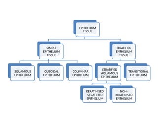
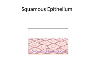
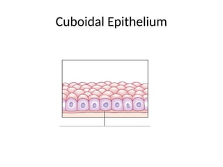
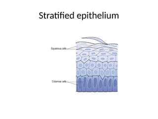
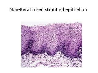
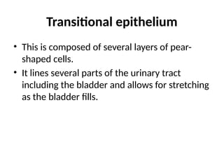
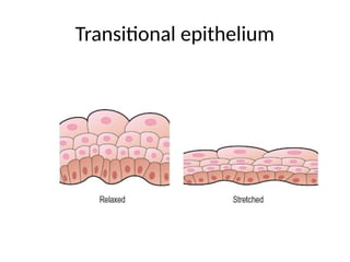
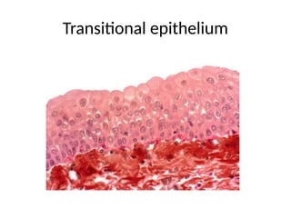
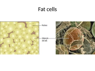
TISSUE
Ms.Shraddha patel
Nursing tutor
SSACON
Introduction
• Tissues consist of large numbers of the same type
of cells and are classified according to the size,
shape and functions of their constituent cells.
• There are four main types of tissue each with
subtypes. They are:
Epithelial tissue or epithelium
Connective tissue
Muscle tissue
Nervous tissue.
Epithelial tissue
• This tissue type covers the body and lines cavities,
hollow organs and tubes. It is also found in glands.
The structure of epithelium is closely related to its
functions, which include:
Protection of underlying structures from, for
example, dehydration, chemical and mechanical
damage.
Secretion.
Absorption.
EPITHELIUM
TISSUE
SIMPLE
EPITHELIUM
TISSUE
SQUAMOUS
EPITHELIUM
CUBOIDAL
EPITHELIUM
COLUMNAR
EPITHELIUM
STRATIFIED
EPITHELIUM
TISSUE
STRATIFIED
AQUAMOUS
EPITHELIUM
KERATINISED
STRATIFIED
EPITHELIUM
NON-
KERATINISED
EPITHELIUM
TRANSITIONAL
EPITHELIUM
• The cells are very closely packed and the
intercellular substance, the matrix, is minimal.
• The cells usually lie on a basement membrane,
which is an inert connective tissue made by the
epithelial cells themselves.
• Epithelial tissue may be:
• simple: a single layer of cells
• stratified: several layers of cells.
Simple epithelium
• Simple epithelium consists of a single layer of
identical cells and is divided into three main types.
• It is usually found on absorptive or secretory
surfaces, where the single layer enhances these
processes, and seldom on surfaces subject to
stress.
• The types are named according to the shape of the
cells, which differs according to their functions.
The more active the tissue, the taller the cells.
Simple Epithelium
Squamous (pavement) epithelium
• This is composed of a single layer of flattened
cells.
• The cells fit closely together like flat stones,
forming a thin and very smooth membrane
across which diffusion occurs easily.
Squamous Epithelium
• It forms the lining of the following structures:
Heart – where it is known as endocardium
Blood vessels & lymph vessels- where it is also
known as endothelium
Alveoli of the lungs
Lining the collecting ducts of nephrons in the
kidneys.
Cuboidal epithelium
• This consists of cube-shaped cells fitting closely
together lying on a basement membrane.
• It forms the kidney tubules and is found in
some glands such as the thyroid.
• Cuboidal epithelium is actively involved in
secretion, absorption and/or excretion.
Cuboidal Epithelium
Columnar epithelium
• This is formed by a single layer of cells, rectangular
in shape, on a basement membrane.
• It lines many organs and often has adaptations that
make it well suited to a specific function.
• The lining of the stomach is formed from simple
columnar epithelium without surface structures.
• The free surface of the columnar epithelium lining
the small intestine is covered with microvilli.
Columnar Epithelium
• Microvilli provide a very large surface area for
absorption of nutrients from the small intestine.
• In the trachea, columnar epithelium is ciliated
and also contains goblet cells that secrete mucus.
• This means that inhaled particles that stick to the
mucus layer are moved towards the throat by
cilia in the respiratory tract.
• In the uterine tubes, ova are propelled along by
ciliary action towards the uterus.
Stratified epithelium
• Stratified epithelia consist of several layers of cells of
various shapes.
• Continual cell division in the lower (basal) layers
pushes cells above nearer and nearer to the surface,
where they are shed.
• Basement membranes are usually absent.
• The main function of stratified epithelium is to protect
underlying structures from mechanical wear and tear.
• There are two main types: stratified squamous and
transitional.
Stratified epithelium
Stratified squamous epithelium
• This is composed of several layers of cells.
• In the deepest layers the cells are mainly
columnar and, as they grow towards the
surface, they become flattened and are then
shed.
Keratinised stratified epithelium
• This is found on dry surfaces subjected to wear
and tear, i.e. skin, hair and nails. The surface layer
consists of dead epithelial cells that have lost their
nuclei and contain the protein keratin.
• This forms a tough, relatively waterproof
protective layer that prevents drying of the live
cells underneath. The surface layer of skin is
rubbed off and is replaced from below.
Non-keratinised stratified epithelium
• This protects moist surfaces subjected to wear
and tear, and prevents them from drying out,
e.g. the conjunctiva of the eyes, the lining of
the mouth, the pharynx, the oesophagus and
the vagina.
Non-Keratinised stratified epithelium
Transitional epithelium
• This is composed of several layers of pear-
shaped cells.
• It lines several parts of the urinary tract
including the bladder and allows for stretching
as the bladder fills.
Transitional epithelium
Connective tissue
• Connective tissue is the most abundant tissue in the
body.
• The connective tissue cells are more widely
separated from each other than in epithelial tissues,
and intercellular substance (matrix) is present in
considerably larger amounts.
• There are usually fibres present in the matrix, which
may be of a semisolid jelly-like consistency or dense
and rigid, depending upon the position and function
of the tissue.
• The fibres form a supporting network for the
cells to attach to.
• Most types of connective tissue have a good
blood supply. Major functions of connective
tissue are:
Binding and structural support
Protection
Transport
Insulation.
Cells in connective tissue
• Connective tissue, excluding blood, is found in
all organs supporting the specialised tissue.
• The different types of cell involved include:
fibroblasts, fat cells, macrophages,
leukocytes and mast cells.
Fibroblasts
• Fibroblasts are large cells with irregular
processes.
• They manufacture collagen and elastic fibres and
a matrix of extracellular material.
• Very fine collagen fibres, sometimes called
reticulin fibres, are found in highly active tissue,
such as the liver and reticular tissue.
Fibroblast
Collagen
• Fibroblasts are particularly active in tissue repair
(wound healing) where they may bind together
the cut surfaces of wounds or form granulation
tissue following tissue destruction.
• The collagen fibres formed during wound healing
shrink as they age, sometimes interfering with
the functions of the organ involved and with
adjacent structures.
Fat cells
• Also known as adipocytes, these cells occur
singly or in groups in many types of connective
tissue and are especially abundant in adipose
tissue.
• They vary in size and shape according to the
amount of fat they contain.
Fat cells
Macrophages
• These are large irregular-shaped cells with
granules in the cytoplasm.
• Some are fixed, i.e. attached to connective tissue
fibres, and others are motile.
• They are an important part of the body’s defence
mechanisms because they are actively phagocytic,
engulfing and digesting cell debris, bacteria and
other foreign bodies.
• Their activities are typical of those of the
monocyte–macrophage defence system, e.g.
monocytes in blood, Kupffer cells in liver
sinusoids, sinus-lining cells in lymph nodes
and spleen, and microglial cells in the brain.
Leukocytes
• White blood cells are normally found in small
numbers in healthy connective tissue but
neutrophils migrate in significant numbers during
infection when they play an important part in
tissue defence.
• Plasma cells develop from B-lymphocytes, a type
of white blood cell. They synthesise and secrete
specific defensive antibodies into the blood and
tissues.
Mast cells
• These are similar to basophil leukocytes.
• They are found in loose connective tissue, under the
fibrous capsule of some organs, e.g. liver and
spleen, and in considerable numbers round blood
vessels.
• Their cytoplasm is packed with granules containing
heparin, histamine and other substances, which are
released when the cells are damaged by disease or
injury.
• Release of the granular contents is called
degranulation.
• Histamine is involved in local and general
inflammatory reactions, it stimulates secretion of
gastric juice and is associated with development
of allergies and hypersensitivity states.
• Heparin prevents coagulation of blood, which
helps to maintain blood flow through inflamed
tissues, supplying cells with oxygen and glucose
and bringing additional protective leukocytes to
the area.
Mast Cells
CONNECTIVE
TISSUE
Loose (areolar) connective tissue
• This is the most generalised type of connective
tissue.
• The matrix is semisolid with many fibroblasts and
some fat cells (adipocytes), mast cells and
macrophages widely separated by elastic and
collagen fibres.
• It is found in almost every part of the body,
providing elasticity and tensile strength.
Loose Connective tissue
• It connects and supports other tissues, for
example:
• Under the skin
• Between muscles
• Supporting blood vessels and nerves
• In the alimentary canal
• In glands supporting secretory cells.
Adipose tissue
• Adipose tissue consists of fat cells adipocytes,
containing large fat globules, in a matrix of areolar
tissue.
• There are two types: white and brown.
Fat cells
White adipose tissue
• This makes up 20–25% of body weight in adults with
a normal body mass index; more is present in
obesity and less in those who are underweight.
• Adipose tissue secretes the hormone leptin.
• The kidneys and eyeballs are supported by adipose
tissue, which is also found between muscle fibres
and under the skin, where it acts as a thermal
insulator and energy store.
Brown adipose tissue
• This is present in the newborn.
• It has a more extensive capillary network than
white adipose tissue.
• When brown tissue is metabolised, it produces less
energy and considerably more heat than other fat,
contributing to the maintenance of body
temperature.
• Sometimes small amounts are present in adults.
Reticular tissue
• Reticular tissue has a semisolid matrix with fine
branching reticulin fibres.
• It contains reticular cells and white blood cells
(monocytes and lymphocytes).
• Reticular tissue is found in lymph nodes and all
organs of the lymphatic system.
Reticular Tissue
Dense connective tissue
• This contains more fibres and fewer cells than
loose connective tissue.
• They are of two types: Fibrous tissue and
Elastic tissue
Fibrous tissue
• This tissue is made up mainly of closely packed
bundles of collagen fibres with very little matrix.
• Fibrocytes (old and inactive fibroblasts) are few in
number and lie in rows between the bundles of
fibres.
• Fibrous tissue is found:
forming ligaments, which bind bones together
As an outer protective covering for bone, called
periosteum
As an outer protective covering of some organs,
e.g. The kidneys, lymph nodes and the brain
Forming muscle sheaths, called muscle fascia
(see fig. 16.61, p. 426), which extend beyond the
muscle to become the tendon that attaches the
muscle to bone.
Fibrous Tissue
Elastic tissue
• Elastic tissue is capable of considerable extension
and recoil.
• There are few cells and the matrix consists
mainly of masses of elastic fibres secreted by
fibroblasts.
• It is found in organs where stretching or
alteration of shape is required, e.g. in large blood
vessel walls, the trachea and bronchi, and the
lungs.
Elastic Tissue
Blood
• This is a fluid connective tissue that contains
various types of cells inside it like RBC, WBC,
Platelets etc.
Cartilage
• Cartilage is firmer than other connective tissues.
• The cells (chondrocytes) are sparse and lie
embedded in matrix reinforced by collagen and
elastic fibres.
• There are three types: hyaline cartilage,
fibrocartilage and elastic fibrocartilage.
Hyaline cartilage
• Hyaline cartilage is a smooth bluish-white tissue.
• The chondrocytes are arranged in small groups
within cell nests and the matrix is solid and smooth.
• Hyaline cartilage provides flexibility, support and
smooth surfaces for movement at joints. It is found:
On the ends of long bones that form joints
Forming the costal cartilages, which attach the ribs
to the sternum
Forming part of the larynx, trachea and bronchi.
Hyaline cartilage
Fibrocartilage
• This consists of dense masses of white collagen fibres in a
matrix similar to that of hyaline cartilage with the cells
widely dispersed.
• It is a tough, slightly flexible, supporting tissue found:
As pads between the bodies of the vertebrae, the
intervertebral discs
Between the articulating surfaces of the bones of the
knee joint, called semilunar cartilages
On the rim of the bony sockets of the hip and shoulder
joints, deepening the cavities without restricting
movement.
Fibrocartilage
Elastic fibrocartilage
• This flexible tissue consists of yellow elastic
fibres lying in a solid matrix with chondrocytes
lying between the fibres.
• It provides support and maintains shape of, e.g.
the pinna or lobe of the ear, the epiglottis and
part of the tunica media of blood vessel walls.
Elastic Fibrocartilage
Bone
• Bone cells (osteocytes) are surrounded by a matrix
of collagen fibres strengthened by inorganic salts,
especially calcium and phosphate.
• This provides bones with their characteristic
strength and rigidity.
• Bone also has considerable capacity for growth in
the first two decades of life and for regeneration
throughout life.
• Two types of bone can be identified by the
naked eye:
compact bone – solid or dense appearance
spongy or cancellous bone –’spongy’ or fine
honeycomb appearance.
Muscle tissue
• This tissue is able to contract and relax, providing
movement within the body and of the body itself.
• Muscle contraction requires a blood supply that
will provide sufficient oxygen, calcium and
nutrients and remove waste products.
• There are three types of specialised contractile
cells, also known as fibres: skeletal muscle, smooth
muscle and cardiac muscle.
Skeletal muscle
• This type is described as skeletal because it forms
those muscles that move the bones (of the skeleton),
striated because striations (stripes) can be seen on
microscopic examination and voluntary as it is under
conscious control.
• Although most skeletal muscle moves bones, the
diaphragm is made from this type of muscle to
accommodate a degree of voluntary control in
breathing.
• In reality, many movements can be finely coordinated,
e.g. writing, but may also be controlled subconsciously.
Skeletal Muscle
• For example, maintaining an upright posture
does not normally require thought unless a new
locomotor skill is being learned, e.g. skating or
cycling, and the diaphragm maintains breathing
while asleep.
• These fibres (cells) are cylindrical, contain
several nuclei and can be up to 35 cm long.
• Skeletal muscle contraction is stimulated by
motor nerve impulses originating in the brain or
spinal cord and ending at the neuromuscular
junction.
Smooth muscle
• Smooth muscle is also described as non-striated,
visceral or involuntary. It does not have striations
and is not under conscious control.
• Some smooth muscle has the intrinsic ability to
initiate its own contractions (automaticity), e.g.
peristalsis.
• It is innervated by the autonomic nervous system.
• Additionally, autonomic nerve impulses, some
hormones and local metabolites stimulate its
contraction.
• It is found in the walls of hollow organs:
Regulating the diameter of blood vessels and
parts of the respiratory tract
Propelling contents along, e.G. The ureters,
ducts of glands and the alimentary tract
Expelling contents of the urinary bladder and
uterus.
Smooth Muscle
• A degree of muscle tone is always present,
meaning that smooth muscle is only completely
relaxed for short periods.
• Contraction of smooth muscle is slower and
more sustained than skeletal muscle.
• When examined under a microscope, the cells
are seen to be spindle shaped with only one
central nucleus.
• Bundles of fibres form sheets of muscle, such as
those found in the walls of the above structures.
Cardiac muscle
• This is only found only in the heart wall.
• It is not under conscious control but, when viewed
under a microscope, cross-stripes (striations)
characteristic of skeletal muscle can be seen.
• Each fibre(cell) has a nucleus and one or more
branches.
• The ends of the cells and their branches are in very
close contact with the ends and branches of
adjacent cells.
Cardiac Muscle
• Microscopically these ‘joints’, or intercalated discs, appear
as lines that are thicker and darker than the ordinary cross-
stripes.
• This arrangement gives cardiac muscle the appearance of a
sheet of muscle rather than a very large number of
individual fibres.
• This is significant when the heart contracts as a wave of
contraction spreads from cell to cell across the intercalated
discs, which means that the cardiac muscle fibres do not
need to be stimulated individually.
• The heart has an intrinsic pacemaker system, which means
that it beats in a coordinated manner without external
nerve stimulation, although the rate at which it beats is
influenced by autonomic nerve impulses, some hormones,
local metabolites and other substances.
Nervous tissue
• Two types of tissue are found in the nervous
system:
Excitable cells – these are called neurones and
they initiate, receive, conduct and transmit
information
Non-excitable cells – also known as glial cells,
these support the neurones.
Tissue regeneration
• The extent to which regeneration is possible
depends on the normal rate of turnover of
particular types of cell.
• Those with a rapid turnover regenerate most
effectively. There are three general categories:
Tissues in which cell replication is a continuous
process regenerate quickly – these include epithelial
cells of, for example, the skin, mucous membrane,
secretory glands, uterine lining and reticular tissue
Other tissues retain the ability to replicate, but do
so infrequently; these include the liver, kidney,
fibroblasts and smooth muscle cells. These tissues
take longer to regenerate
Some cells are normally unable to replicate
including nerve cells (neurones) and skeletal and
cardiac muscle cells meaning that damaged tissue
cannot be replaced. Extensively damaged tissue is
usually replaced by fibrous tissue, meaning that the
functions of the original tissue are lost.
Membranes
1. Epithelial membranes
• These membranes are sheets of epithelial tissue
and supporting connective tissue that cover or
line many internal structures or cavities.
• The main ones are mucous membrane, serous
membrane and the skin (cutaneous membrane).
• A. Mucous membrane
• This is the moist lining of the alimentary, respiratory
and genitourinary tracts and is sometimes referred
to as the mucosa.
• The membrane surface consists of epithelial cells,
some of which produce a secretion called mucus, a
slimy tenacious fluid.
• As it accumulates the cells become distended and
finally burst, discharging the mucus onto the free
surface.
• As the cells fill up with mucus they have the
appearance of a goblet or flask and are known as
goblet cells.
• Organs lined by mucous membrane have a moist
slippery surface.
• Mucus protects the lining membrane from drying,
and mechanical and chemical injury.
• In the respiratory tract it traps inhaled particles,
preventing them from entering the alveoli of the
lungs.
B. Serous membrane
• Serous membranes, or serosa, secrete serous
watery fluid.
• They consist of a double layer of loose areolar
connective tissue lined by simple squamous
epithelium.
• The parietal layer lines a cavity and the visceral
layer surrounds organs (the viscera) within the
cavity.
• The two layers are separated by serous fluid
secreted by the epithelium.
• There are three sites where serous membranes
are found:
The pleura lining the thoracic cavity and
surrounding The lungs
The pericardium lining the pericardial cavity and
Surrounding the heart
The peritoneum lining the abdominal cavity and
Surrounding abdominal organs.
• The serous fluid between the visceral and parietal
layers enables an organ to glide freely within the
cavity without being damaged by friction between
it and adjacent organs.
• For example, the heart changes its shape and size
during each beat and friction damage is prevented
by the arrangement of pericardium and its serous
fluid.
Synovial membrane
• This membrane lines the cavities of moveable joints
and surrounds tendons that could be injured by
rubbing against bones, e.g. over the wrist joint.
• It is not an epithelial membrane, but instead
consists of areolar connective tissue and elastic
fibres.
• Synovial membrane secretes clear, sticky, oily
synovial fluid, which lubricates and nourishes the
joints.
Glands
• Glands are groups of epithelial cells that produce
specialised secretions.
• Those that discharge their secretion onto the
epithelial surface of hollow organs, either directly or
through a duct, are called exocrine glands and vary
considerably in size, shape and complexity.
• Their secretions include mucus, saliva, digestive
juices and earwax.
• Other glands discharge their secretions into blood
and lymph. These are called endocrine glands
(ductless glands) and they secrete hormones.
Gland

Tissue Perfusionrelated products.pptx

  • 1.
  • 2.
    Introduction • Tissues consistof large numbers of the same type of cells and are classified according to the size, shape and functions of their constituent cells. • There are four main types of tissue each with subtypes. They are: Epithelial tissue or epithelium Connective tissue Muscle tissue Nervous tissue.
  • 3.
    Epithelial tissue • Thistissue type covers the body and lines cavities, hollow organs and tubes. It is also found in glands. The structure of epithelium is closely related to its functions, which include: Protection of underlying structures from, for example, dehydration, chemical and mechanical damage. Secretion. Absorption.
  • 4.
  • 5.
    • The cellsare very closely packed and the intercellular substance, the matrix, is minimal. • The cells usually lie on a basement membrane, which is an inert connective tissue made by the epithelial cells themselves. • Epithelial tissue may be: • simple: a single layer of cells • stratified: several layers of cells.
  • 6.
    Simple epithelium • Simpleepithelium consists of a single layer of identical cells and is divided into three main types. • It is usually found on absorptive or secretory surfaces, where the single layer enhances these processes, and seldom on surfaces subject to stress. • The types are named according to the shape of the cells, which differs according to their functions. The more active the tissue, the taller the cells.
  • 7.
  • 8.
    Squamous (pavement) epithelium •This is composed of a single layer of flattened cells. • The cells fit closely together like flat stones, forming a thin and very smooth membrane across which diffusion occurs easily.
  • 9.
  • 10.
    • It formsthe lining of the following structures: Heart – where it is known as endocardium Blood vessels & lymph vessels- where it is also known as endothelium Alveoli of the lungs Lining the collecting ducts of nephrons in the kidneys.
  • 11.
    Cuboidal epithelium • Thisconsists of cube-shaped cells fitting closely together lying on a basement membrane. • It forms the kidney tubules and is found in some glands such as the thyroid. • Cuboidal epithelium is actively involved in secretion, absorption and/or excretion.
  • 12.
  • 13.
    Columnar epithelium • Thisis formed by a single layer of cells, rectangular in shape, on a basement membrane. • It lines many organs and often has adaptations that make it well suited to a specific function. • The lining of the stomach is formed from simple columnar epithelium without surface structures. • The free surface of the columnar epithelium lining the small intestine is covered with microvilli.
  • 14.
  • 15.
    • Microvilli providea very large surface area for absorption of nutrients from the small intestine. • In the trachea, columnar epithelium is ciliated and also contains goblet cells that secrete mucus. • This means that inhaled particles that stick to the mucus layer are moved towards the throat by cilia in the respiratory tract. • In the uterine tubes, ova are propelled along by ciliary action towards the uterus.
  • 16.
    Stratified epithelium • Stratifiedepithelia consist of several layers of cells of various shapes. • Continual cell division in the lower (basal) layers pushes cells above nearer and nearer to the surface, where they are shed. • Basement membranes are usually absent. • The main function of stratified epithelium is to protect underlying structures from mechanical wear and tear. • There are two main types: stratified squamous and transitional.
  • 17.
  • 18.
    Stratified squamous epithelium •This is composed of several layers of cells. • In the deepest layers the cells are mainly columnar and, as they grow towards the surface, they become flattened and are then shed.
  • 19.
    Keratinised stratified epithelium •This is found on dry surfaces subjected to wear and tear, i.e. skin, hair and nails. The surface layer consists of dead epithelial cells that have lost their nuclei and contain the protein keratin. • This forms a tough, relatively waterproof protective layer that prevents drying of the live cells underneath. The surface layer of skin is rubbed off and is replaced from below.
  • 20.
    Non-keratinised stratified epithelium •This protects moist surfaces subjected to wear and tear, and prevents them from drying out, e.g. the conjunctiva of the eyes, the lining of the mouth, the pharynx, the oesophagus and the vagina.
  • 21.
  • 22.
    Transitional epithelium • Thisis composed of several layers of pear- shaped cells. • It lines several parts of the urinary tract including the bladder and allows for stretching as the bladder fills.
  • 23.
  • 24.
  • 25.
    Connective tissue • Connectivetissue is the most abundant tissue in the body. • The connective tissue cells are more widely separated from each other than in epithelial tissues, and intercellular substance (matrix) is present in considerably larger amounts. • There are usually fibres present in the matrix, which may be of a semisolid jelly-like consistency or dense and rigid, depending upon the position and function of the tissue.
  • 26.
    • The fibresform a supporting network for the cells to attach to. • Most types of connective tissue have a good blood supply. Major functions of connective tissue are: Binding and structural support Protection Transport Insulation.
  • 27.
    Cells in connectivetissue • Connective tissue, excluding blood, is found in all organs supporting the specialised tissue. • The different types of cell involved include: fibroblasts, fat cells, macrophages, leukocytes and mast cells.
  • 28.
    Fibroblasts • Fibroblasts arelarge cells with irregular processes. • They manufacture collagen and elastic fibres and a matrix of extracellular material. • Very fine collagen fibres, sometimes called reticulin fibres, are found in highly active tissue, such as the liver and reticular tissue.
  • 29.
  • 30.
  • 31.
    • Fibroblasts areparticularly active in tissue repair (wound healing) where they may bind together the cut surfaces of wounds or form granulation tissue following tissue destruction. • The collagen fibres formed during wound healing shrink as they age, sometimes interfering with the functions of the organ involved and with adjacent structures.
  • 32.
    Fat cells • Alsoknown as adipocytes, these cells occur singly or in groups in many types of connective tissue and are especially abundant in adipose tissue. • They vary in size and shape according to the amount of fat they contain.
  • 33.
  • 34.
    Macrophages • These arelarge irregular-shaped cells with granules in the cytoplasm. • Some are fixed, i.e. attached to connective tissue fibres, and others are motile. • They are an important part of the body’s defence mechanisms because they are actively phagocytic, engulfing and digesting cell debris, bacteria and other foreign bodies.
  • 35.
    • Their activitiesare typical of those of the monocyte–macrophage defence system, e.g. monocytes in blood, Kupffer cells in liver sinusoids, sinus-lining cells in lymph nodes and spleen, and microglial cells in the brain.
  • 36.
    Leukocytes • White bloodcells are normally found in small numbers in healthy connective tissue but neutrophils migrate in significant numbers during infection when they play an important part in tissue defence. • Plasma cells develop from B-lymphocytes, a type of white blood cell. They synthesise and secrete specific defensive antibodies into the blood and tissues.
  • 37.
    Mast cells • Theseare similar to basophil leukocytes. • They are found in loose connective tissue, under the fibrous capsule of some organs, e.g. liver and spleen, and in considerable numbers round blood vessels. • Their cytoplasm is packed with granules containing heparin, histamine and other substances, which are released when the cells are damaged by disease or injury.
  • 38.
    • Release ofthe granular contents is called degranulation. • Histamine is involved in local and general inflammatory reactions, it stimulates secretion of gastric juice and is associated with development of allergies and hypersensitivity states. • Heparin prevents coagulation of blood, which helps to maintain blood flow through inflamed tissues, supplying cells with oxygen and glucose and bringing additional protective leukocytes to the area.
  • 39.
  • 40.
  • 41.
    Loose (areolar) connectivetissue • This is the most generalised type of connective tissue. • The matrix is semisolid with many fibroblasts and some fat cells (adipocytes), mast cells and macrophages widely separated by elastic and collagen fibres. • It is found in almost every part of the body, providing elasticity and tensile strength.
  • 42.
  • 43.
    • It connectsand supports other tissues, for example: • Under the skin • Between muscles • Supporting blood vessels and nerves • In the alimentary canal • In glands supporting secretory cells.
  • 44.
    Adipose tissue • Adiposetissue consists of fat cells adipocytes, containing large fat globules, in a matrix of areolar tissue. • There are two types: white and brown.
  • 45.
  • 46.
    White adipose tissue •This makes up 20–25% of body weight in adults with a normal body mass index; more is present in obesity and less in those who are underweight. • Adipose tissue secretes the hormone leptin. • The kidneys and eyeballs are supported by adipose tissue, which is also found between muscle fibres and under the skin, where it acts as a thermal insulator and energy store.
  • 47.
    Brown adipose tissue •This is present in the newborn. • It has a more extensive capillary network than white adipose tissue. • When brown tissue is metabolised, it produces less energy and considerably more heat than other fat, contributing to the maintenance of body temperature. • Sometimes small amounts are present in adults.
  • 48.
    Reticular tissue • Reticulartissue has a semisolid matrix with fine branching reticulin fibres. • It contains reticular cells and white blood cells (monocytes and lymphocytes). • Reticular tissue is found in lymph nodes and all organs of the lymphatic system.
  • 49.
  • 50.
    Dense connective tissue •This contains more fibres and fewer cells than loose connective tissue. • They are of two types: Fibrous tissue and Elastic tissue
  • 51.
    Fibrous tissue • Thistissue is made up mainly of closely packed bundles of collagen fibres with very little matrix. • Fibrocytes (old and inactive fibroblasts) are few in number and lie in rows between the bundles of fibres.
  • 52.
    • Fibrous tissueis found: forming ligaments, which bind bones together As an outer protective covering for bone, called periosteum As an outer protective covering of some organs, e.g. The kidneys, lymph nodes and the brain Forming muscle sheaths, called muscle fascia (see fig. 16.61, p. 426), which extend beyond the muscle to become the tendon that attaches the muscle to bone.
  • 53.
  • 54.
    Elastic tissue • Elastictissue is capable of considerable extension and recoil. • There are few cells and the matrix consists mainly of masses of elastic fibres secreted by fibroblasts. • It is found in organs where stretching or alteration of shape is required, e.g. in large blood vessel walls, the trachea and bronchi, and the lungs.
  • 55.
  • 56.
    Blood • This isa fluid connective tissue that contains various types of cells inside it like RBC, WBC, Platelets etc.
  • 57.
    Cartilage • Cartilage isfirmer than other connective tissues. • The cells (chondrocytes) are sparse and lie embedded in matrix reinforced by collagen and elastic fibres. • There are three types: hyaline cartilage, fibrocartilage and elastic fibrocartilage.
  • 58.
    Hyaline cartilage • Hyalinecartilage is a smooth bluish-white tissue. • The chondrocytes are arranged in small groups within cell nests and the matrix is solid and smooth. • Hyaline cartilage provides flexibility, support and smooth surfaces for movement at joints. It is found: On the ends of long bones that form joints Forming the costal cartilages, which attach the ribs to the sternum Forming part of the larynx, trachea and bronchi.
  • 59.
  • 60.
    Fibrocartilage • This consistsof dense masses of white collagen fibres in a matrix similar to that of hyaline cartilage with the cells widely dispersed. • It is a tough, slightly flexible, supporting tissue found: As pads between the bodies of the vertebrae, the intervertebral discs Between the articulating surfaces of the bones of the knee joint, called semilunar cartilages On the rim of the bony sockets of the hip and shoulder joints, deepening the cavities without restricting movement.
  • 61.
  • 62.
    Elastic fibrocartilage • Thisflexible tissue consists of yellow elastic fibres lying in a solid matrix with chondrocytes lying between the fibres. • It provides support and maintains shape of, e.g. the pinna or lobe of the ear, the epiglottis and part of the tunica media of blood vessel walls.
  • 63.
  • 64.
    Bone • Bone cells(osteocytes) are surrounded by a matrix of collagen fibres strengthened by inorganic salts, especially calcium and phosphate. • This provides bones with their characteristic strength and rigidity. • Bone also has considerable capacity for growth in the first two decades of life and for regeneration throughout life.
  • 65.
    • Two typesof bone can be identified by the naked eye: compact bone – solid or dense appearance spongy or cancellous bone –’spongy’ or fine honeycomb appearance.
  • 66.
    Muscle tissue • Thistissue is able to contract and relax, providing movement within the body and of the body itself. • Muscle contraction requires a blood supply that will provide sufficient oxygen, calcium and nutrients and remove waste products. • There are three types of specialised contractile cells, also known as fibres: skeletal muscle, smooth muscle and cardiac muscle.
  • 67.
    Skeletal muscle • Thistype is described as skeletal because it forms those muscles that move the bones (of the skeleton), striated because striations (stripes) can be seen on microscopic examination and voluntary as it is under conscious control. • Although most skeletal muscle moves bones, the diaphragm is made from this type of muscle to accommodate a degree of voluntary control in breathing. • In reality, many movements can be finely coordinated, e.g. writing, but may also be controlled subconsciously.
  • 68.
  • 69.
    • For example,maintaining an upright posture does not normally require thought unless a new locomotor skill is being learned, e.g. skating or cycling, and the diaphragm maintains breathing while asleep. • These fibres (cells) are cylindrical, contain several nuclei and can be up to 35 cm long. • Skeletal muscle contraction is stimulated by motor nerve impulses originating in the brain or spinal cord and ending at the neuromuscular junction.
  • 70.
    Smooth muscle • Smoothmuscle is also described as non-striated, visceral or involuntary. It does not have striations and is not under conscious control. • Some smooth muscle has the intrinsic ability to initiate its own contractions (automaticity), e.g. peristalsis. • It is innervated by the autonomic nervous system. • Additionally, autonomic nerve impulses, some hormones and local metabolites stimulate its contraction.
  • 71.
    • It isfound in the walls of hollow organs: Regulating the diameter of blood vessels and parts of the respiratory tract Propelling contents along, e.G. The ureters, ducts of glands and the alimentary tract Expelling contents of the urinary bladder and uterus.
  • 72.
  • 73.
    • A degreeof muscle tone is always present, meaning that smooth muscle is only completely relaxed for short periods. • Contraction of smooth muscle is slower and more sustained than skeletal muscle. • When examined under a microscope, the cells are seen to be spindle shaped with only one central nucleus. • Bundles of fibres form sheets of muscle, such as those found in the walls of the above structures.
  • 74.
    Cardiac muscle • Thisis only found only in the heart wall. • It is not under conscious control but, when viewed under a microscope, cross-stripes (striations) characteristic of skeletal muscle can be seen. • Each fibre(cell) has a nucleus and one or more branches. • The ends of the cells and their branches are in very close contact with the ends and branches of adjacent cells.
  • 75.
  • 76.
    • Microscopically these‘joints’, or intercalated discs, appear as lines that are thicker and darker than the ordinary cross- stripes. • This arrangement gives cardiac muscle the appearance of a sheet of muscle rather than a very large number of individual fibres. • This is significant when the heart contracts as a wave of contraction spreads from cell to cell across the intercalated discs, which means that the cardiac muscle fibres do not need to be stimulated individually. • The heart has an intrinsic pacemaker system, which means that it beats in a coordinated manner without external nerve stimulation, although the rate at which it beats is influenced by autonomic nerve impulses, some hormones, local metabolites and other substances.
  • 77.
    Nervous tissue • Twotypes of tissue are found in the nervous system: Excitable cells – these are called neurones and they initiate, receive, conduct and transmit information Non-excitable cells – also known as glial cells, these support the neurones.
  • 78.
    Tissue regeneration • Theextent to which regeneration is possible depends on the normal rate of turnover of particular types of cell. • Those with a rapid turnover regenerate most effectively. There are three general categories: Tissues in which cell replication is a continuous process regenerate quickly – these include epithelial cells of, for example, the skin, mucous membrane, secretory glands, uterine lining and reticular tissue
  • 79.
    Other tissues retainthe ability to replicate, but do so infrequently; these include the liver, kidney, fibroblasts and smooth muscle cells. These tissues take longer to regenerate Some cells are normally unable to replicate including nerve cells (neurones) and skeletal and cardiac muscle cells meaning that damaged tissue cannot be replaced. Extensively damaged tissue is usually replaced by fibrous tissue, meaning that the functions of the original tissue are lost.
  • 80.
    Membranes 1. Epithelial membranes •These membranes are sheets of epithelial tissue and supporting connective tissue that cover or line many internal structures or cavities. • The main ones are mucous membrane, serous membrane and the skin (cutaneous membrane).
  • 81.
    • A. Mucousmembrane • This is the moist lining of the alimentary, respiratory and genitourinary tracts and is sometimes referred to as the mucosa. • The membrane surface consists of epithelial cells, some of which produce a secretion called mucus, a slimy tenacious fluid. • As it accumulates the cells become distended and finally burst, discharging the mucus onto the free surface. • As the cells fill up with mucus they have the appearance of a goblet or flask and are known as goblet cells.
  • 82.
    • Organs linedby mucous membrane have a moist slippery surface. • Mucus protects the lining membrane from drying, and mechanical and chemical injury. • In the respiratory tract it traps inhaled particles, preventing them from entering the alveoli of the lungs.
  • 83.
    B. Serous membrane •Serous membranes, or serosa, secrete serous watery fluid. • They consist of a double layer of loose areolar connective tissue lined by simple squamous epithelium. • The parietal layer lines a cavity and the visceral layer surrounds organs (the viscera) within the cavity. • The two layers are separated by serous fluid secreted by the epithelium.
  • 84.
    • There arethree sites where serous membranes are found: The pleura lining the thoracic cavity and surrounding The lungs The pericardium lining the pericardial cavity and Surrounding the heart The peritoneum lining the abdominal cavity and Surrounding abdominal organs.
  • 85.
    • The serousfluid between the visceral and parietal layers enables an organ to glide freely within the cavity without being damaged by friction between it and adjacent organs. • For example, the heart changes its shape and size during each beat and friction damage is prevented by the arrangement of pericardium and its serous fluid.
  • 86.
    Synovial membrane • Thismembrane lines the cavities of moveable joints and surrounds tendons that could be injured by rubbing against bones, e.g. over the wrist joint. • It is not an epithelial membrane, but instead consists of areolar connective tissue and elastic fibres. • Synovial membrane secretes clear, sticky, oily synovial fluid, which lubricates and nourishes the joints.
  • 87.
    Glands • Glands aregroups of epithelial cells that produce specialised secretions. • Those that discharge their secretion onto the epithelial surface of hollow organs, either directly or through a duct, are called exocrine glands and vary considerably in size, shape and complexity. • Their secretions include mucus, saliva, digestive juices and earwax. • Other glands discharge their secretions into blood and lymph. These are called endocrine glands (ductless glands) and they secrete hormones.
  • 88.