TISSUES
Tissue is acellular organizational level intermediate
between cells and a complete organism. A tissue is an ensemble of
similar cells from the same origin that together carry out a specific
function. Organs are then formed by the functional grouping
together of multiple tissues.
The study of tissue is known as histology or, in connection with
disease, histopathology. The classical tools for studying tissues are
the paraffin block in which tissue is embedded and then sectioned,
the histological stain, and the optical microscope. In the last couple of
decades, developments in electron microscopy,immunofluorescence,
and the use of frozen tissue sections have enhanced the detail that
can be observed in tissues. With these tools, the classical
appearances of tissues can be examined in health and disease,
enabling considerable refinement of clinical diagnosis and prognosis.
Meristematic tissue consistsof actively dividing cells,
and leads to increase in length and thickness of the
plant. The primary growth of a plant occurs only in
certain, specific regions, such as in the tips of stems or
roots. It is in these regions that meristematic tissue is
present. Cells in these tissues are roughly spherical or
polyhedral, to rectangular in shape, and have thin cell
walls. New cells produced by meristem are initially those
of meristem itself, but as the new cells grow and
mature, their characteristics slowly change and they
become differentiated as components of the region of
occurrence of meristimatic tissues
Meristematic tissues
7.
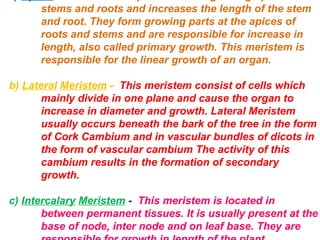
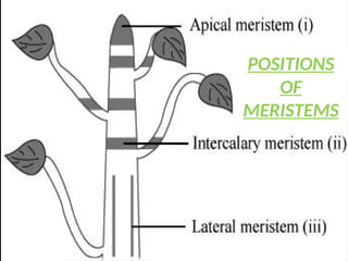
stems and rootsand increases the length of the stem
and root. They form growing parts at the apices of
roots and stems and are responsible for increase in
length, also called primary growth. This meristem is
responsible for the linear growth of an organ.
b) Lateral Meristem - This meristem consist of cells which
mainly divide in one plane and cause the organ to
increase in diameter and growth. Lateral Meristem
usually occurs beneath the bark of the tree in the form
of Cork Cambium and in vascular bundles of dicots in
the form of vascular cambium. The activity of this
cambium results in the formation of secondary
growth.
c) Intercalary Meristem - This meristem is located in
between permanent tissues. It is usually present at the
base of node, inter node and on leaf base. They are
stems and roots and increases the length of the stem
and root. They form growing parts at the apices of
roots and stems and are responsible for increase in
length, also called primary growth. This meristem is
responsible for the linear growth of an organ.
b) Lateral Meristem - This meristem consist of cells which
mainly divide in one plane and cause the organ to
increase in diameter and growth. Lateral Meristem
usually occurs beneath the bark of the tree in the form
of Cork Cambium and in vascular bundles of dicots in
the form of vascular cambium The activity of this
cambium results in the formation of secondary
growth.
c) Intercalary Meristem - This meristem is located in
between permanent tissues. It is usually present at the
base of node, inter node and on leaf base. They are
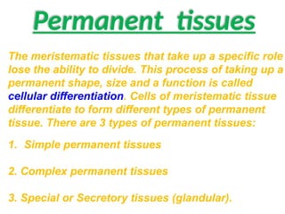
Permanent tissues
The meristematictissues that take up a specific role
lose the ability to divide. This process of taking up a
permanent shape, size and a function is called
cellular differentiation. Cells of meristematic tissue
differentiate to form different types of permanent
tissue. There are 3 types of permanent tissues:
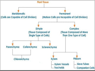
1. Simple permanent tissues
2. Complex permanent tissues
3. Special or Secretory tissues (glandular).
10.
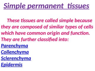
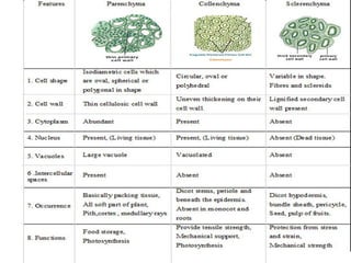
Simple permanent tissues
Thesetissues are called simple because
they are composed of similar types of cells
which have common origin and function.
They are further classified into:
Parenchyma
Collenchyma
Sclerenchyma
Epidermis
12.
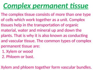
Complex permanent tissue
Thecomplex tissue consists of more than one type
of cells which work together as a unit. Complex
tissues help in the transportation of organic
material, water and mineral up and down the
plants. That is why it is also known as conducting
and vascular tissue. The common types of complex
permanent tissue are:
1. Xylem or wood
2. Phloem or bast.
Xylem and phloem together form vascular bundles.
13.
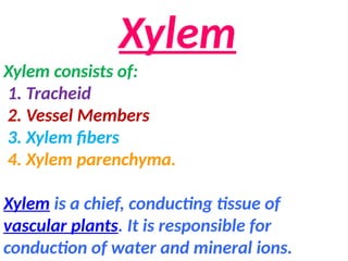
Xylem
Xylem consists of:
1.Tracheid
2. Vessel Members
3. Xylem fibers
4. Xylem parenchyma.
Xylem is a chief, conducting tissue of
vascular plants. It is responsible for
conduction of water and mineral ions.
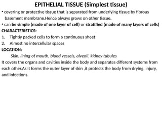
EPITHELIAL TISSUE (Simplesttissue)
• covering or protective tissue that is separated from underlying tissue by fibrous
basement membrane.Hence always grows on other tissue.
• can be simple (made of one layer of cell) or stratified (made of many layers of cells)
CHARACTERISTICS:
1. Tightly packed cells to form a continuous sheet
2. Almost no intercellular spaces
LOCATION:
Skin, lining of mouth, blood vessels, alveoli, kidney tubules
It covers the organs and cavities inside the body and separates different systems from
each other.As it forms the outer layer of skin ,it protects the body from drying, injury,
and infections.
18.
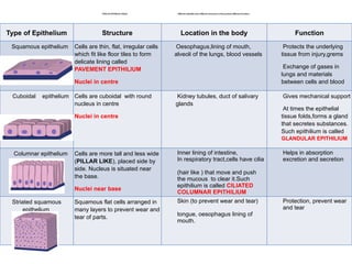
TYPES OF EPITHELIALTISSUE Different epithelia show different structures as they perform different functions
Type of Epithelium Structure Location in the body Function
Squamous epithelium Cells are thin, flat, irregular cells
which fit like floor tiles to form
delicate lining called
PAVEMENT EPITHILIUM
Nuclei in centre
Oesophagus,lining of mouth,
alveoli of the lungs, blood vessels
Protects the underlying
tissue from injury,grems
Exchange of gases in
lungs and materials
between cells and blood
Cuboidal epithelium Cells are cuboidal with round
nucleus in centre
Nuclei in centre
Kidney tubules, duct of salivary
glands
Gives mechanical support
At times the epithelial
tissue folds,forms a gland
that secretes substances.
Such epithilium is called
GLANDULAR EPITHILIUM
Columnar epithelium Cells are more tall and less wide
(PILLAR LIKE), placed side by
side. Nucleus is situated near
the base.
Nuclei near base
Inner lining of intestine,
In respiratory tract,cells have cilia
(hair like ) that move and push
the mucous to clear it.Such
epithilium is called CILIATED
COLUMNAR EPITHILIUM
Helps in absorption
excretion and secretion
Striated squamous
epithelium
Squamous flat cells arranged in
many layers to prevent wear and
tear of parts.
Skin (to prevent wear and tear)
tongue, oesophagus lining of
mouth.
Protection, prevent wear
and tear
19.
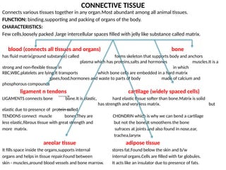
CONNECTIVE TISSUE
Connects varioustissues together in any organ.Most abundant among all animal tissues.
FUNCTION: binding,supporting and packing of organs of the body.
CHARACTERISTICS:
Few cells,loosely packed ,large intercellular spaces filled with jelly like substance called matrix.
blood (connects all tissues and organs) bone
has fluid matrix(ground substance) called forms skeleton that supports body and anchors
plasma which has proteins,salts and hormones muscles.It is a
strong and non-flexible tissue in in which
RBC,WBC,platelets are lying.It transports which bone cells are embedded in a hard matrix
gases,food,hormones and waste to parts of body made of calcium and
phosphorous compounds
ligament n tendons cartilage (widely spaced cells)
LIGAMENTS connects bone bone.It is elastic, hard elastic tissue softer than bone.Matrix is solid
has strength and very less matrix. but
elastic due to presence of protein called
TENDONS connect muscle bones.They are CHONDRIN which is why we can bend a cartilage
less elastic,fibrous tissue with great strength and but not the bone.It smoothens the bone
more matrix. sufraces at joints and also found in nose,ear,
trachea,larynx
areolar tissue adipose tissue
It fills space inside the organs,supports internal stores fat.Found below the skin and b/w
organs and helps in tissue repair.Found between internal organs.Cells are filled with far globules.
skin - muscles,around blood vessels and bone marrow. It acts like an insulator due to presence of fats.
20.
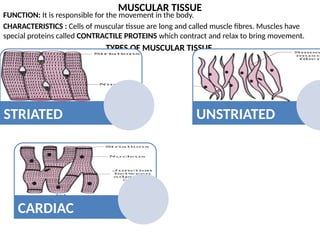
MUSCULAR TISSUE
FUNCTION: Itis responsible for the movement in the body.
CHARACTERISTICS : Cells of muscular tissue are long and called muscle fibres. Muscles have
special proteins called CONTRACTILE PROTEINS which contract and relax to bring movement.
TYPES OF MUSCULAR TISSUE
STRIATED UNSTRIATED
CARDIAC
21.
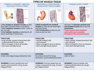
TYPES OF MUSCLETISSUE
STRIPED or STRIATED/ SKELETAL/
VOLUNTARY MUSCLE FIBRES
UNSTRIPED or NON-STRIATED /
INVOLUNTARY MUSCLE FIBRES
CARDIAC MUSCLE FIBRES
Called striped or striated muscles as they
have light and dark bands or striations.
Called voluntary muscles as they move as
per our will.
Called skeletal muscles as attached to all
bones and help in movement.
Called unstriped/non striated
muscles as they do not show light
and dark bands.
Called involuntary as they do not
move as per our will.
Structure in between striated and
non-striated muscle fibres and are
involuntary.
These muscles show rhythmic
contraction and relaxation
throughout life and pump blood.
STRUCTURE
• long cylinder shaped unbranched cells
• multinucleated (many nuclei)
• fibres arranged in form of bundles
• have dark and light bands
STRUCTURE
• spindle shaped cells
• uninucleated
• fibres arranged in form of sheets
• dark and light bands absent
STRUCTURE
• short cylinder shaped,branched cells
• uninucleated
• fibres arranged in form of network
• faint dark and light bands
LOCATION
Limbs (arms,legs),tongue,body,face neck
LOCATION
Iris,ureters,bronchi of lungs,
alimentary canal wall,blood vessels
LOCATION
walls of heart
WORKING Contract quickly but can't
remain contracted for a long .So get
fatigued/tired.
WORKING Contract slowly but can
remain contracted for long .So don’t
get fatigued/tired.
WORKING Contract quickly and
rhythmically.Therefore do not get
fatigued/tired.
22.
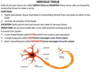
NERVOUS TISSUE
Cells ofnervous tissue are called NERVE CELLS or NEURONS.Many nerve cells are bound by
connective tissue to make a nerve.
FUNCTION:
1. highly specialised tissue that helps in transmitting stimuli from one place to other in the
body.
2. controls all activities of the body.
LOCATION: Brain,spinal cord and nerves are made of nervous tissue.
STRUCTURE: Its structure looks like a tree with branches coming out of it.
A neuron has 3 parts –
3. a star shaped body called CYTON which has nucleus and cytoplasm
4. a single long part called AXON(carry messages away from cyton)
5. short ,branched part called DENDRITES(carry messages towards cyton).