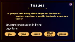
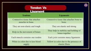
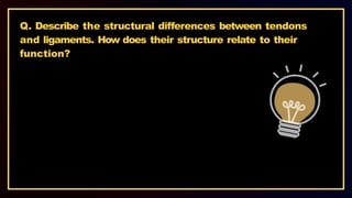
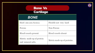
‘A group ofcells having similar shape and function act
together to perform a specific function is known as a
tissue.’
Tissues
CELL
S
TISSUE
S
ORGANS
ORGAN
SYSTE
M
ORGANIS
M
Structural organisation in living
organisms:
4.
Q. What isthe utility of tissues in multicellular
organisms?
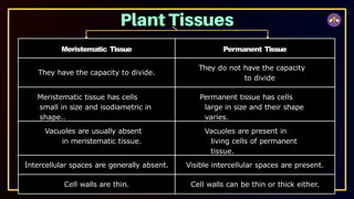
Meristematic Tissue PermanentTissue
They have the capacity to divide.
They do not have the capacity
to divide
Meristematic tissue has cells
small in size and isodiametric in
shape..
Permanent tissue has cells
large in size and their shape
varies.
Vacuoles are usually absent
in meristematic tissue.
Vacuoles are present in
living cells of permanent
tissue.
Intercellular spaces are generally absent. Visible intercellular spaces are present.
Cell walls are thin. Cell walls can be thin or thick either.
7.
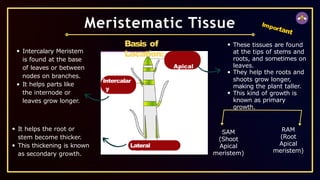
Intercalar
y
Meristem
Lateral
Meristem
Meristematic Tissue
Basis of
Location:
Apical
Meriste
m
Thesetissues are found
at the tips of stems and
roots, and sometimes on
leaves.
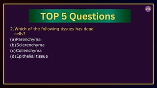
They help the roots and
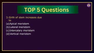
shoots grow longer,
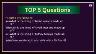
making the plant taller.
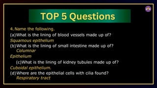
This kind of growth is
known as primary
growth.
Intercalary Meristem
is found at the base
of leaves or between
nodes on branches.
It helps parts like
the internode or
leaves grow longer.
It helps the root or
stem become thicker.
This thickening is known
as secondary growth.
Important
SAM
(Shoot
Apical
meristem)
RAM
(Root
Apical
meristem)
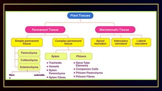
The permanent tissuesare composed of those cells which have lost
their capability to divide.
They have definite shape, size and thickness. The permanent tissue may
be dead or living.
The division & differentiation of the cells of meristematic tissues give rise
to permanent tissues.
In cell differentiation, developing tissue and organs change from simple
to more complex forms to become specialized for specific functions.
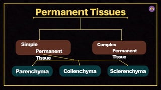
Permanent tissues are classified into two types on the basis of Structure
and Composition i.e. Simple Permanent Tissues and Complex Permanent
Tissues.
Permanent Tissue
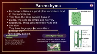
Chlorenchyma
Tissues
Parenchyma tissues supportplants and store food
in roots and stems.
They form the basic packing tissue in
plants. The cells are simple and not very
specialized. These cells have thin walls and
are alive.
They have large gaps between them
because they
are loosely packed.
Parenchyma
Parenchyma tissues which
contain chlorophyll and can
perform photosynthesis.
Aerenchyma Tissues
Parenchyma tissues with large air spaces
help plants float by making them buoyant.
13.
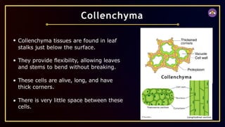
Collenchyma tissues arefound in leaf
stalks just below the surface.
They provide flexibility, allowing leaves
and stems to bend without breaking.
These cells are alive, long, and have
thick corners.
There is very little space between these
cells.
Collenchyma
14.
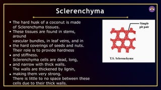
The hard huskof a coconut is made
of Sclerenchyma tissues.
These tissues are found in stems,
around
vascular bundles, in leaf veins, and in
the hard coverings of seeds and nuts.
Their role is to provide hardness
and stiffness.
Sclerenchyma cells are dead, long,
and narrow with thick walls.
The walls are thickened by lignin,
making them very strong.
There is little to no space between these
cells due to their thick walls.
Sclerenchyma
15.
Q. How aresimple tissues different from complex tissues
in plants?
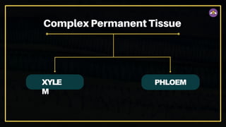
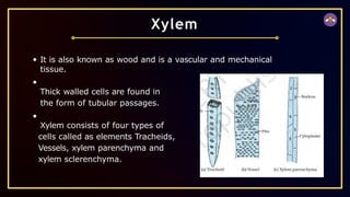
It is alsoknown as wood and is a vascular and mechanical
tissue.
Thick walled cells are found in
the form of tubular passages.
Xylem consists of four types of
cells called as elements Tracheids,
Vessels, xylem parenchyma and
xylem sclerenchyma.
Xylem
18.
TRACHEIDS
They are elongatedangular
dead cells (primitive elements)
mainly involved in conduction
of water and minerals in
gymnosperms.
VESSELS
They are advance element
(generally found in
angiosperms).
Vessels are cylindrical tube like
structures placed one above
the other end to end which
form a continuous channel for
efficient conduction of water.
XYLEM PARENCHYMA
They are small & thick walled parenchymatous cells subjected
for storage of starch (food).
19.
XYLEM SCLERENCHYMA (XYLEM
FIBRE)
Theyare non-living fibres with thick walls and narrow cavities
provide mechanical support.
Except xylem parenchyma all other xylem elements are
dead. The annual rings present in the trunk of a tree are
xylem rings.
By counting the number of annual rings, we can determine the age
of a tree.
20.
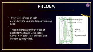
They also consistof both
parenchymatous and sclerenchymatous
cells.
Phloem consists of four types of
element which are Sieve tubes,
Companion cells, Phloem fibre and
Phloem parenchyma.
PHLOEM
Important
21.
SIEVE
TUBES
Sieve tubes areslender tube like structures made up of elongated,
thin walled cells placed end to end.
The end walls of sieve tube cells are perforated by numerous
pores, called as sieve plates.
Nucleus of sieve cell degenerates at maturity. However,
cytoplasm persists, because of protoplasmic continuation of
sieve tube with companion cell through plasmodesmata.
Sieve cells possess slime protein or protein which is concerned
with growth and repair of sieve cells.
22.
COMPANION
CELLS
Companion cells havedense cytoplasm and prominent nuclei.
Sieve cells & companion cells are so called sister cells because
they originate from single mother cell.
PHLOEM FIBRE
They give mechanical support to sieve tubes.
PHLOEM PARENCHYMA
They store food and help in radial conduction of food.
23.
Feature
Function
Direction of
Conduction
Conducting Channels
LivingElements
Dead Elements
Mechanical
Support
Xylem
Conducts water and minerals
Mostly unidirectional (from roots
to apical parts)
Tracheids and vessels
(dead elements)
Xylem parenchyma (only
living element)
Tracheids, vessels, and fibers
Provides mechanical strength
to the plant
Phloem
Conducts organic solutes or
food materials
Bidirectional (from leaves to
storage/growing parts or vice
versa)
Sieve tubes (living elements)
Sieve tubes, companion cells,
and phloem parenchyma
Phloem fibers
Does not perform a
mechanical function
Important
The epidermis protectsplants from injury, pathogens, and water loss.
In desert plants (xerophytes), the thick cuticle of the epidermis
reduces water loss by lowering the rate of transpiration.
The epidermis in certain areas also allows for gas exchange
during respiration and photosynthesis.
In roots, epidermal cells increase the surface area, aiding in
better absorption of water and nutrients.
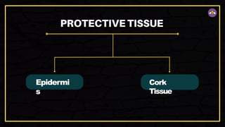
Epidermis
26.
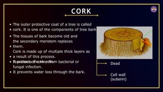
CORK
The outer protectivecoat of a tree is called
cork. It is one of the components of tree bark.
The tissues of bark become old and
the secondary meristem replaces
them.
Cork is made up of multiple thick layers as
a result of this process.
Functions of cork cells:
It protects the tree from bacterial or
fungal infection.
It prevents water loss through the bark.
Dead
Cell wall
(suberin)
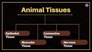
Epithelial Tissue
Always growson some other types of tissue.
Cells of epithelium are set very close to each other and the tissue rests
on a non-cellular basement membrane.
Consists of single layer of cells.
Blood vessels are absent and non-nervous in nature.
It covers all the organs and lines the cavities of hollow organs like
stomach. It is primarily protective in function.
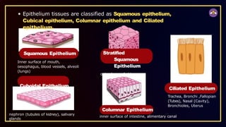
29.
Epithelium tissues areclassified as Squamous epithelium,
Cubical epithelium, Columnar epithelium and Ciliated
epithelium.
Squamous Epithelium
Inner surface of mouth,
oesophagus, blood vessels, alveoli
(lungs)
Cuboidal Epithelium
Stratified
Squamous
Epithelium
outer layer of skin
nephron (tubules of kidney), salivary
glands
Ciliated Epithelium
Trachea, Bronchi ,Fallopian
(Tubes), Nasal (Cavity),
Bronchioles, Uterus
Columnar Epithelium
inner surface of intestine, alimentary canal
30.
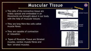
The cells ofthe connective tissue are
widely spaced and embedded in an
Movements are brought about in our body
with the help of muscular tissues.
They are long fibre-like cells called
muscle fibres.
They are capable of contraction
or relaxation.
Types of Muscular Tissue are Striated
muscles, cardiac muscle fibres and
Non- striated muscles.
Muscular Tissue Important
striated muscle
smooth
muscle
cardiac
muscle
31.
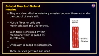
Striated Muscles/ Skeletal
muscles
Theyare also called as voluntary muscles because these are under
the control of one’s will.
Muscle fibres or cells are
multinucleated and unbranched.
Each fibre is enclosed by thin
membrane which is called as
sarcolemma.
Cytoplasm is called as sarcoplasm.
These muscles get tired and need
32.
Cardiac Muscle
Fibres
They areonly involuntary muscles.
Only found in the walls of heart.
Their structure is in between the striated and non-striated
muscles.
They are uninucleated and branched. Branches are united
by intercalated disc.
In these muscles rhythmic contraction and relaxation
occurs throughout the life.
33.
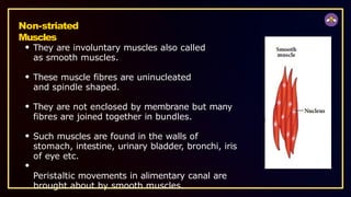
Non-striated
Muscles
They are involuntarymuscles also called
as smooth muscles.
These muscle fibres are uninucleated
and spindle shaped.
They are not enclosed by membrane but many
fibres are joined together in bundles.
Such muscles are found in the walls of
stomach, intestine, urinary bladder, bronchi, iris
of eye etc.
Peristaltic movements in alimentary canal are
brought about by smooth muscles.
34.
Feature
Control
Locatio
n
Structu
re
Membr
ane
Cytopla
sm
Striated Muscle (Voluntary
Muscles)
Voluntary(under the control
of one’s will)
Attached to the skeleton
Multinucleated and
unbranched fibers
Enclosed by a thin membrane
called sarcolemma
Called sarcoplasm
Get tired and need rest
Striated (striped appearance)
Responsible for
voluntary movements
Non-Striated Muscle (Smooth
Muscles)
Involuntary
Walls of stomach, intestine,
urinary bladder, bronchi, iris,
etc.
Uninucleated, spindle-shaped
fibers
Not enclosed by a membrane,
fibers joined in bundles
Called sarcoplasm
Do not get tired easily
Non-striated (smooth
appearance)
Controls involuntary movements
like peristalsis
Cardiac Muscle
Involuntary
Walls of the heart
Uninucleated and branched
fibers
Enclosed by sarcolemma
with intercalated discs
Called sarcoplasm
Do not get tired, rhythmic
contraction and relaxation
occur
Intermediate structure
between striated and non-
striated
Controls rhythmic contraction
Important
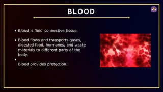
BLOOD
Blood is fluidconnective tissue.
Blood flows and transports gases,
digested food, hormones, and waste
materials to different parts of the
body.
Blood provides protection.
37.
Plasma
It forms 55%part of blood. It consistsof 90-91% of water, 7%
of protein (Albumin, fibrinogen, globulin), 0.9% of inorganic salt
etc.
Corpuscles
Forms 45% part of blood.
RBCs
They are also called as erthyrocytes, containing red coloured
respiratory pigment called haemoglobin that helps in transportation
of oxygen.
38.
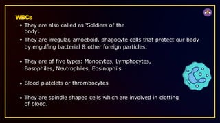
WBCs
They are alsocalled as ‘Soldiers of the
body’.
They are irregular, amoeboid, phagocyte cells that protect our body
by engulfing bacterial & other foreign particles.
They are of five types: Monocytes, Lymphocytes,
Basophiles, Neutrophiles, Eosinophils.
Blood platelets or thrombocytes
They are spindle shaped cells which are involved in clotting
of blood.
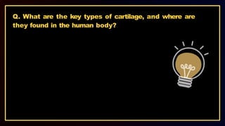
Q. What arethe key types of cartilage, and where are
they found in the human body?
43.
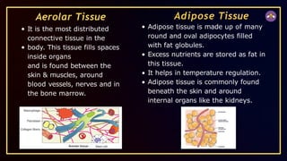
Aerolar Tissue
It isthe most distributed
connective tissue in the
body. This tissue fills spaces
inside organs
and is found between the
skin & muscles, around
blood vessels, nerves and in
the bone marrow.
Adipose Tissue
Adipose tissue is made up of many
round and oval adipocytes filled
with fat globules.
Excess nutrients are stored as fat in
this tissue.
It helps in temperature regulation.
Adipose tissue is commonly found
beneath the skin and around
internal organs like the kidneys.
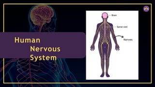
They are highlyspecialized tissue due to which the animals are able
to perceive and respond to the stimuli.
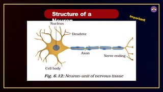
Their functional unit is called as nerve cell or
neuron. Structural and functional unit of nervous
system.
Longest cell in body.
Cell body is called cyton which is covered by
Nervous Tissue
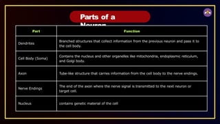
Parts of a
Neuron
PartFunction
Dendrites
Branched structures that collect information from the previous neuron and pass it to
the cell body.
Cell Body (Soma)
Contains the nucleus and other organelles like mitochondria, endoplasmic reticulum,
and Golgi body.
Axon Tube-like structure that carries information from the cell body to the nerve endings.
Nerve Endings
The end of the axon where the nerve signal is transmitted to the next neuron or
target cell.
Nucleus contains genetic material of the cell
48.
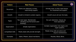
Feature Plant TissuesAnimal Tissues
Movement
Plants are stationary, so they
need strong support.
Animals move, so they need tissues
for movement and support.
Growth Growth is limited to certain regions. Growth occurs all over the body.
Types
Two main types: Meristematic
(dividing) and Permanent (non-
dividing).
No such distinction; tissues are
more specialized.
Organization Simple organization.
Complex organization into organs and
organ systems.
Living/Dead Cells Mostly dead cells provide strength.
Mostly living cells are active and
require maintenance.
Examples Xylem, Phloem, Apical meristems. Muscles, Blood, Bone.
49.
Q. Describe thestructure and functions of the central
nervous system (CNS).
50.
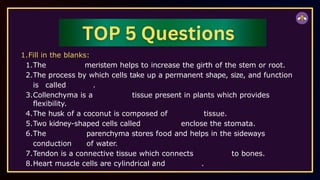
1.Fill in theblanks:
1.The meristem helps to increase the girth of the stem or root.
2.The process by which cells take up a permanent shape, size, and function
is called .
3.Collenchyma is a tissue present in plants which provides
flexibility.
4.The husk of a coconut is composed of tissue.
5.Two kidney-shaped cells called enclose the stomata.
6.The parenchyma stores food and helps in the sideways
conduction of water.
7.Tendon is a connective tissue which connects to bones.
8.Heart muscle cells are cylindrical and .
51.
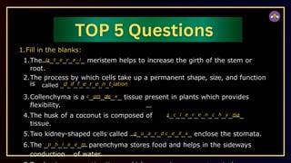
1.Fill in theblanks:
1.The la_t_e_r_a_l_ meristem helps to increase the girth of the stem or
root.
2.The process by which cells take up a permanent shape, size, and function
is called _d_if_f_e_r_e_n_t.iation
3.Collenchyma is a c_om_ple_x_ tissue present in plants which provides
flexibility.
4.The husk of a coconut is composed of s_c_l_e_r_e_n_c_h_y_ma_
tissue.
5.Two kidney-shaped cells called g_u_a_r_d c_e_ll_s_ enclose the stomata.
6.The _p_h_l_o_e_m parenchyma stores food and helps in the sideways
conduction of water.
52.
2.Which of thefollowing tissues has dead
cells?
(a)Parenchyma
(b)Sclerenchyma
(c)Collenchyma
(d)Epithelial tissue
53.
3.Girth of stemincreases due
to
(a)Apical meristem
(b)Lateral meristem
(c)Intercalary meristem
(d)Vertical meristem
54.
4.Name the following.
(a)Whatis the lining of blood vessels made up
of?
(b)What is the lining of small intestine made up
of?
(c)What is the lining of kidney tubules made up
of?
(d)Where are the epithelial cells with cilia found?
55.
4.Name the following.
(a)Whatis the lining of blood vessels made up of?
Squamous epithelium
(b)What is the lining of small intestine made up of?
Columnar
Epithelium
(c)What is the lining of kidney tubules made up of?
Cuboidal epithelium.
(d)Where are the epithelial cells with cilia found?
Respiratory tract
56.
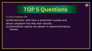
5.Give reasons for
(a)Meristematiccells have a prominent nucleus and
dense cytoplasm but they lack vacuole.
(b)Intercellular spaces are absent in sclerenchymatous
tissues.
57.
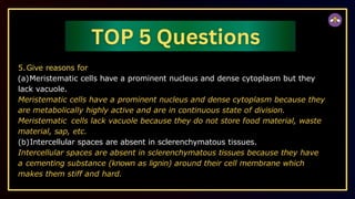
5.Give reasons for
(a)Meristematiccells have a prominent nucleus and dense cytoplasm but they
lack vacuole.
Meristematic cells have a prominent nucleus and dense cytoplasm because they
are metabolically highly active and are in continuous state of division.
Meristematic cells lack vacuole because they do not store food material, waste
material, sap, etc.
(b)Intercellular spaces are absent in sclerenchymatous tissues.
Intercellular spaces are absent in sclerenchymatous tissues because they have
a cementing substance (known as lignin) around their cell membrane which
makes them stiff and hard.