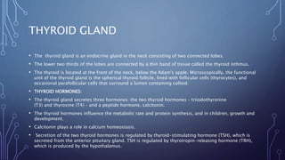
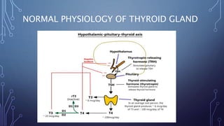
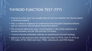
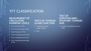
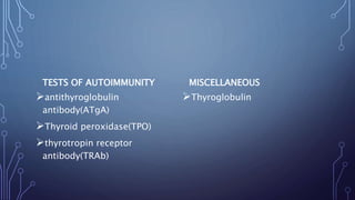
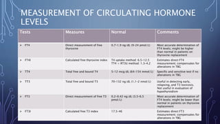
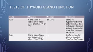
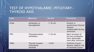
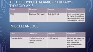
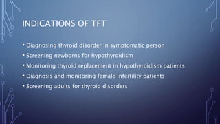
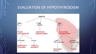
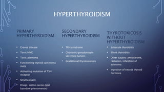
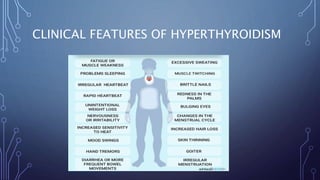
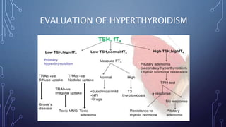
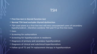
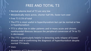
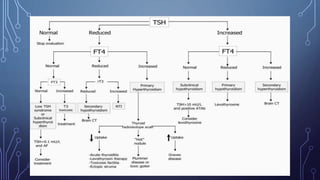
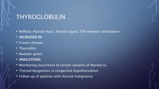
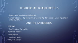
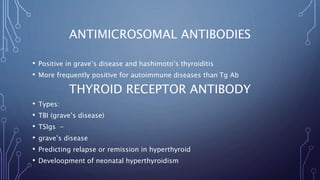
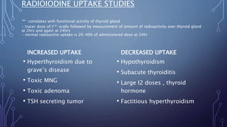
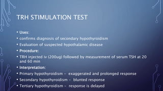
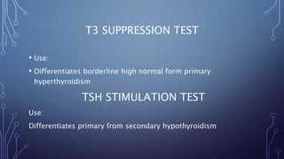
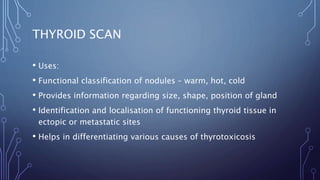
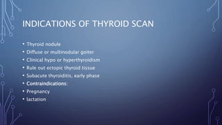
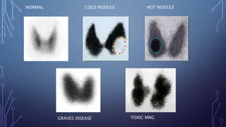
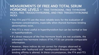
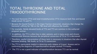
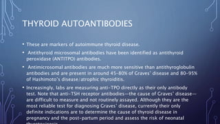
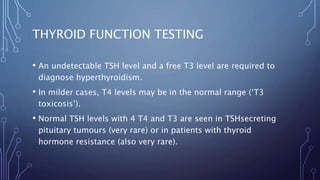
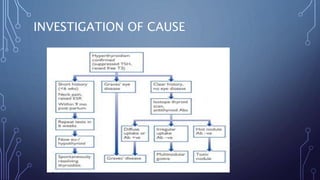
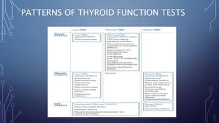
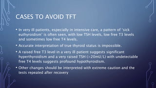
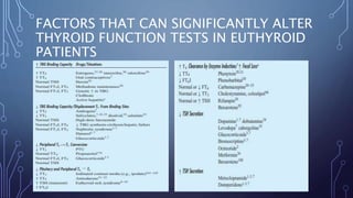
THYROID
FUNCTION TEST
PRESENTED BY:
SHAISTA SUMAYYA
PHARMD
SULTAN UL ULOOM COLLEGE OF PHARMACY, HYDERABAD.
THYROID GLAND
• The thyroid gland is an endocrine gland in the neck consisting of two connected lobes.
• The lower two thirds of the lobes are connected by a thin band of tissue called the thyroid isthmus.
• The thyroid is located at the front of the neck, below the Adam's apple. Microscopically, the functional
unit of the thyroid gland is the spherical thyroid follicle, lined with follicular cells (thyrocytes), and
occasional parafollicular cells that surround a lumen containing colloid.
• THYROID HORMONES:
• The thyroid gland secretes three hormones: the two thyroid hormones – triiodothyronine
(T3) and thyroxine (T4) – and a peptide hormone, calcitonin.
• The thyroid hormones influence the metabolic rate and protein synthesis, and in children, growth and
development.
• Calcitonin plays a role in calcium homeostasis.
• Secretion of the two thyroid hormones is regulated by thyroid-stimulating hormone (TSH), which is
secreted from the anterior pituitary gland. TSH is regulated by thyrotropin-releasing hormone (TRH),
which is produced by the hypothalamus.
NORMAL PHYSIOLOGY OF THYROID GLAND
THYROID FUNCTION TEST (TFT)
• Thyroid function tests are usually done to find out whether the thyroid gland
is working properly.
• This is mainly to diagnose an underactive thyroid gland (hypothyroidism)
and an overactive thyroid gland (hyperthyroidism).
• The principal laboratory tests recommended in the initial evaluation of
thyroid disorders are the TSH and the FT4 levels.
• Positive thyroid antibodies indicate an autoimmune thyroid etiology.
• Adjuncts to the previous tests include the total T3 (TT3), free T3 (FT3) or
FT3 index (FT3I), RAIU and scan, TRAb, ultrasound, and FNA biopsy
TFT CLASSIFICATION
MEASUREMENT OF
CIRCULATING
HORMONE LEVELS
 Free Thyroxine (FT4)
 Free Thyroxine Index (FT4I)
 Total Thyroxine (TT4)
 Total Triiodothyronine (TT3)
 Free Triiodothyronine (FT3)
 Free Triiodothyronine Index
(FT3I)
TESTS OF THYROID
GLAND FUNCTION
 radioactive iodine
uptake (RAIU)
 Scan
TEST OF
HYPOTHALAMIC-
PITUITARY-THYROID
AXIS
 thyroid-stimulating hormone
(TSH)
TESTS OF AUTOIMMUNITY
antithyroglobulin
antibody(ATgA)
Thyroid peroxidase(TPO)
thyrotropin receptor
antibody(TRAb)
MISCELLANEOUS
Thyroglobulin
MEASUREMENT OF CIRCULATING HORMONE
LEVELS
Tests Measures Normal Comments
 FT4 Direct measurement of free
thyroxine
0.7–1.9 ng/dL (9–24 pmol/L) Most accurate determination of
FT4 levels; might be higher
than normal in patients on
thyroxine replacement
 FT4I Calculated free thyroxine index T4 uptake method: 6.5–12.5
TT4 × RT3U method: 1.3–4.2
Estimates direct FT4
measurement; compensates for
alterations in TBG
 TT4 Total free and bound T4 5–12 mcg/dL (64–154 mmol/L) Specific and sensitive test if no
alterations in TBG
 TT3 Total free and bound T3 70–132 ng/dL (1.1–2 nmol/L) Useful in detecting early,
relapsing, and T3 toxicosis.
Not useful in evaluation of
hypothyroidism
 FT3 Direct measurement of free T3 0.2–0.42 ng/dL (3.5–6.5
pmol/L)
Most accurate determination of
FT4 levels; might be lower than
normal in patients on thyroxine
replacement
 FT3I Calculated free T3 index 17.5–46 Estimates direct FT3
measurement; compensates for
alterations in TBG
TESTS OF THYROID GLAND FUNCTION
Tests Measures Normals Comments
RAIU Gland’s use of
iodine after trace
dose of either 123I or
131I
5%–35% Useful in
hyperthyroidism to
determine RAI dose
in Graves’; does not
provide information
regarding hormone
synthesis
Scan Gland size, shape,
and tissue activity
after 123I or 99mTc
— Useful in nodular
disease to detect
“cold” or “hot” areas
TEST OF HYPOTHALAMIC-PITUITARY-
THYROID AXIS
Tests Measures Normal Comments
ATgA Antibodies to
thyroglobulin
<1 IU/mL Present in
autoimmune thyroid
disease;
undetectable during
remission
TPO Thyroperoxidase
antibodies
<1 IU/mL More sensitive of
the two antibodies;
titers detectable
even after remission
TRAb Thyroid receptor
stimulating
antibody
<125% Confirms Graves’
disease; detects risk
of neonatal Graves’
TEST OF HYPOTHALAMIC-PITUITARY-
THYROID AXIS
Tests Measures Normal Comments
TSH Pituitary TSH level 0.5–5 μU/mL Most sensitive index
for hyperthyroidism,
hypothyroidism, and
replacement therapy
MISCELLANEOUS
Tests Measures Normal Comments
Thyroglobulin Colloid protein of
normal thyroid
gland
<56 ng/mL Marker for recurrent
thyroid cancer or
metastases in
thyroidectomized
patients
INDICATIONS OF TFT
• Diagnosing thyroid disorder in symptomatic person
• Screening newborns for hypothyroidism
• Monitoring thyroid replacement in hypothyroidism patients
• Diagnosis and monitoring female infertility patients
• Screening adults for thyroid disorders
ABNORMALITIES OF
THYROID
HYPOTHYROIDISM
• Causes:
• PRIMARY(high TSH)
• Autoimmune: Hashimoto’s, Atrophic
• Iatrogenic: subtotal/total thyroidectomy, external neck irradiation
• Drugs: lithium, antithyroid drugs, p-amino salicylic acid
• Congenital hypothyroidism: absent/ectopic thyroid gland, dyshormonogenesis, TSHR mutation
• Iodine deficiency
• Infiltrative disorders
• SECONDARY(low TSH)
• Hypopitutarism: tumors, surgery, irradiation, infiltrative disorders, Sheehan’s syndrome, trauma
• Isolated TSh deficiency or inactivity
• TERTIARY(low TSH , low TRH)
• Diseases of hypothalamus
• TRANSIENT:
• Silent/postpartum thyroiditis, subacute thyroiditis
CLINICAL FEATURES OF HYPOTHYROIDISM
EVALUATION OF HYPOTHYROIDISM
HYPERTHYROIDISM
PRIMARY
HYPERTHYROIDISM
• Graves disease
• Toxic MNG
• Toxic adenoma
• Functioning thyroid carcinoma
mets
• Activating mutation of TSH
receptor
• Struma ovarii
• Drugs: iodine excess (jod
basedow phenomenon)
SECONDARY
HYPERTHYROIDISM
• TRH syndrome
• Chorionic gonadotropin
secreting tumors
• Gestational thyrotoxicosis
THYROTOXICOSIS
WITHOUT
HYPERTHYROIDISM
• Subacute thyroiditis
• Silent thyroiditis
• Other causes: amiodarone,
radiation, infarction of
adenoma
• Ingestion of excess thyroid
hormone
CLINICAL FEATURES OF HYPERTHYROIDISM
EVALUATION OF HYPERTHYROIDISM
TSH
• First line test in thyroid function test
• Normal TSH level excludes thyroid dysfunction
• TSH used alone as a first line test will miss unsuspected cases of secondary
hypothyroidism , therefore combine TSH and T4 as first line tests.
• Uses:
• Screening for euthyroidism
• Screening for hypothyroidism in newborns
• Diagnosis of primary and secondary hypothyroidism
• Diagnosis of clinical and subclinical hyperthyroidism
• Follow up of T3 and T4 replacement therapy in hypothyroidism
TSH
INCREASE
• Primary hypothyroidism
• Addison’s disease
• Anti TSH antibodies
• Pitutary adenoma
• Postoperative
• Thyroiditis
• Drugs: amiodarone, bensaarazide,
clomiphene, lithium, methimazole,
metochlorpramide, propylthiouracil
DECREASE
• Primary hyperthyroidism
• Hashimoto’s thyroiditis
• Drugs: ASA, heparin,
ketoconazole, T3, dopamine,
glucocorticoids
TOTAL THYROXINE
• Includes free thyroxine and protein bound thyroxine
• The FT4 are the most reliable tests for the evaluation of
hormone concentrations, especially when thyroid hormone
binding abnormalities exist.
• falsely elevated levels of TT4 are common in the euthyroid
pregnant woman .
THYROXINE
INCREASE
• Hyperthyroidism
• Pitutary TSH secreting
pituitary tumor
• Raised TBG
DECREASE
• Primary hypothyroidism
• Secondary/pituitary
hypothyroidism
• Severe non thyroidal illness
• Decreased TBG
THYROID BINDING GLOBULIN (TBG)
Normal levels: 13 – 39 ug/dl
INCREASE
• Drugs: clofibrate, estrogen,
oral contraceptives, heroin,
methadone
• Genetic
• Pregnancy
• Acute intermittent porphyria
• hyperproteinemia
DECREASE
• Drugs: androgens,
glucocorticoids, phenytoin,
large doses of salicylates
• Malnutrition
• Hypoproteinemia, nephrotic
syndrome
• Cushing’s syndrome
FREE AND TOTAL T3
• Normal plasma level of T3 are very low
• Metabolically more active, shorter half life, faster turn over
• Free T3 0.5% of total
• The FT3 is most useful in hyperthyroidism but can be normal or low
in hypothyroidism
• TT3 is often low in older patients and in many acute and chronic
nonthyroidal illnesses because the peripheral conversion of T4 to T3
is decreased.
• The TT3 is particularly helpful in detecting early relapse of Graves’
disease and in confirming the diagnosis of hyperthyroidism despite
normal TT4 levels.
• Uses:
THYROGLOBULIN
• Reflects thyroid mass, thyroid injury, TSH receptor stimulation
• INCREASED IN:
• Graves disease
• Thyroiditis
• Nodular goiter
• INDICATIONS:
• Monitoring recurrence of certain variants of thyroid ca
• Thyroid dysgenesis in congenital hypothyroidism
• Follow up of patients with thyroid malignancy
THYROID AUTOANTIBODIES
• Diagnosing autoimmune diseases
• Autoantibodies : Tg, thyroid microsomal Ag, TSH receptor, non Tg colloid
antigen, TSH, T4
ANTI Tg ANTIBODIES
POSITIVE:
• hashimoto’s thyroiditis
• grave’s disease
• myxedema
• nontoxic goitre
• thyroid cancer
ANTIMICROSOMAL ANTIBODIES
• Positive in grave’s disease and hashimoto’s thyroiditis
• More frequently positive for autoimmune diseases than Tg Ab
THYROID RECEPTOR ANTIBODY
• Types:
• TBI (grave’s disease)
• TSIgs -
• grave’s disease
• Predicting relapse or remission in hyperthyroid
• Develoopment of neonatal hyperthyroidism
RADIOIODINE UPTAKE STUDIES
- correlates with functional activity of thyroid gland
- tracer dose of I131 orally followed by measurement of amount of radioactivity over thyroid gland
at 2hrs and again at 24hrs
- normal radioactive uptake is 20-40% of administered dose at 24hr
INCREASED UPTAKE
• Hyperthyroidism due to
grave’s disease
• Toxic MNG
• Toxic adenoma
• TSH secreting tumor
DECREASED UPTAKE
• Hypothyroidism
• Subacute thyroiditis
• Large I2 doses , thyroid
hormone
• Factitious hyperthyroidism
TRH STIMULATION TEST
• Uses:
• confirms diagnosis of secondary hypothyroidism
• Evaluation of suspected hypothalamic disease
• Procedure:
• TRH injected iv (200ug) followed by measurement of serum TSH at 20
and 60 min
• Interpretation:
• Primary hypothyroidism - exaggerated and prolonged response
• Secondary hypothyroidism - blunted response
• Tertiary hypothyroidism - response is delayed
T3 SUPPRESSION TEST
• Use:
• Differentiates borderline high normal form primary
hyperthyroidism
TSH STIMULATION TEST
Use:
Differentiates primary from secondary hypothyroidism
THYROID SCAN
• Uses:
• Functional classification of nodules – warm, hot, cold
• Provides information regarding size, shape, position of gland
• Identification and localisation of functioning thyroid tissue in
ectopic or metastatic sites
• Helps in differentiating various causes of thyrotoxicosis
INDICATIONS OF THYROID SCAN
• Thyroid nodule
• Diffuse or multinodular goiter
• Clinical hypo or hyperthyroidism
• Rule out ectopic thyroid tissue
• Subacute thyroiditis, early phase
• Contraindications:
• Pregnancy
• lactation
NORMAL COLD NODULE HOT NODULE
GRAVES DISEASE TOXIC MNG
MEASUREMENTS OF FREE AND TOTAL SERUM
HORMONE LEVELS - FREE THYROXINE, FREE THYROXINE
INDEX, FREE TRIIODOTHYRONINE, AND FREE TRIIODOTHYRONINE
INDEX
• The FT4 and FT3 are the most reliable tests for the evaluation of
hormone concentrations, especially when thyroid hormone binding
abnormalities exist.
• The FT3 is most useful in hyperthyroidism but can be normal or low
in hypothyroidism.
• If a direct measure of the free hormone levels are not available, the
estimated free hormone indices (FT4I, FT3I) can provide comparable
information.
• However, these indices do not correct for changes observed in
patients with “euthyroid sick” nonthyroidal illnesses whose TBG
binding affinity is altered. In these circumstances, the FT4 and FT3
TOTAL THYROXINE AND TOTAL
TRIIODOTHYRONINE
• The total thyroxine (TT4) and total triiodothyronine (TT3) measure both free and bound
(total) serum T4 and T3.
• Because the bound fraction is the major fraction measured, situations that change the
hormone’s affinity for TBG or the TBG level will influence the results.
• For example, falsely elevated levels of TT4 and TT3 are common in the euthyroid
pregnant woman .
• In addition, the TT3 is often low in older patients and in many acute and chronic
nonthyroidal illnesses because the peripheral conversion of T4 to T3 is decreased.
• Therefore, careful interpretation of these tests is necessary in situations that alter thyroid
hormone binding, TBG levels, or T4 to T3 conversion.
• The TT3 is particularly helpful in detecting early relapse of Graves’ disease and in
confirming the diagnosis of hyperthyroidism despite normal TT4 levels.
• The TT3 is not a good indicator of hypothyroidism because TT3 can be normal.
THYROID AUTOANTIBODIES
• These are markers of autoimmune thyroid disease.
• Antithyroid microsomal antibodies have been identified as antithyroid
peroxidase (ANTITPO) antibodies.
• Antimicrosomal antibodies are much more sensitive than antithyroglobulin
antibodies and are present in around 45–80% of Graves’ disease and 80–95%
of Hashimoto’s disease/atrophic thyroiditis.
• Increasingly, labs are measuring anti-TPO directly as their only antibody
test. Note that anti-TSH receptor antibodies—the cause of Graves’ disease—
are difficult to measure and not routinely assayed. Although they are the
most reliable test for diagnosing Graves’ disease, currently their only
definite indications are to determine the cause of thyroid disease in
pregnancy and the post-partum period and assess the risk of neonatal
THYROID FUNCTION TESTING
• An undetectable TSH level and a free T3 level are required to
diagnose hyperthyroidism.
• In milder cases, T4 levels may be in the normal range (‘T3
toxicosis’).
• Normal TSH levels with 4 T4 and T3 are seen in TSHsecreting
pituitary tumours (very rare) or in patients with thyroid
hormone resistance (also very rare).
INVESTIGATION OF CAUSE
ANTI-TSH RECEPTOR ANTIBODY TESTING
• This test is not routinely available in most labs. Although it is
positive in >90% of cases of Graves’ disease, in most cases it
does not alter clinical management. Indications include
distinguishing gestational thyrotoxicosis or post-partum
thyroiditis from Graves’ disease, indicating the risk of neonatal
thyrotoxicosis and (controversial) predicting recurrence after a
course of thionamide drug therapy
BIOCHEMICAL DIAGNOSIS
• 4 TSH with T4 in the normal range is referred to as subclinical
hypothyroidism. 4 TSH with 5 T4 is overt hypothyroidism. 5 T4
with TSH in the normal range may be due to pituitary failure (2°
hypothyroidism) and if Repeat tests in 6 weeks Now eu-/
hypothyroid Spontaneously resolving thyroiditis
Hyperthyroidism confirmed (suppressed TSH, raised free T3)
Short history (
DIFFERENTIAL DIAGNOSIS (CAUSES)
• n iodine sufficient countries, the vast majority of spontaneous
hypothyroidism is due to autoimmune thyroiditis (Hashimoto’s
disease if goitre present, atrophic thyroiditis if goitre absent)—
antithyroid antibodies present in 80–90% of cases. Other common
causes are post-thyroidectomy, post-radioiodine therapy and side
effects of amiodarone or lithium. Rarer causes include treatment with
cytokines (e.g. interferons, GM-CSF, interleukin-2), vast excess
iodine intake (iodine drops, water purifying tablets), congenital
hypothyroidism (caused by a variety of genetic defects, should be
detected by neonatal screening programme), iodine deficiency
(urinary iodide excretion
DIAGNOSTIC CATCHES
• 4 TSH and 5 T4 always represents hypothyroidism. If the TSH alone is 4 and
the T4 is not even slightly low, a heterophile antibody interfering in the TSH
assay may be present in the patient’s serum. This is especially likely if there
is no change in TSH level after thyroxine treatment but the T4 level rises
(confirming compliance with tablets). For unusual patterns of thyroid
function tests, see Fig. 2.14. Note that within the first 1–3 months (or
longer) after treatment of hyperthyroidism, profound hypothyroidism may
develop with a 5 T4 but the TSH may still be suppressed or only mildly
raised due to the long period of TSH suppression prior to treatment. Raised
TSH alone with disproportionate symptoms of lethargy may be seen in
hypoadrenalism. If suspected treat with steroids first as thyroxine may
precipitate an Addisonian crisis.
• Transient hypothyroidism
• Transient or self-resolving hypothyroidism, often preceded by
hyperthyroidism, is seen in viral thyroiditis, after pregnancy
(post-partum thyroiditis) and in some individuals with
autoimmune thyroiditis (positive antithyroid antibodies).
Treatment temporarily with thyroxine is only required if the
patient is very symptomatic. Thyroid function should return to
normal within 6 months. Hypothyroidism may also be transient
in the first 6 months after radioiodine therapy.
SUBCLINICAL HYPOTHYROIDISM
• A raised TSH (<20mU/L) with normal T4/T3 is very common and
seen in 5–10% of women and ~2% of males. It is usually due to
subclinical autoimmune thyroid disease and is frequently discovered
on routine testing. In randomised trials, ~20% of patients obtain
psychological benefit from beginning T4 therapy, in many others it is
probably truly asymptomatic. If antithyroid antibodies are detectable,
the rate of progression to overt hypothyroidism is ~50% at 20 years,
but higher than this with higher initial TSH levels. If the TSH alone is
raised with negative antibodies (or the TSH is normal with raised
antibodies alone), overt hypothyroidism develops in 25% at 20 years.
A reasonable approach is a trial of thyroxine
HYPOTHYROIDISM AND PREGNANCY
• Overt hypothyroidism is associated with poor obstetric outcomes
• subclinical hypothyroidism is associated with a slight reduction in the baby’s IQ and
should be treated.
• Many authorities advocate screening for hypothyroidism in all antenatal patients as early
as possible in pregnancy.
• Patients on T4 need to increase their dose by 50g from the first trimester of pregnancy.
• Maternal thyroxine can compensate for fetal thyroid failure in utero but congenital
hypothyroidism must be detected at birth (screening test) to avoid mental retardation.
• Where the mother and fetus are both hypothyroid—most commonly due to iodine
deficiency—mental retardation can develop in utero (cretinisim).
• Mothers with positive antithyroid antibodies and/or subclinical hypothyroidism have a
50% chance of developing (transient) post-partum thyroiditis.
THYROID FUNCTION TEST RESULTS IN
DIFFERENT THYROID CONDITIONS
Total
T4
Free T4 Total T3 T3 Resin
Uptake
Free
Thyroxine
Index
TSH
Normal 4.5–
10.9
mcg/dL
0.8–2.7
ng/dL
60–181
ng/dL
22% to 34% 1.0–4.3
units
0.5–
4.7
mIU/L
Hyperthyroi
d
↑↑ ↑↑ ↑↑↑ ↑ ↑↑↑ ↓↓
Hypothyroid ↓↓ ↓↓ ↓ ↓↓ ↓↓↓ ↑↑
Increased
TBG
↑ Normal ↑ ↓ Normal Normal
PATTERNS OF THYROID FUNCTION TESTS
CASES TO AVOID TFT
• In very ill patients, especially in intensive care, a pattern of ‘sick
euthyroidism’ is often seen, with low TSH levels, low free T3 levels
and sometimes low free T4 levels.
• Accurate interpretation of true thyroid status is impossible.
• A raised free T3 level in a very ill patient suggests significant
hyperthyroidism and a very raised TSH (>20mU/L) with undetectable
free T4 levels suggests profound hypothyroidism.
• Other changes should be interpreted with extreme caution and the
tests repeated after recovery
FACTORS THAT CAN SIGNIFICANTLY ALTER
THYROID FUNCTION TESTS IN EUTHYROID
PATIENTS
THANK YOU

THYROID FUNCTION TEST

  • 1.
    THYROID FUNCTION TEST PRESENTED BY: SHAISTASUMAYYA PHARMD SULTAN UL ULOOM COLLEGE OF PHARMACY, HYDERABAD.
  • 2.
    THYROID GLAND • Thethyroid gland is an endocrine gland in the neck consisting of two connected lobes. • The lower two thirds of the lobes are connected by a thin band of tissue called the thyroid isthmus. • The thyroid is located at the front of the neck, below the Adam's apple. Microscopically, the functional unit of the thyroid gland is the spherical thyroid follicle, lined with follicular cells (thyrocytes), and occasional parafollicular cells that surround a lumen containing colloid. • THYROID HORMONES: • The thyroid gland secretes three hormones: the two thyroid hormones – triiodothyronine (T3) and thyroxine (T4) – and a peptide hormone, calcitonin. • The thyroid hormones influence the metabolic rate and protein synthesis, and in children, growth and development. • Calcitonin plays a role in calcium homeostasis. • Secretion of the two thyroid hormones is regulated by thyroid-stimulating hormone (TSH), which is secreted from the anterior pituitary gland. TSH is regulated by thyrotropin-releasing hormone (TRH), which is produced by the hypothalamus.
  • 3.
    NORMAL PHYSIOLOGY OFTHYROID GLAND
  • 4.
    THYROID FUNCTION TEST(TFT) • Thyroid function tests are usually done to find out whether the thyroid gland is working properly. • This is mainly to diagnose an underactive thyroid gland (hypothyroidism) and an overactive thyroid gland (hyperthyroidism). • The principal laboratory tests recommended in the initial evaluation of thyroid disorders are the TSH and the FT4 levels. • Positive thyroid antibodies indicate an autoimmune thyroid etiology. • Adjuncts to the previous tests include the total T3 (TT3), free T3 (FT3) or FT3 index (FT3I), RAIU and scan, TRAb, ultrasound, and FNA biopsy
  • 5.
    TFT CLASSIFICATION MEASUREMENT OF CIRCULATING HORMONELEVELS  Free Thyroxine (FT4)  Free Thyroxine Index (FT4I)  Total Thyroxine (TT4)  Total Triiodothyronine (TT3)  Free Triiodothyronine (FT3)  Free Triiodothyronine Index (FT3I) TESTS OF THYROID GLAND FUNCTION  radioactive iodine uptake (RAIU)  Scan TEST OF HYPOTHALAMIC- PITUITARY-THYROID AXIS  thyroid-stimulating hormone (TSH)
  • 6.
    TESTS OF AUTOIMMUNITY antithyroglobulin antibody(ATgA) Thyroidperoxidase(TPO) thyrotropin receptor antibody(TRAb) MISCELLANEOUS Thyroglobulin
  • 7.
    MEASUREMENT OF CIRCULATINGHORMONE LEVELS Tests Measures Normal Comments  FT4 Direct measurement of free thyroxine 0.7–1.9 ng/dL (9–24 pmol/L) Most accurate determination of FT4 levels; might be higher than normal in patients on thyroxine replacement  FT4I Calculated free thyroxine index T4 uptake method: 6.5–12.5 TT4 × RT3U method: 1.3–4.2 Estimates direct FT4 measurement; compensates for alterations in TBG  TT4 Total free and bound T4 5–12 mcg/dL (64–154 mmol/L) Specific and sensitive test if no alterations in TBG  TT3 Total free and bound T3 70–132 ng/dL (1.1–2 nmol/L) Useful in detecting early, relapsing, and T3 toxicosis. Not useful in evaluation of hypothyroidism  FT3 Direct measurement of free T3 0.2–0.42 ng/dL (3.5–6.5 pmol/L) Most accurate determination of FT4 levels; might be lower than normal in patients on thyroxine replacement  FT3I Calculated free T3 index 17.5–46 Estimates direct FT3 measurement; compensates for alterations in TBG
  • 8.
    TESTS OF THYROIDGLAND FUNCTION Tests Measures Normals Comments RAIU Gland’s use of iodine after trace dose of either 123I or 131I 5%–35% Useful in hyperthyroidism to determine RAI dose in Graves’; does not provide information regarding hormone synthesis Scan Gland size, shape, and tissue activity after 123I or 99mTc — Useful in nodular disease to detect “cold” or “hot” areas
  • 9.
    TEST OF HYPOTHALAMIC-PITUITARY- THYROIDAXIS Tests Measures Normal Comments ATgA Antibodies to thyroglobulin <1 IU/mL Present in autoimmune thyroid disease; undetectable during remission TPO Thyroperoxidase antibodies <1 IU/mL More sensitive of the two antibodies; titers detectable even after remission TRAb Thyroid receptor stimulating antibody <125% Confirms Graves’ disease; detects risk of neonatal Graves’
  • 10.
    TEST OF HYPOTHALAMIC-PITUITARY- THYROIDAXIS Tests Measures Normal Comments TSH Pituitary TSH level 0.5–5 μU/mL Most sensitive index for hyperthyroidism, hypothyroidism, and replacement therapy MISCELLANEOUS Tests Measures Normal Comments Thyroglobulin Colloid protein of normal thyroid gland <56 ng/mL Marker for recurrent thyroid cancer or metastases in thyroidectomized patients
  • 11.
    INDICATIONS OF TFT •Diagnosing thyroid disorder in symptomatic person • Screening newborns for hypothyroidism • Monitoring thyroid replacement in hypothyroidism patients • Diagnosis and monitoring female infertility patients • Screening adults for thyroid disorders
  • 12.
  • 13.
    HYPOTHYROIDISM • Causes: • PRIMARY(highTSH) • Autoimmune: Hashimoto’s, Atrophic • Iatrogenic: subtotal/total thyroidectomy, external neck irradiation • Drugs: lithium, antithyroid drugs, p-amino salicylic acid • Congenital hypothyroidism: absent/ectopic thyroid gland, dyshormonogenesis, TSHR mutation • Iodine deficiency • Infiltrative disorders • SECONDARY(low TSH) • Hypopitutarism: tumors, surgery, irradiation, infiltrative disorders, Sheehan’s syndrome, trauma • Isolated TSh deficiency or inactivity • TERTIARY(low TSH , low TRH) • Diseases of hypothalamus • TRANSIENT: • Silent/postpartum thyroiditis, subacute thyroiditis
  • 14.
    CLINICAL FEATURES OFHYPOTHYROIDISM
  • 15.
  • 16.
    HYPERTHYROIDISM PRIMARY HYPERTHYROIDISM • Graves disease •Toxic MNG • Toxic adenoma • Functioning thyroid carcinoma mets • Activating mutation of TSH receptor • Struma ovarii • Drugs: iodine excess (jod basedow phenomenon) SECONDARY HYPERTHYROIDISM • TRH syndrome • Chorionic gonadotropin secreting tumors • Gestational thyrotoxicosis THYROTOXICOSIS WITHOUT HYPERTHYROIDISM • Subacute thyroiditis • Silent thyroiditis • Other causes: amiodarone, radiation, infarction of adenoma • Ingestion of excess thyroid hormone
  • 17.
    CLINICAL FEATURES OFHYPERTHYROIDISM
  • 18.
  • 19.
    TSH • First linetest in thyroid function test • Normal TSH level excludes thyroid dysfunction • TSH used alone as a first line test will miss unsuspected cases of secondary hypothyroidism , therefore combine TSH and T4 as first line tests. • Uses: • Screening for euthyroidism • Screening for hypothyroidism in newborns • Diagnosis of primary and secondary hypothyroidism • Diagnosis of clinical and subclinical hyperthyroidism • Follow up of T3 and T4 replacement therapy in hypothyroidism
  • 20.
    TSH INCREASE • Primary hypothyroidism •Addison’s disease • Anti TSH antibodies • Pitutary adenoma • Postoperative • Thyroiditis • Drugs: amiodarone, bensaarazide, clomiphene, lithium, methimazole, metochlorpramide, propylthiouracil DECREASE • Primary hyperthyroidism • Hashimoto’s thyroiditis • Drugs: ASA, heparin, ketoconazole, T3, dopamine, glucocorticoids
  • 21.
    TOTAL THYROXINE • Includesfree thyroxine and protein bound thyroxine • The FT4 are the most reliable tests for the evaluation of hormone concentrations, especially when thyroid hormone binding abnormalities exist. • falsely elevated levels of TT4 are common in the euthyroid pregnant woman .
  • 22.
    THYROXINE INCREASE • Hyperthyroidism • PitutaryTSH secreting pituitary tumor • Raised TBG DECREASE • Primary hypothyroidism • Secondary/pituitary hypothyroidism • Severe non thyroidal illness • Decreased TBG
  • 23.
    THYROID BINDING GLOBULIN(TBG) Normal levels: 13 – 39 ug/dl INCREASE • Drugs: clofibrate, estrogen, oral contraceptives, heroin, methadone • Genetic • Pregnancy • Acute intermittent porphyria • hyperproteinemia DECREASE • Drugs: androgens, glucocorticoids, phenytoin, large doses of salicylates • Malnutrition • Hypoproteinemia, nephrotic syndrome • Cushing’s syndrome
  • 24.
    FREE AND TOTALT3 • Normal plasma level of T3 are very low • Metabolically more active, shorter half life, faster turn over • Free T3 0.5% of total • The FT3 is most useful in hyperthyroidism but can be normal or low in hypothyroidism • TT3 is often low in older patients and in many acute and chronic nonthyroidal illnesses because the peripheral conversion of T4 to T3 is decreased. • The TT3 is particularly helpful in detecting early relapse of Graves’ disease and in confirming the diagnosis of hyperthyroidism despite normal TT4 levels. • Uses:
  • 26.
    THYROGLOBULIN • Reflects thyroidmass, thyroid injury, TSH receptor stimulation • INCREASED IN: • Graves disease • Thyroiditis • Nodular goiter • INDICATIONS: • Monitoring recurrence of certain variants of thyroid ca • Thyroid dysgenesis in congenital hypothyroidism • Follow up of patients with thyroid malignancy
  • 27.
    THYROID AUTOANTIBODIES • Diagnosingautoimmune diseases • Autoantibodies : Tg, thyroid microsomal Ag, TSH receptor, non Tg colloid antigen, TSH, T4 ANTI Tg ANTIBODIES POSITIVE: • hashimoto’s thyroiditis • grave’s disease • myxedema • nontoxic goitre • thyroid cancer
  • 28.
    ANTIMICROSOMAL ANTIBODIES • Positivein grave’s disease and hashimoto’s thyroiditis • More frequently positive for autoimmune diseases than Tg Ab THYROID RECEPTOR ANTIBODY • Types: • TBI (grave’s disease) • TSIgs - • grave’s disease • Predicting relapse or remission in hyperthyroid • Develoopment of neonatal hyperthyroidism
  • 29.
    RADIOIODINE UPTAKE STUDIES -correlates with functional activity of thyroid gland - tracer dose of I131 orally followed by measurement of amount of radioactivity over thyroid gland at 2hrs and again at 24hrs - normal radioactive uptake is 20-40% of administered dose at 24hr INCREASED UPTAKE • Hyperthyroidism due to grave’s disease • Toxic MNG • Toxic adenoma • TSH secreting tumor DECREASED UPTAKE • Hypothyroidism • Subacute thyroiditis • Large I2 doses , thyroid hormone • Factitious hyperthyroidism
  • 30.
    TRH STIMULATION TEST •Uses: • confirms diagnosis of secondary hypothyroidism • Evaluation of suspected hypothalamic disease • Procedure: • TRH injected iv (200ug) followed by measurement of serum TSH at 20 and 60 min • Interpretation: • Primary hypothyroidism - exaggerated and prolonged response • Secondary hypothyroidism - blunted response • Tertiary hypothyroidism - response is delayed
  • 31.
    T3 SUPPRESSION TEST •Use: • Differentiates borderline high normal form primary hyperthyroidism TSH STIMULATION TEST Use: Differentiates primary from secondary hypothyroidism
  • 32.
    THYROID SCAN • Uses: •Functional classification of nodules – warm, hot, cold • Provides information regarding size, shape, position of gland • Identification and localisation of functioning thyroid tissue in ectopic or metastatic sites • Helps in differentiating various causes of thyrotoxicosis
  • 33.
    INDICATIONS OF THYROIDSCAN • Thyroid nodule • Diffuse or multinodular goiter • Clinical hypo or hyperthyroidism • Rule out ectopic thyroid tissue • Subacute thyroiditis, early phase • Contraindications: • Pregnancy • lactation
  • 34.
    NORMAL COLD NODULEHOT NODULE GRAVES DISEASE TOXIC MNG
  • 35.
    MEASUREMENTS OF FREEAND TOTAL SERUM HORMONE LEVELS - FREE THYROXINE, FREE THYROXINE INDEX, FREE TRIIODOTHYRONINE, AND FREE TRIIODOTHYRONINE INDEX • The FT4 and FT3 are the most reliable tests for the evaluation of hormone concentrations, especially when thyroid hormone binding abnormalities exist. • The FT3 is most useful in hyperthyroidism but can be normal or low in hypothyroidism. • If a direct measure of the free hormone levels are not available, the estimated free hormone indices (FT4I, FT3I) can provide comparable information. • However, these indices do not correct for changes observed in patients with “euthyroid sick” nonthyroidal illnesses whose TBG binding affinity is altered. In these circumstances, the FT4 and FT3
  • 36.
    TOTAL THYROXINE ANDTOTAL TRIIODOTHYRONINE • The total thyroxine (TT4) and total triiodothyronine (TT3) measure both free and bound (total) serum T4 and T3. • Because the bound fraction is the major fraction measured, situations that change the hormone’s affinity for TBG or the TBG level will influence the results. • For example, falsely elevated levels of TT4 and TT3 are common in the euthyroid pregnant woman . • In addition, the TT3 is often low in older patients and in many acute and chronic nonthyroidal illnesses because the peripheral conversion of T4 to T3 is decreased. • Therefore, careful interpretation of these tests is necessary in situations that alter thyroid hormone binding, TBG levels, or T4 to T3 conversion. • The TT3 is particularly helpful in detecting early relapse of Graves’ disease and in confirming the diagnosis of hyperthyroidism despite normal TT4 levels. • The TT3 is not a good indicator of hypothyroidism because TT3 can be normal.
  • 37.
    THYROID AUTOANTIBODIES • Theseare markers of autoimmune thyroid disease. • Antithyroid microsomal antibodies have been identified as antithyroid peroxidase (ANTITPO) antibodies. • Antimicrosomal antibodies are much more sensitive than antithyroglobulin antibodies and are present in around 45–80% of Graves’ disease and 80–95% of Hashimoto’s disease/atrophic thyroiditis. • Increasingly, labs are measuring anti-TPO directly as their only antibody test. Note that anti-TSH receptor antibodies—the cause of Graves’ disease— are difficult to measure and not routinely assayed. Although they are the most reliable test for diagnosing Graves’ disease, currently their only definite indications are to determine the cause of thyroid disease in pregnancy and the post-partum period and assess the risk of neonatal
  • 38.
    THYROID FUNCTION TESTING •An undetectable TSH level and a free T3 level are required to diagnose hyperthyroidism. • In milder cases, T4 levels may be in the normal range (‘T3 toxicosis’). • Normal TSH levels with 4 T4 and T3 are seen in TSHsecreting pituitary tumours (very rare) or in patients with thyroid hormone resistance (also very rare).
  • 39.
  • 40.
    ANTI-TSH RECEPTOR ANTIBODYTESTING • This test is not routinely available in most labs. Although it is positive in >90% of cases of Graves’ disease, in most cases it does not alter clinical management. Indications include distinguishing gestational thyrotoxicosis or post-partum thyroiditis from Graves’ disease, indicating the risk of neonatal thyrotoxicosis and (controversial) predicting recurrence after a course of thionamide drug therapy
  • 41.
    BIOCHEMICAL DIAGNOSIS • 4TSH with T4 in the normal range is referred to as subclinical hypothyroidism. 4 TSH with 5 T4 is overt hypothyroidism. 5 T4 with TSH in the normal range may be due to pituitary failure (2° hypothyroidism) and if Repeat tests in 6 weeks Now eu-/ hypothyroid Spontaneously resolving thyroiditis Hyperthyroidism confirmed (suppressed TSH, raised free T3) Short history (
  • 42.
    DIFFERENTIAL DIAGNOSIS (CAUSES) •n iodine sufficient countries, the vast majority of spontaneous hypothyroidism is due to autoimmune thyroiditis (Hashimoto’s disease if goitre present, atrophic thyroiditis if goitre absent)— antithyroid antibodies present in 80–90% of cases. Other common causes are post-thyroidectomy, post-radioiodine therapy and side effects of amiodarone or lithium. Rarer causes include treatment with cytokines (e.g. interferons, GM-CSF, interleukin-2), vast excess iodine intake (iodine drops, water purifying tablets), congenital hypothyroidism (caused by a variety of genetic defects, should be detected by neonatal screening programme), iodine deficiency (urinary iodide excretion
  • 43.
    DIAGNOSTIC CATCHES • 4TSH and 5 T4 always represents hypothyroidism. If the TSH alone is 4 and the T4 is not even slightly low, a heterophile antibody interfering in the TSH assay may be present in the patient’s serum. This is especially likely if there is no change in TSH level after thyroxine treatment but the T4 level rises (confirming compliance with tablets). For unusual patterns of thyroid function tests, see Fig. 2.14. Note that within the first 1–3 months (or longer) after treatment of hyperthyroidism, profound hypothyroidism may develop with a 5 T4 but the TSH may still be suppressed or only mildly raised due to the long period of TSH suppression prior to treatment. Raised TSH alone with disproportionate symptoms of lethargy may be seen in hypoadrenalism. If suspected treat with steroids first as thyroxine may precipitate an Addisonian crisis.
  • 44.
    • Transient hypothyroidism •Transient or self-resolving hypothyroidism, often preceded by hyperthyroidism, is seen in viral thyroiditis, after pregnancy (post-partum thyroiditis) and in some individuals with autoimmune thyroiditis (positive antithyroid antibodies). Treatment temporarily with thyroxine is only required if the patient is very symptomatic. Thyroid function should return to normal within 6 months. Hypothyroidism may also be transient in the first 6 months after radioiodine therapy.
  • 45.
    SUBCLINICAL HYPOTHYROIDISM • Araised TSH (<20mU/L) with normal T4/T3 is very common and seen in 5–10% of women and ~2% of males. It is usually due to subclinical autoimmune thyroid disease and is frequently discovered on routine testing. In randomised trials, ~20% of patients obtain psychological benefit from beginning T4 therapy, in many others it is probably truly asymptomatic. If antithyroid antibodies are detectable, the rate of progression to overt hypothyroidism is ~50% at 20 years, but higher than this with higher initial TSH levels. If the TSH alone is raised with negative antibodies (or the TSH is normal with raised antibodies alone), overt hypothyroidism develops in 25% at 20 years. A reasonable approach is a trial of thyroxine
  • 46.
    HYPOTHYROIDISM AND PREGNANCY •Overt hypothyroidism is associated with poor obstetric outcomes • subclinical hypothyroidism is associated with a slight reduction in the baby’s IQ and should be treated. • Many authorities advocate screening for hypothyroidism in all antenatal patients as early as possible in pregnancy. • Patients on T4 need to increase their dose by 50g from the first trimester of pregnancy. • Maternal thyroxine can compensate for fetal thyroid failure in utero but congenital hypothyroidism must be detected at birth (screening test) to avoid mental retardation. • Where the mother and fetus are both hypothyroid—most commonly due to iodine deficiency—mental retardation can develop in utero (cretinisim). • Mothers with positive antithyroid antibodies and/or subclinical hypothyroidism have a 50% chance of developing (transient) post-partum thyroiditis.
  • 47.
    THYROID FUNCTION TESTRESULTS IN DIFFERENT THYROID CONDITIONS Total T4 Free T4 Total T3 T3 Resin Uptake Free Thyroxine Index TSH Normal 4.5– 10.9 mcg/dL 0.8–2.7 ng/dL 60–181 ng/dL 22% to 34% 1.0–4.3 units 0.5– 4.7 mIU/L Hyperthyroi d ↑↑ ↑↑ ↑↑↑ ↑ ↑↑↑ ↓↓ Hypothyroid ↓↓ ↓↓ ↓ ↓↓ ↓↓↓ ↑↑ Increased TBG ↑ Normal ↑ ↓ Normal Normal
  • 48.
    PATTERNS OF THYROIDFUNCTION TESTS
  • 49.
    CASES TO AVOIDTFT • In very ill patients, especially in intensive care, a pattern of ‘sick euthyroidism’ is often seen, with low TSH levels, low free T3 levels and sometimes low free T4 levels. • Accurate interpretation of true thyroid status is impossible. • A raised free T3 level in a very ill patient suggests significant hyperthyroidism and a very raised TSH (>20mU/L) with undetectable free T4 levels suggests profound hypothyroidism. • Other changes should be interpreted with extreme caution and the tests repeated after recovery
  • 50.
    FACTORS THAT CANSIGNIFICANTLY ALTER THYROID FUNCTION TESTS IN EUTHYROID PATIENTS
  • 51.