Anatomy of the Male External genitalia.pptx
•Download as PPTX, PDF•
1 like•357 views

Major Function: Makes sperm cells (gametes) and transfer the sperm into the female reproductive system in order to fertilize the female gametes to produce a zygote.

Recommended
More Related Content
What's hot
What's hot (20)
Similar to Anatomy of the Male External genitalia.pptx
Similar to Anatomy of the Male External genitalia.pptx (20)
More from Dr Ndayisaba Corneille
More from Dr Ndayisaba Corneille (20)
Recently uploaded
Recently uploaded (20)
Anatomy of the Male External genitalia.pptx
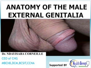
- 1. Dr. NDAYISABA CORNEILLE CEO of CHG MBChB,DCM,BCSIT,CCNA Supported BY
- 2. Male Reproductive System Major Function: Makes sperm cells (gametes) and transfer the sperm into the female reproductive system in order to fertilize the female gametes to produce a zygote. Dr Ndayisaba Corneille 2
- 3. MALE EXTERNAL GENITAL • Scrotum – Main content is the testis • Penis • Penile Urethra Dr Ndayisaba Corneille 3
- 4. Scrotum • A pouch of skin that hangs from the body below the perineum. • It is a saccular extension of the anterior abdominal wall • Contains two testes and their Spermatic cords. Dr Ndayisaba Corneille 4
- 5. Scrotum • . • The scrotum has a temperature of about 93.2F (the rest of the body is usually 98.6F) • Sperm formation occurs most rapidly at this cooler temp • Inside the scrotum are coverings of the testes. They are continuous with the anterior abdominal wall and going from superficial to deep these layers are: – External spermatic fascia – from the External oblique muscle – Cremasteric muscle and fascia – from the internal oblique muscle – Internal spermatic fascia- from the ttansversalis fascia – Tunica vaginalis is the peritoneal sac that partially encloses the testes Dr Ndayisaba Corneille 5
- 7. Scrotum – The skin is wrinkled and pigmented. Its superficial fascia is devoid of fat – Two muscles associated with the scrotum include – Dartos muscle • Elevates testes and wrinkles scrotal surface when the temperature is low – Cremaster muscle • Pulls testes closer to body during sexual arousal or cold Dr Ndayisaba Corneille 7
- 8. Blood supply • Posterior scrotal branches of the perineal artery: Arise from the internal pudendal artery which is a branch on the internal iliac artery. • Anterior scrotal branches of the external pudendal artery: The external pudendal artery branches directly from the femoral artery, a continuation of the external iliac artery. • Cremasteric artery: A branch of the inferior epigastric artery, which arises from the external iliac artery. Dr Ndayisaba Corneille 8
- 9. Nerve supply • Genital branch of the genitofemoral nerve: Arising from the lumbar plexus, supplies the anterolateral surface of the scrotum. • Anterior scrotal nerves: These are branches of the ilioinguinal nerve (L1) from the lumbar plexus. They supply the anterior surface of the scrotum. • Posterior scrotal nerves: Arise from the perineal branch of the pudendal nerve (S2-4) which forms from the sacral plexus. They supply the posterior surface of the scrotum. • Perineal branch of the Posterior cutaneous nerve of thigh, it suppleis the inferior surface Dr Ndayisaba Corneille 9
- 10. Dissection view showing superficial and deeper features of the scrotum, testes, and related structures Inguinal ligament Superficial inguinal ring Cremaster muscle Scrotum containing the dartos muscle Inguinal canal Nerve Artery Venous plexus Ductus deferens Spermatic cords Scrotal septum Scrotal cavity Raphe Dr Ndayisaba Corneille 10
- 11. Dr Ndayisaba Corneille 11
- 12. Dr Ndayisaba Corneille 12
- 13. TESTIS • The testis is primary reproductive organs of the male reproductive system. • The testis is the male gonad which is homologous with the ovary of the female. • They produce sex hormones called ANDROGENS • Each testis is an oval structure about 5 cm long and 3 cm in diameter • An adult testis weight about 10 to 15 gm. Dr Ndayisaba Corneille 13
- 14. The testis……….. • The testes develops retroperitoneal on the posterior abdominal wall and descend into the scrotum just before birth • During development its decent is believed to be guided by the Posterior gonad ligament (Gubernaculum) – as the body grows the gubernaculum does not, thus testis is drawn downward through the inguinal canal and finally into the scrotum. • The mullerian inhibiting hormone produced by the developing testes is Involved in this transabdominal migration • If the testes fail to descend the production of testosterone persists but spermatozoa cannot be produced Gubernac ulum testes Dr Ndayisaba Corneille 14
- 15. The Testes…………………… • A fibrous capsule which covers each testis is called the tunica albuginea. • Each testes is enclosed by the tunica vaginalis, a continuation of the peritoneum that lines the abdominopelvic cavity. • The testis is finally suspended in the scrotum by the spermatic cord and anchored to the floor of the scrotum by the scrotal ligament which is the remnant of the gubernaculum testes. Dr Ndayisaba Corneille 15
- 16. Testicle………. 16 The tunica albuginea gives rise to septa (partitions) that divide the testis into lobules (about 250 to 400) Each lobule contains 3 or 4 highly coiled seminiferous tubules These converge to become rete testis – Rete testis (collecting area outside of lobules) – Efferent ductules (lead from rete testis to epididymis) Dr Ndayisaba Corneille
- 17. Vascular Supply • The main arterial supply to the testes and epididymis is via – the paired testicular arteries, which arise directly from the abdominal aorta. They descend down the abdomen, and pass into the scrotum via the inguinal canal, contained within the spermatic cord. – the cremasteric artery from the inferior epigastric artery and – the artery of the vas deferens (from the inferior vesical artery). These branches give anastomoses to the main testicular artery. Dr Ndayisaba Corneille 17
- 18. Venous drainage • Venous drainage is achieved via the paired testicular veins. They are formed from the pampiniform plexus in the scrotum • The left testicular vein drains into the left renal vein, while the right testicular vein drains directly into the inferior vena cava. Dr Ndayisaba Corneille 18
- 19. Dr Ndayisaba Corneille 19
- 20. Abnormalities in testis migration: Cryptorchism • Cryptorchidism is failure of the testis to completely descend into the scrotum. • The term is derived from the Greek words kryptos and orchis, meaning “hidden testis.” • Cryptorchism “abdominal” testis –is detrimental to spermatogenesis and normal testicular metabolism • Leads to arrested spermatogenesis Dr Ndayisaba Corneille 20
- 21. Dr Ndayisaba Corneille 21
- 22. Seminiferous Tubules • Sperm is produced in this network of tubules called seminiferous tubules by the process of spermatogenesis • Interstitial cells (cells of Leydig), which produce male sex hormones, are located between the seminiferous tubules within a lobule. Dr Ndayisaba Corneille 22
- 23. Dr Ndayisaba Corneille 23
- 24. Epididymis • The epididymis holds the testes in place and connects the testes to the Vas Deferens. • Consists anatomically : * Head (CAPUT) * Body (CORPUS) * Tail (CAUDA) Located the upper , posterior and lower portions of the testis Dr Ndayisaba Corneille 24
- 25. Epididymis • a long tube (about 6 meters) • Its head joins the efferent ductules and caps the superior aspect of the testis • Its tail continues with the vas deferens Consists histologically : *Its epithelium is pseudostratified stereo- ciliated columnar with many smooth muscle fibers and elastic C.T. with fibrous connective tissue stroma. (Stereo cilia are more like giant microvilli they are not true cilia. Stereo means solid) Dr Ndayisaba Corneille 25
- 26. 26 Epididymis………………….. Sperm that leave the testes are immature and incapable of fertilizing ova. They complete their maturation process and become fertile as they move through the epididymis. Mature sperm are stored in the lower portion, or tail, of the epididymis Upon ejaculation the epididymis contracts, expelling sperm into the ductus deferens Dr Ndayisaba Corneille
- 27. Function • Sperm transport • Sperm storage : Mainly in the tail (up to 60 days motile and fertile) • Sperm maturation :Sperm undergo functional maturation here for 2 weeks • Sperm absorption : In prolonged sexual rest • Secrets glyceryl- phosphorylcholine which metabolized by sperm in female genitalia for gaining more energy during capacitation Dr Ndayisaba Corneille 27
- 28. Vas Deferens • Cord like structure from end of ductus epididymis to ejaculatory duct at Cilliculua Seminalis in the anterior portion of pelvic urethra dorsal to the neck of the urinary bladder • Each ductus is 30-45 cm in length • Drains the testes and epididymis, by carrying sperm to the pelvic cavity. • The epithelium is pseudo stratified columnar Dr Ndayisaba Corneille 28
- 29. The anatomical course of the vas deferens is as follows: • It is continuous with the tail of the epididymis. • Travels through the inguinal canal, as part of the spermatic cord. • Moves down the lateral pelvic wall close to the ischial spine. • Turns medially to pass between the bladder and the ureter and then travels downward on the posterior surface of the bladder. • The inferior narrow part of the ampulla joins the duct from the seminal vesicle to form the ejaculatory duct. Dr Ndayisaba Corneille 29
- 30. Function of V D • Transport sperm at the time of ejaculation. The smooth muscle in its wall is arranged in 3 layers Inner and outer longitudinal and a middle circular • Partial reservoir in the ampulla • Nutrition to stored sperm • stores viable sperm for up to 3 months. • May absorb dead sperm • May contain large number of sperm equal to an ejaculate but after vasotomy, It will take about 20 ejaculations for sperm to be completely flushed out of the VD. Dr Ndayisaba Corneille 30
- 31. ACCESSORY GLANDS • The accessory glands include – the seminal vesicles, – prostate gland, and – the bulbourethral glands. • These glands secrete fluids that form the semen. Dr Ndayisaba Corneille 31
- 32. Seminal Vesicles • The seminal vesicles are sac- shaped glands located next to the ampullae of the ductus deferentia. • Each gland is about 5 cm long and tapers into a short duct that joins the ductus deferens to form the ejaculatory duct. • The epithelium is pseudostratified columnar or simple columnar Dr Ndayisaba Corneille 32
- 33. Function of Seminal Vesicle • Secrete main volume of seminal plasma (65%) which act as a vehicle to sperm activity • Secrete fructose, for energy to sperm • Secrete citric acid as buffer to sperm • Secrete potassium and sodium ion to control the equilibrium of osmotic pressure • Secrete Flavin which give yellow coloration sometimes to normal ejaculate • Stimulates flagellum movement in spermatozoa » First step of capacitation Dr Ndayisaba Corneille 33
- 34. Prostate • It is a firm, dense structure located just inferior to the urinary bladder. • It encircles the urethra as it leaves the urinary bladder. • Numerous short ducts from the prostate gland empty into the prostatic urethra. Dr Ndayisaba Corneille 34
- 35. Function of Prostate : • The secretions of the prostate are thin and milky colored. • They enhance the motility of the sperm. • Secrete large amount of minerals that regulate the buffering system of seminal plasma • Secrete amino acids and other elements for sperm nutrition • Its alkaline secretion helps to neutralize the acidic environment of the vagina • Proteolytic enzymes: acts to "decoagulate" the semen that was coagulated by seminal vesicle secretions, which helps the sperm begin their journey once inside the vagina Dr Ndayisaba Corneille 35
- 36. Bulbourethral Glands (Cowper's) • Small and oval in shape, located near the base of the penis. • A short duct which enters the proximal end of the penile urethra. • In response to sexual stimulation, it secretes a clean alkaline mucus-like fluid which is also a natural lubricant • Its secretion neutralize traces of acidic urine in the penile urethra prior to ejaculation Dr Ndayisaba Corneille 36
- 37. Produced: Seminiferous tubules Stored: Epididymis Transported through epididymis by rhythmic peristaltic contractions as they mature Epididymis Vas Deferens Ejaculatory duct (ampulla of vas deferens fuses with duct of seminal vesicle “ejaculatory duct”) prostate prostatic urethra (then passes the bulbourethral gland) membranous urethra penile urethra Sperm Summary Dr Ndayisaba Corneille 37
- 38. Seminal Fluid or Semen • a slightly alkaline mixture of sperm cells and secretions from the accessory glands. • The volume of semen in a single ejaculation may vary from 1.5 to 6.0 ml. • There are between 50 to 150 million sperm per milliliter of semen. • Sperm counts below 10 to 20 million spermatozoa per milliliter usually present fertility problems. Dr Ndayisaba Corneille 38
- 39. Penis • Penis (conducts urine to exterior and semen to female vagina during intercourse) – Root (fixed portion attached to body wall) – Body or shaft (movable, tubular part) – Glans or head (expanded end around urethral opening) • Neck (between shaft and glans) • Prepuce (foreskin) – Smegma (waxy secretion) Dr Ndayisaba Corneille 39
- 40. Figure 25.5 2 Penis layers (superficial to deep): Outer skin (similar to scrotum), Dermis has smooth muscle continuous with dartos, Underlying areolar tissue allows skin to move, Elastic tissue (encircling internal structures) Superficial and deep dorsal veins of penis Deep artery of penis Erectile tissues Urethra Skin Dermis Areolar tissue Dense network of elastic fibers Corpora cavernosa Corpus spongiosum Tissue Layers of the Penis Dr Ndayisaba Corneille 40
- 41. Penis………. Three cylindrical bodies: • Corpora cavernosa – two erectile bodies enclosed by a dense white fibrous capsule: tunica albuginea. • Corpus spongiosum – ventrally (corpus cavernosum urethrae) containing the spongy urethra, ending in the glans penis • Crura – attached to the conjoint rami of pubis and ischium. Root: bulb+crura+bulbospongiosus m.) body Dr Ndayisaba Corneille 41
- 42. Blood supply of the penis • Two dorsalis penile aa. –terminal branch of the internal pudendal artery • Deep arteries (cavernous artery) – paired, principal artery supplying the cavernous spaces • Bulbourethral artery– spongy urtethra (internal pudendal) Dr Ndayisaba Corneille 42
- 43. Dr Ndayisaba Corneille 43
- 44. Venous drainage • Superficial – superficial dorsal vein • Deep – deep dorsal vein sinuses – emissary veins - circumflex veins Dr Ndayisaba Corneille 44
- 45. ANATOMY OF THE ERECTION Erection is as a result of various sexual stimuli:Touch and mechanical stimulation of the penis (reflexogen) Erotic view, sound, and smell (central or psychogenic) The erection can also be inhibited by any emotional or higher mental activity The third type of the erection is the so called nocturnal erection associated to sleeping period. Dr Ndayisaba Corneille 45
- 46. Sexual stimulus Relaxation of the smooth muscle in cavernous spaces and arterioles. Blood inflow to the caveornous spaces. Occlusion of the venous dranaige. Intracavernous blood pressure > systemic blood pressure ERECTION Dr Ndayisaba Corneille 46
- 47. Erection of the penis (and the clitoris) Dr Ndayisaba Corneille 47
- 48. Erection of the penis (and the clitoris) Continuous inflow of blood with decreased out flow lead to full erection and hardness. Dr Ndayisaba Corneille 48
- 49. Erection………….. • Bulbo-urethral glands secrete to lubricate penis tip • Erection and copulation controlled with parasympathatic nerve fibers • 10-15 minutes are required for erection and copulation normally Dr Ndayisaba Corneille 49
- 50. Ejaculation (male orgasm) Ejaculation (male orgasm) • Bulbocavernosus muscles (at base) push semen toward external urethral orifice • Ischiocavernosus muscles (along sides) stiffen erect penis • –contraction of urethra and penis: expulsion: semen expelled • Sensation experienced = Ejaculatory Inevitability • Accompanied by increased heart rate, blood pressure and breathing rate • Intense myotonia Dr Ndayisaba Corneille 50
- 51. Seminalis emission and ejaculation. Bulbospongiosus muscles contract fast and serially; The semen is released (ejaculated) form the urethra (propulsion). Sphincter of the bladder sontracts, preventing expulsion of the urine. Dr Ndayisaba Corneille 51
- 52. Never forget these concepts •POINT and SHOOT •PARA and SYMPA –THANK U Dr Ndayisaba Corneille 52
- 53. END Dr Ndayisaba Corneille THANKS FOR LISTENING By DR NDAYISABA CORNEILLE MBChB,DCM,BCSIT,CCNA Contact us: amentalhealths@gmail.com/ ndayicoll@gmail.com whatsaps :+256772497591 /+250788958241 53