Anatomy of Female internal genitalia.pptx
•Download as PPTX, PDF•
1 like•587 views

This document summarizes the internal female genitalia, including the ovaries, fallopian tubes, uterus, cervix, and upper part of the vagina. It describes the location, structure, blood supply, functions, and common disorders of each organ. The ovaries produce eggs and sex hormones. The fallopian tubes receive eggs from the ovaries, provide a site for fertilization, and transport fertilized eggs to the uterus. The uterus receives and nourishes a fertilized egg. The cervix connects the uterus to the vagina, which acts as a birth canal. Common disorders like ovarian cysts, ovarian cancer, and ectopic pregnancies are also discussed.

Recommended
More Related Content
What's hot
What's hot (20)
Similar to Anatomy of Female internal genitalia.pptx
Similar to Anatomy of Female internal genitalia.pptx (20)
More from Dr Ndayisaba Corneille
More from Dr Ndayisaba Corneille (20)
Recently uploaded
Recently uploaded (20)
Anatomy of Female internal genitalia.pptx
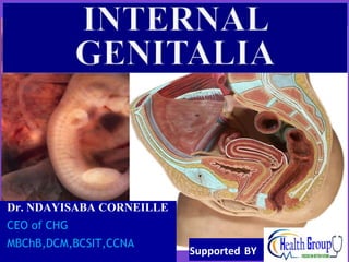
- 1. Dr. NDAYISABA CORNEILLE CEO of CHG MBChB,DCM,BCSIT,CCNA Supported BY
- 4. Contents Ovaries Fallopian tubes Uterus Cervix Upper part of vagina Dr Ndayisaba Corneille
- 6. Ovaries Female gonads Almond shaped, 4 x 2 cm Located in ovarian fossa which is bounded above by external iliac artery and behind by internal iliac artery Covered by a true capsule, the tunica albuginea and a false capsule, the germinal epithelium. Dr Ndayisaba Corneille
- 7. Supports for the ovaries Mesovarium that attaches it to the back of the broad ligament Round ligament that runs from the medial border of the ovaries to the uterus Suspensory ligament that runs from lateral aspect of the ovaries to the pelvic wall. Dr Ndayisaba Corneille
- 9. CONT… Before puberty, the ovary is smooth but after puberty, it becomes scarry due to continous release of mature ova. Blood supply: ovarian artery, a branch of abdominal aorta at L1 Veins are ovarian vein that join inferior venacava on the right and left renal vein on the left. Dr Ndayisaba Corneille
- 10. Ovaries before puberty Dr Ndayisaba Corneille
- 11. Ovaries after puberty Dr Ndayisaba Corneille
- 12. Functions of the ovaries: Production of gametes Production of female sex hormones in a sexually mature female. Dr Ndayisaba Corneille
- 13. Disorders of ovaries Ovarian cyst Ovarian cancer Ovarian pregnancy Dr Ndayisaba Corneille
- 14. Ovarian cyst Dr Ndayisaba Corneille
- 15. Ovarian cancer Dr Ndayisaba Corneille
- 16. Ovarian pregnancy Dr Ndayisaba Corneille
- 17. Fallopian tubes Tubular muscular organs, 10 cm long, that extend from the ovaries to the uterus Composed of the infundibulum, ampulla, isthmus and the intramural part Infundibulum: funnel shaped with numerous fimbriae overlying the ovaries Ampulla: widest part. Site for fertilisation Isthmus: narrowest part, lies lateral to the uterus Dr Ndayisaba Corneille
- 18. CONT… Intramural part: segment that pierces the uterine wall. Functions of the fallopian tubes Receives ova Site of fertilization Nourishment of fertilized ova Transport of fertilized ova to the uterus. Dr Ndayisaba Corneille
- 19. Blood supply of fallopian tubes Ovarian artery from abdominal aorta and uterine artery from internal iliac. Dr Ndayisaba Corneille
- 20. Fallopian tubes Dr Ndayisaba Corneille
- 21. Ectopic pregnancy Dr Ndayisaba Corneille
- 22. Disorders of uterine tubes Ectopic pregnancy Occluded oviducts due to Pelvic inflammatory disease. Dr Ndayisaba Corneille
- 23. Uterus Hollow pear shaped organ, 8X5X2.5cm Divided into fundus, body and cervix Fundus: part of uterus above entrance of fallopian tubes Body: part below entrance of fallopian tubes Cervix: part next to vagina. Dr Ndayisaba Corneille
- 26. Relations of the uterus Anteriorly: urinary bladder. Between uterus and bladder is the uterovesicle pouch. Posteriorly: rectum.between uterus and rectum is the puch of douglas Laterally: broad ligament, uterine artery, uterine tubes, round ligament Dr Ndayisaba Corneille
- 27. CONT… Blood supply: uterine artery from internal iliac artery. Veins follow arteries. Supports of the uterus: Levator ani muscles, transverse cervical, pubocervical, sacrocervical ligaments and broad ligament. Most important support is transverse cervical ligament. Dr Ndayisaba Corneille
- 28. CONT… Functions of the uterus Reception, retention and nutrition of fertilized ova. Positions of the uterus Anteversion: long axis of uterus bent at 90 degrees to long axis of vagina. Commonest position. Anteflexion: long axis of uterus bent at 90 degrees to long axis of cervix. Dr Ndayisaba Corneille
- 29. CONT… Retroversion: long axis of uterus bent backwards to long axis of vagina Retroflexion: long axis of uterus bent backwards to long axis of cervix. Dr Ndayisaba Corneille
- 30. Pregnant uterus Dr Ndayisaba Corneille
- 35. Vagina A muscular tube, 8 cm long with an anterior and posterior walls in apposition Extends from the vaginal orifice to the cervix Has an upper half that lies within the pelvis and a lower half that lies below the pelvis Dr Ndayisaba Corneille
- 36. CONT… Functions of the vagina Copulation Birth canal Excretory organ for menstural blood. Relations of the vagina Anteriorly: bladder, urethra Posteriorly; sigmoid, rectum, perineal body Laterally: ureter, levator ani muscles, vestibule Dr Ndayisaba Corneille
- 37. CONT… Blood supply: vaginal artery and vaginal branch of uterine artery, Lymphatics: upper half, internal ilaic nodes, lover half, superficial inguinal nodes Supports of the vagina: levator ani muscles, uterine ligaments, perineal body, urogenital diaphragm. Dr Ndayisaba Corneille
- 38. Uterus bicornis Dr Ndayisaba Corneille
- 39. END Dr Ndayisaba Corneille THANKS FOR LISTENING By DR NDAYISABA CORNEILLE MBChB,DCM,BCSIT,CCNA Contact us: amentalhealths@gmail.com/ ndayicoll@gmail.com whatsaps :+256772497591 /+250788958241