This document discusses nodular hyperplasia of the liver, also known as focal nodular hyperplasia (FNH). It describes FNH as the second most common benign liver tumor, predominantly affecting women in their third to fourth decades. Imaging techniques, especially contrast-enhanced ultrasound and MRI with gadoxetic acid, can diagnose FNH in 90% of cases based on characteristic hypervascular appearance. While usually asymptomatic, large FNH lesions can sometimes cause pain or compress nearby structures, requiring surgical resection. Otherwise, asymptomatic FNH generally does not require treatment.
Hepatic hemangioma is the most common benign liver tumor. It is composed of vascular channels of various sizes lined with endothelium and fibrous tissue. Imaging plays an important role in diagnosis. On ultrasound, hemangiomas appear hyper echoic but can also appear hypoechoic, especially in fatty livers. On CT and MRI, hemangiomas enhance gradually from the periphery inward over time following contrast administration. While typically benign, giant hemangiomas over 4cm can potentially cause complications and may require treatment with transarterial embolization. Differential diagnoses include other liver lesions that appear similar on imaging.
Hepatocellular carcinoma is the most common liver cancer worldwide. Risk factors include hepatitis B and C infections, cirrhosis, and certain toxins and inherited conditions. Clinically, patients may present with an abdominal mass, jaundice, ascites, or gastrointestinal bleeding. Histologically, tumors can be well, moderately, or poorly differentiated. Treatment options include surgical resection or liver transplantation for early stage disease. Other local ablative therapies like radiofrequency ablation may be used if surgery is not possible. Systemic chemotherapy has a limited role and is rarely effective for hepatocellular carcinoma.
Benign Neoplasms of Liver
This document provides an overview of benign liver tumors, including their epidemiology, etiology, classification, clinical features, diagnosis and treatment. The main benign liver tumors discussed are hepatocellular adenoma, cavernous hemangioma, and focal nodular hyperplasia. Hepatocellular adenoma is more common in females using oral contraceptives and can present with pain or rupture. Cavernous hemangioma is the most common benign liver tumor and often asymptomatic. Larger hemangiomas can cause pain, jaundice or complications. Imaging such as MRI is used for diagnosis, while treatment depends on size and symptoms, and may include resection, ablation or e
This lecture was part of an educational course performed by the IATTGI group this August in Buenos Aires and describes novel targets and novel drugs in hepatocellular carcinoma.
This document discusses hepatocellular carcinoma (HCC). It notes that HCC is one of the most common cancers worldwide and is often associated with cirrhosis from hepatitis B, hepatitis C, or other liver diseases. Screening high-risk patients is important for early detection when treatment may be curative. Treatment options include surgical resection or liver transplantation for early stage disease, and ablation, chemoembolization, or targeted molecular therapies for later stages. However, most patients are not candidates for curative therapies at diagnosis due to late stage, highlighting the importance of screening to detect HCC early.
This document provides an overview of hepatocellular carcinoma (HCC). It discusses the anatomy, epidemiology, screening, diagnosis, staging and management of HCC. HCC is commonly caused by viral hepatitis and cirrhosis. Diagnosis involves imaging such as ultrasound, CT or MRI showing characteristic arterial enhancement and venous washout. Staging systems include AJCC, Okuda, CLIP and BCLC which incorporate tumor burden, liver function and performance status. Management options include surgical resection, liver transplantation, ablation and arterially directed therapies like TACE.
- A 65-year-old male presented with a left iliac fossa mass and changes in bowel habits. CT and examination revealed a large sigmoid colon and rectal mass. He underwent exploratory laparotomy and Hartmann's procedure with resection of the mass and nodes. Pathology found a clear cell carcinoma.
- Follow up CT found no evidence of recurrence but a stationary small renal mass. Options for the renal mass include radical nephrectomy or modifying the incision for access. Restoring bowel continuity may be considered with possible loop ileostomy protection.
- Renal cell carcinoma is known for its varied presentations and unpredictable behavior, sometimes presenting as unusual metastases that require individualized management even for solitary or
Benign liver tumors present diagnostic challenges due to overlap between lesions on imaging and clinical features. Hemangiomas are typically the only clearly diagnosed tumors without biopsy. Biopsy or laparoscopy are reasonable invasive approaches for diagnostic uncertainty. The predominant treatment is observation, except for adenomas which often require surgery due to malignant potential. Diagnostic uncertainty is an acceptable indication for surgical intervention.
Hepatocellular carcinoma (HCC) is the most common type of liver cancer. It is the fifth most common cancer worldwide and the third leading cause of cancer death. The main risk factors are hepatitis B, hepatitis C, alcoholism, and cirrhosis. HCC often develops from chronic liver inflammation and regeneration caused by these conditions. Patients may present with non-specific symptoms like abdominal pain, weight loss, and fatigue. Diagnosis involves blood tests, imaging studies, and biopsy. Prognosis is generally poor, with most patients dying within 2 years from cancer progression or liver failure.
Hepatocellular carcinoma (HCC) is the most common type of liver cancer. It has a high worldwide incidence, especially in areas where hepatitis B is prevalent like Southeast Asia. Major risk factors for HCC include hepatitis B and C infections, cirrhosis of the liver from any cause, and alcohol abuse. The disease progresses as hepatocytes undergo repeated cycles of cell death and regeneration due to chronic inflammation and cirrhosis, accumulating mutations over time that can lead to cancer. Diagnosis involves blood tests, imaging like ultrasound or CT scan, and often a biopsy. Staging systems evaluate tumor characteristics, liver function, and physical status to determine prognosis and treatment options. Treatment may include surgical resection, liver transplantation, ablation
This document discusses solitary liver lesions, categorizing them as benign tumours, infections, trauma, malignant tumours or other. It provides detailed information about cavernous haemangioma, including that it is the most common benign liver tumour, often appearing as a well-defined hypodense lesion on imaging with characteristic enhancement. Hepatic abscesses and hydatid cysts are also described, noting ultrasound, CT and MRI findings help differentiate bacterial vs parasitic abscesses and stages of cyst growth.
1) Hepatic hemangiomas are benign liver tumors consisting of blood-filled cavities lined by endothelial cells.
2) They are usually asymptomatic but can sometimes cause pain, nausea, or other digestive symptoms. Complications include bleeding, infection, or mass effect.
3) Diagnosis is usually made using ultrasound, CT, or MRI which show characteristic patterns of enhancement. Treatment is usually conservative but resection may be considered for large or symptomatic hemangiomas.
The document discusses a case of hepatocellular carcinoma (HCC) in a 68-year-old male patient. Imaging revealed a solitary liver lesion with characteristics of HCC on CT scan. Given the patient's good performance status and the localized nature of the disease, he underwent a right posterior sectionectomy. Histopathology confirmed HCC. The patient recovered well post-operatively without major complications. Surgical resection can provide cure for select patients with HCC, especially when the tumor is localized and the patient's liver function is preserved.
This document discusses the management of metastatic liver tumors, focusing on colorectal liver metastases. Some key points:
- The liver is the most common site of metastasis from colorectal cancer. Surgical resection offers the only chance of cure or prolonged survival for resectable colorectal liver metastases, with 5-year survival rates of 40% for margin-negative resection.
- Factors associated with poorer prognosis include short disease-free interval, multiple tumors, bilobar involvement, large tumor size, and elevated CEA levels.
- Preoperative imaging with CT, MRI, and ultrasound is used to evaluate resectability and tumor extent. Laparoscopy can help identify unresectable disease.
Management of Advances Hepatocellular CarcinomaPratap Tiwari
Hepatocellular carcinoma (HCC) is a leading cause of cancer death worldwide. For advanced HCC that cannot be treated with surgery or transplantation, the standard of care has been sorafenib. Lenvatinib and cabozantinib have also shown efficacy in advanced HCC. Immunotherapy with nivolumab has shown promise based on phase II data. Combination therapies and future targeted agents may provide additional treatment options for this difficult to treat cancer.
This document discusses hepatocellular carcinoma (HCC), the most common type of primary liver cancer. It covers the epidemiology, risk factors, pathogenesis, clinical presentation, diagnosis, prognostic factors, and treatment options for HCC. The highest rates are seen in regions where hepatitis B is endemic, and major risk factors include chronic hepatitis B and C infections, cirrhosis, and aflatoxin exposure. Diagnosis involves imaging tests like ultrasound, CT, and MRI along with blood tests. Treatment depends on tumor size and liver function, and may include resection, transplantation, ablation, embolization, or chemotherapy.
Brief description on the benign tumors of liver that includes hemangioma, focal nodular hyperplasia, regenerative nodular hyperplasia, dysplastic foci, dysplastic nodules and focal fatty change.
This document discusses liver lesions and their appearance on various imaging modalities. It covers benign lesions like hemangioma, focal nodular hyperplasia and hepatic adenoma. Malignant primary lesions discussed are hepatocellular carcinoma and hepatoblastoma. Imaging features of hypervascular and hypovascular lesions on multiphasic CT are summarized. Hepatocellular carcinoma risk factors and clinical presentation are outlined. Imaging appearance of HCC on ultrasound, CT and MRI is described in detail. Hepatic metastases are also discussed along with hypervascular metastatic lesions.
This document discusses primary liver tumors, including benign and malignant types. It provides detailed information on hepatocellular carcinoma (HCC), the most common primary liver malignancy. HCC is often associated with liver cirrhosis and viral hepatitis. Diagnosis involves imaging and blood markers. Surgical resection or liver transplantation offer the best chance of survival for eligible patients with early-stage HCC within Milan criteria. Other local and systemic therapies are options for patients who cannot undergo surgery.
Cholangiocarcinoma is a rare cancer that affects the bile ducts. It occurs most often in older adults and risk factors include primary sclerosing cholangitis and liver flukes. The cancer is classified based on location and can be intrahepatic, perihilar, or distal. Surgical resection is the main treatment if the cancer is resectable, while palliative options are used for unresectable cases to relieve symptoms of biliary obstruction. Prognosis is generally poor due to late diagnosis but resection provides the best chance for survival.
The document discusses the workup and diagnosis of liver masses. It describes various benign and malignant liver lesions including hemangioma, focal nodular hyperplasia, hepatic adenoma, liver cysts, hepatocellular carcinoma, and metastases. Multiphasic CT is useful for characterizing lesions based on enhancement patterns in the arterial, portal venous, and delayed phases. Biopsy may be needed to confirm diagnosis of suspicious lesions.
The document summarizes hepatocellular carcinoma (HCC) and examines hepatitis C virus (HCV) as a risk factor. It provides descriptive epidemiology on HCC, identifying it as the 4th most common cancer worldwide. It then analyzes HCV as a major risk factor for HCC. Details are given on the virology, epidemiology, transmission routes, stages of infection, diagnosis and treatment of HCV, as well as its role in leading to HCC. Recommendations for primary prevention include national HBV vaccination programs and screening blood supplies.
This document discusses the approach to evaluating and diagnosing liver masses. It defines a liver mass and explains how imaging techniques are used in the diagnosis. The differential diagnosis for liver masses can range from benign to malignant lesions. Cystic lesions discussed in detail include pyogenic and amoebic liver abscesses. Solid lesions include inflammatory conditions like abscesses as well as benign and malignant tumors. Treatment options for different lesions are outlined.
This document discusses benign and malignant liver tumors. It covers common benign tumors like hepatic adenoma, focal nodular hyperplasia, and hemangioma. Malignant tumors discussed include hepatocellular carcinoma, cholangiocarcinoma, and metastatic cancers. Diagnostic workup and available treatment options are also summarized, including liver resection, ablation, and transplantation. The document concludes by outlining the benefits of minimally invasive robotic and laparoscopic liver surgery approaches.
Multiple Hepatic and Osseous Focal Lesions without Splenomegaly and/or Lymph ...JohnJulie1
Hepatic involvement is a common extranodal manifestation of common and some rare hematologic malignancies. Although the imaging features of more common hepatic diseases such as hepatocellular carcinoma, metastases, and infection may overlap with those of hepatic hematologic malignancies, combining the imaging features with clinical manifestations and laboratory findings can facilitate correct diagnosis. Imaging has an important role in diagnosis of hepatic focal lesions.
Introduction: Diffuse Large B Cell Lymphoma (DLBCL), as a set of heterogeneous aggressive lymphoma, most commonly originated in the germinal center B lymphocytes. Rare cases were from peripheral blood B cell (outside germinal center) or from the inert lymphoma development and transformation. And such tumor with originality outside germinal center was seldom seen in the literature. Even the tumor in combination with chronic gastritis has not been reported before.
The document discusses evaluation and management of liver lesions. It describes common benign and malignant solid and cystic liver lesions. For solid lesions, it recommends following an algorithm including history, exam, labs, imaging like CT/MRI, and potentially biopsy to determine if the lesion is benign or malignant. For cystic lesions, it recommends monitoring asymptomatic simple cysts with ultrasound but surgically treating symptomatic or complicated cysts.
Liver Fibrosis: Difficulties in Diagnostic and Treatment: A Review-Crimson Pu...CrimsonGastroenterology
Early discovery of liver fibrosis and cirrhosis is becoming more relevant because of enhanced incidence of hepatocellular carcinoma. There a many underlying factors in developing liver fibrosis (i.e. viral hepatitis, steatohepatitis). Diagnosis of liver fibrosis is difficult; chronic liver failure and less distinct fibrosis stages can be underestimated, when laboratory routine parameters and native ultrasound of the liver are unsuspicious. Liver biopsy is a common element of diagnostic workup in hepatic cirrhosis, alongside clinical examination and abdominal ultrasound, and is the accepted diagnostic gold standard. But there is no unitary system of histological classification used to evaluate the degree of fibrosis, and individual systems are often validated only for individual disease entities. On the other hand liver biopsy is of less tolerance for patients. In the last years serological markers for detecting liver fibrosis were developed with different validity. Various imaging modalities have been proposed as methods for assessing liver fibrosis
by liver stiffness measurement. They are sufficient to approve the suspicious of liver fibrosis and/or to uncover unknown chronic liver failure. Studies showed the clinical usefulness of acoustic radiation force impulse shear wave elasticity imaging (ARFI-SWEI) is efficient as a preventive screening method to uncover fibrosis. The ARFI-SWEI system is integrated in an ultrasound device has a good accuracy and high reproducibility. Therapy of liver fibrosis depends on underlying disease and degree of liver failure. When liver failure can be cured liver fibrosis can regress. Direct antifibrotic drugs are
actually not available but in progress.
Hepatic carcinoma, also known as hepatocellular carcinoma (HCC), is one of the most common and deadly cancers worldwide, killing over 1 million people per year. Risk factors include hepatitis B and C infections, cirrhosis, alcohol use, and aflatoxin exposure. HCC typically presents in patients with cirrhosis as an asymptomatic liver mass and is diagnosed through blood tests showing elevated AFP levels and imaging exams like ultrasound, CT, or MRI. Treatment depends on the stage but may include surgical resection, liver transplantation, ablation procedures, embolization, or chemotherapy. Long-term surveillance after treatment is important for early detection of recurrence.
Case of a rare inflammatory hepatic hilar mass mimicking cholangiocarcinomaDebdeep Banerjee
This case report describes a rare case of an inflammatory hepatic hilar mass that mimicked cholangiocarcinoma in a 66-year-old man. Imaging and biopsy were initially suggestive of cholangiocarcinoma but surgical exploration revealed the mass to be caused by diffuse fibrotic and inflammatory reactions. A definitive diagnosis of benign inflammatory mass, rather than cholangiocarcinoma, was made based on histopathological examination showing only chronic inflammation. The patient recovered well with conservative management, highlighting the challenges of distinguishing between benign and malignant hilar masses preoperatively.
Renal cell carcinoma (RCC) accounts above 3 percent of all cancers and often diagnosed incidentally. Highest incidence reported in Western countries. Classical tried of RCC now rarely seen, In about 25 percent of cases, patients often present with an advanced disease. Upper gastrointestinal (GI) bleeding due to stomach metastasis of RCC is rare and to the best of our knowledge, only a few cases are reported in the literature. Gastric metastasis of RCC is often associated with poor outcome. Managing such cases can be very tricky; hence it is imperative for surgeons to be very familiar and to have a high grade of suspicion in order to prevent delay and upgrade of tumour staging. We report the case of a patient presented to our centre with upper GI bleeding as the primary presenting complaint in an advanced RCC and our experience managing the condition.
Staging and investigation of hepatobillary caAtulGupta369
This document provides an overview of cancers affecting the liver, gallbladder, and pancreas. It discusses the epidemiology, risk factors, clinical presentation, diagnosis, and staging of liver cancer, gallbladder cancer, and pancreatic cancer. Diagnostic tests include blood tests, imaging like ultrasound, CT, MRI, and PET scans. Biopsy may be done for unresectable tumors. The document also provides details on noninvasive diagnosis criteria and treatment guidelines for liver cancer put forth by professional associations.
This patient presented with rectal bleeding and weight loss and was found to have stage III adenocarcinoma. Given his family history of colorectal cancer in a first-degree relative at a young age, he is at high risk for hereditary non-polyposis colorectal cancer (HNPCC). HNPCC accounts for 5-7% of colorectal cancers and results from a mutation in DNA mismatch repair genes. Individuals with HNPCC have an increased lifetime risk of colorectal and other cancers. The patient was counseled on genetic testing and increased screening for relatives is recommended.
This document discusses the classification and treatment of liver tumors. It outlines four main categories of liver tumors: primary solid benign tumors, primary solid malignant neoplasms, cystic neoplasms, and metastatic tumors. Hepatocellular carcinoma and focal nodular hyperplasia are described as two of the most common primary benign and malignant liver tumors respectively. Surgical resection or liver transplantation are identified as the primary treatments for hepatocellular carcinoma when the liver function and extent of disease are suitable.
This document summarizes the management of hepatocellular carcinoma. It begins by introducing HCC as the most common form of liver cancer. It then discusses risk factors, surveillance methods, diagnosis via imaging and biopsy, and the BCLC staging system. The BCLC system links stage of disease to treatment options and expected survival outcomes. For early stages, curative treatments like resection, transplantation, and ablation are recommended. Intermediate stages receive transarterial chemoembolization, while advanced stages are treated with sorafenib. End-stage disease involves symptomatic support only.
A 26-year-old male presented with a 3-month history of abdominal pain and a 3-week history of an enlarging epigastric swelling. On examination, a large firm mass was found in the epigastric region extending to the left side. Imaging revealed a left testicular mass with enlarged abdominal and supraclavicular lymph nodes. A biopsy showed a left testis seminoma. The patient underwent a left radical orchidectomy and was referred for chemotherapy. Testicular seminoma commonly presents as a painless testicular lump but can spread through the lymphatic system to cause abdominal or supraclavicular masses. Staging involves tumor markers, imaging, and pathological examination to determine the extent of the primary
This document discusses non-alcoholic fatty liver disease (NAFLD), which ranges from simple steatosis to non-alcoholic steatohepatitis (NASH) and cirrhosis. It occurs without significant alcohol use or other known causes. Risk factors include obesity, dyslipidemia, and type 2 diabetes. Due to the rise in metabolic syndrome, NAFLD is now the most common cause of chronic liver disease. Genetic and metabolic factors like insulin resistance and inflammation contribute to its development and progression. Liver biopsy is still the gold standard for diagnosis but carries risks, so noninvasive imaging methods are being developed and studied.
This document summarizes primary and secondary liver malignancies, their management, and principles of liver resection. It covers hepatocellular carcinoma (HCC), the most common primary liver cancer, risk factors, presentation, diagnosis, staging, and treatment options. Intrahepatic cholangiocarcinoma and metastatic tumors to the liver are also discussed. Surgical resection is the main curative treatment for early-stage HCC and intrahepatic cholangiocarcinoma when possible.
Primary Follicular Lymphoma of the spleen: A Case report and literature reviewiosrjce
IOSR Journal of Dental and Medical Sciences is one of the speciality Journal in Dental Science and Medical Science published by International Organization of Scientific Research (IOSR). The Journal publishes papers of the highest scientific merit and widest possible scope work in all areas related to medical and dental science. The Journal welcome review articles, leading medical and clinical research articles, technical notes, case reports and others.
This document provides information about lymphoma, including Hodgkin lymphoma and non-Hodgkin lymphoma. It discusses the workup, tests, staging, prognostic factors like the International Prognostic Index, treatments, and management of lymphoma in both inpatient and outpatient settings. Key points covered include diagnostic testing like biopsies, immunophenotyping, common genetic translocations, associations with infections, and treatment approaches depending on factors like tumor stage and grade.
RCC typically presents in the 6th and 7th decade of life and accounts for 2-3% of adult malignancies. Clear cell RCC arises from the proximal convoluted tubules and is associated with VHL gene mutations. Established risk factors include tobacco, obesity, and hypertension. CT scan is the preferred imaging modality and can identify enhancing renal masses. Surgical resection is the main treatment, with partial nephrectomy preferred for smaller tumors when possible to preserve renal function. Follow up involves history, exam, bloodwork and imaging depending on pathologic stage.
A 69-year-old man presented with abdominal and shoulder pain. Imaging showed a large pleural-based mass in his left lung. Biopsy of the mass revealed diffuse large B-cell lymphoma. He was treated with R-CHOP chemotherapy and achieved complete remission. Primary lymphoma of the pleura is extremely rare but can occur without associated conditions like HIV or prior tuberculosis. Biopsy is needed to diagnose pleural tumors due to lack of specific imaging features.
This case report describes a 61-year-old male patient who presented with chronic abdominal pain and was found to have xanthogranulomatous cholecystitis (XGC), a rare inflammatory disease of the gallbladder, along with gallstones. Imaging studies revealed thickening of the gallbladder wall and a mass, concerning for possible gallbladder carcinoma. The patient underwent cholecystectomy and was found to have XGC pathology, characterized by lipid-laden macrophages and chronic inflammatory cells infiltrating the gallbladder wall. XGC is a benign condition that can be confused for gallbladder cancer. The patient's surgery and recovery were uncomplicated.
Staging and investigation of ca kidney and bladderAtulGupta369
This document discusses staging and investigations for kidney and bladder cancer. It provides details on:
- Risk factors, pathological subtypes, and epidemiology of kidney cancer
- Genetic and non-genetic risk factors for bladder cancer
- Evaluations for diagnosis of both cancers including lab tests, imaging like CT, MRI, and pathology examination
- Presenting signs, symptoms, and classifications of bladder cancer
It is an informative overview of kidney and bladder cancers covering their risk factors, diagnostic workup, classifications, and epidemiology.
Similar to Texas chapter of the ACP annual meeting 2016 poster (20)
share - Lions, tigers, AI and health misinformation, oh my!.pptxTina Purnat
• Pitfalls and pivots needed to use AI effectively in public health
• Evidence-based strategies to address health misinformation effectively
• Building trust with communities online and offline
• Equipping health professionals to address questions, concerns and health misinformation
• Assessing risk and mitigating harm from adverse health narratives in communities, health workforce and health system
Integrating Ayurveda into Parkinson’s Management: A Holistic ApproachAyurveda ForAll
Explore the benefits of combining Ayurveda with conventional Parkinson's treatments. Learn how a holistic approach can manage symptoms, enhance well-being, and balance body energies. Discover the steps to safely integrate Ayurvedic practices into your Parkinson’s care plan, including expert guidance on diet, herbal remedies, and lifestyle modifications.
Travel vaccination in Manchester offers comprehensive immunization services for individuals planning international trips. Expert healthcare providers administer vaccines tailored to your destination, ensuring you stay protected against various diseases. Conveniently located clinics and flexible appointment options make it easy to get the necessary shots before your journey. Stay healthy and travel with confidence by getting vaccinated in Manchester. Visit us: www.nxhealthcare.co.uk
Local Advanced Lung Cancer: Artificial Intelligence, Synergetics, Complex Sys...Oleg Kshivets
Overall life span (LS) was 1671.7±1721.6 days and cumulative 5YS reached 62.4%, 10 years – 50.4%, 20 years – 44.6%. 94 LCP lived more than 5 years without cancer (LS=2958.6±1723.6 days), 22 – more than 10 years (LS=5571±1841.8 days). 67 LCP died because of LC (LS=471.9±344 days). AT significantly improved 5YS (68% vs. 53.7%) (P=0.028 by log-rank test). Cox modeling displayed that 5YS of LCP significantly depended on: N0-N12, T3-4, blood cell circuit, cell ratio factors (ratio between cancer cells-CC and blood cells subpopulations), LC cell dynamics, recalcification time, heparin tolerance, prothrombin index, protein, AT, procedure type (P=0.000-0.031). Neural networks, genetic algorithm selection and bootstrap simulation revealed relationships between 5YS and N0-12 (rank=1), thrombocytes/CC (rank=2), segmented neutrophils/CC (3), eosinophils/CC (4), erythrocytes/CC (5), healthy cells/CC (6), lymphocytes/CC (7), stick neutrophils/CC (8), leucocytes/CC (9), monocytes/CC (10). Correct prediction of 5YS was 100% by neural networks computing (error=0.000; area under ROC curve=1.0).
Adhd Medication Shortage Uk - trinexpharmacy.comreignlana06
The UK is currently facing a Adhd Medication Shortage Uk, which has left many patients and their families grappling with uncertainty and frustration. ADHD, or Attention Deficit Hyperactivity Disorder, is a chronic condition that requires consistent medication to manage effectively. This shortage has highlighted the critical role these medications play in the daily lives of those affected by ADHD. Contact : +1 (747) 209 – 3649 E-mail : sales@trinexpharmacy.com
Muktapishti is a traditional Ayurvedic preparation made from Shoditha Mukta (Purified Pearl), is believed to help regulate thyroid function and reduce symptoms of hyperthyroidism due to its cooling and balancing properties. Clinical evidence on its efficacy remains limited, necessitating further research to validate its therapeutic benefits.
REGULATION FOR COMBINATION PRODUCTS AND MEDICAL DEVICES.pptx
Texas chapter of the ACP annual meeting 2016 poster
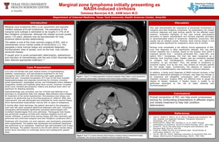
1. Marginal zone lymphoma initially presenting as
NASH-induced cirrhosis
Debdeep Banerjee A.B., ASM Islam M.D.
Department of Internal Medicine, Texas Tech University Health Sciences Center, Amarillo
Case Presentation
Marginal zone lymphoma (MZL) is an uncommon and typically
indolent subtype of B-cell lymphomas. The prevalence of the
marginal zone subtype is estimated to be roughly 5-17% of all
Non-Hodgkins Lymphomas. Although the median survival usually
spans >10 years, patients may be asymptomatic for long
durations before acutely deteriorating.
The spleen is one of the most common origins of MZL, with a
characteristic tumor marker profile of CD19/20/22 (+). This
alongside a bone marrow biopsy are considered diagnostic.
Extralymphatic sites of spread, such as the liver, may indicate
advanced disease.
If caught prior to acute symptomatic deterioration, splenectomy
and induction of chemotherapy with the anti-CD20 rituximab have
been deemed appropriate treatment.1-2
Introduction
This case demonstrates the importance of extranodal involvement in
marginal zone Non-Hodgkins Lymphoma. Its presentation can initially
confound diagnosis and lead workup specific for the affected organ
systems. Important highlights of this case include pancytopenia
secondary to splenic sequestration and cirrhosis stigmata secondary
to decompensated status of lymphoma. Relatively uncommon, MZL
usually has an indolent course. However, bone marrow biopsy in this
patient became significant for malignancy within only 8 months.
Perhaps most remarkable is the difficult clinical appearance of MZL,
such that diagnosis is often significantly delayed. MZL has been
further classified into 3 entities, based on the location from which it
originally derives from – including extranodal MZL, nodal MZL, and
splenic MZL.3 Increasingly, the liver has been recognized as a
distinctly local lymphoid organ. Hepatic cells are constantly exposed
to antigens and hematogenous introduction, via splanchnic
circulation, to gut microbes.4 Thus, the spread of lymphoma to
lymphoid organs, such as the liver, may present as clinically specific
for the target organ. Furthermore, the absence of regional or
peripheral lymphadenopathy may additionally confound diagnosis.
In addition, a faulty quick diagnosis of NASH-induced cirrhosis, in the
absence of alternative etiologies of cirrhosis, may steer the clinician in
an expensive and altogether unnecessary path. Ultrasound or
magnetic resonance imaging (MRI) are effective tools to gauge the
degree of hepatic steatosis, if hepatic biopsy is unable to be
performed secondary to the risk of major bleeding with
thrombocytopenia.5
Discussion
Prompt recognition of MZL can help avoid unnecessary
workup for confounding presentations in affected organs
and initiate treatment to help halt condition
deterioration.
Conclusions
References
1. Joshi M., Sheikh H., Abbi K., et. al. (2012). Marginal zone lymphoma: old,
new, targeted, and epigenetic therapies. Therapeutic Advances in
Hematology, 3(5), 275–290.
2. McDermott RS., Maher MM., Dunleavy K., et. al. (2002) Unusual
presentations of lymphoma. Journal of Clinical Oncology, 20(7), 1943-1946.
3. Zinzani PL. (2012) The many faces of marginal zone lymphoma. ASH
Education Book, 2012(1), 426-432.
4. Crispe IN. (2009) The liver as a lymphoid organ. Annual Review of
Immunology 2009(27), 147-163.
5. Lee SS., Park SH. (2014). Radiologic evaluation of nonalcoholic fatty liver
disease. World Journal of Gastroenterology, 20(23), 7392–7402.
A 63-year-old female with past medical history of hyperlipidemia,
diabetes, hypertension, and pancytopenia presented to her first
emergency room (ER) visit with worsening right upper quadrant
abdominal pain. Computed tomography (CT) scan at the time revealed
hepatosplenomegaly and stigmata concerning for cirrhosis, including
portal hypertension, esophageal varices, and intrahepatic biliary dilatation.
Coagulation studies and hepatitis panel were normal; however, complete
blood count revealed pancytopenia. History and physical exam were not
significant for bleeding episodes.
Gastroenterology was consulted, and her cirrhosis was believed to be
secondary to nonalcoholic fatty liver disease. Beta-blockers were initiated
for treatment of her portal hypertension. Percutaneous liver biopsy was
unable to be performed due to thrombocytopenia of <60K/ml. After pain
improvement, she was discharged and scheduled for bone marrow biopsy,
which demonstrated hypercellular marrow with no signs of malignancy.
8 months after initial discharge, the patient returned to the emergency
room with left upper quadrant abdominal distension with pain. Computed
tomography demonstrated significantly worsened splenomegaly with
consistent hepatomegaly from prior admission. Transjugular liver biopsy
was performed revealing cirrhosis with interface hepatitis and B-cell
marker infiltrates. A second bone marrow biopsy with flow cytometry was
performed and confirmed marginal zone Non-Hodgkins Lymphoma (NHL).
Treatment with rituximab, uric acid reducers, and neutropenic precautions
were implemented. The patient was discharged and portacath placement
was performed as an outpatient. Treatment was well-tolerated until 9 days
after discharge. The patient returned to the ER with altered mental status,
severe shock secondary to multiorgan failure and subsequently passed
away.
Figure 1. Axial CT on initial presentation revealing intrahepatic biliary dilation, portal hypertension,
and significant splenomegaly. Subsequent bone marrow biopsy revealed no malignancy.
Figure 2. Axial CT 8 months following initial admission revealing massive hepatosplenomegaly.
Subsequent liver and bone marrow biopsies confirmed marginal zone NHL.