Download to read offline
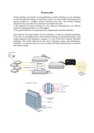


Western blot is a technique used to detect specific proteins in a complex mixture. It involves separating protein mixtures by size using gel electrophoresis, transferring the separated proteins to a solid support, and detecting target proteins using appropriately matched antibodies. Once detected, the target protein will appear as a band that is easy to interpret and unambiguous. Southern blot is a technique used to detect DNA sequences in a sample. It involves separating DNA fragments by size using gel electrophoresis, transferring the fragments to a membrane, and using probes to detect DNA sequences of interest by binding to their complementary sequences. Multiple probes can be used to detect different sequences in the same sample.


