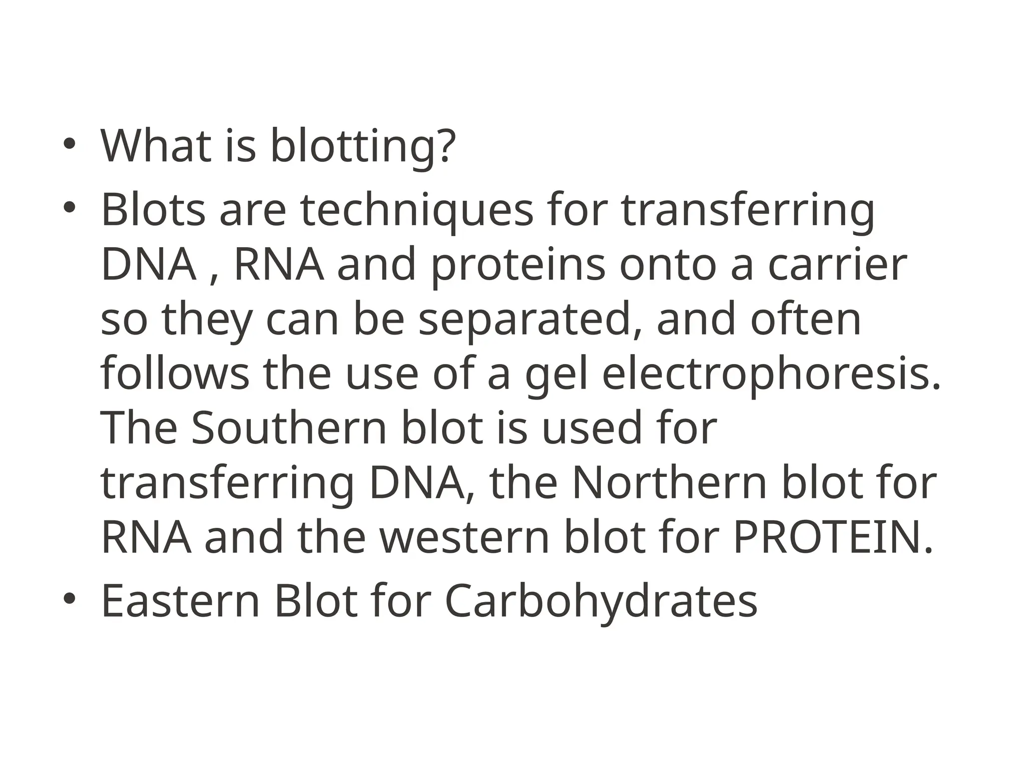
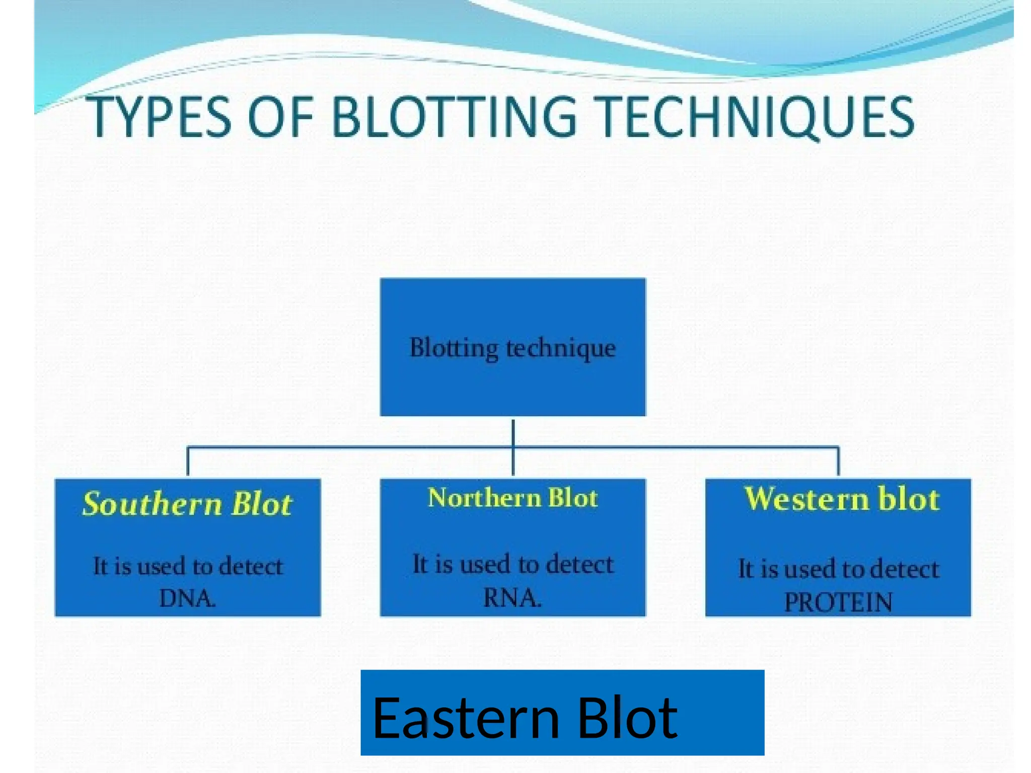
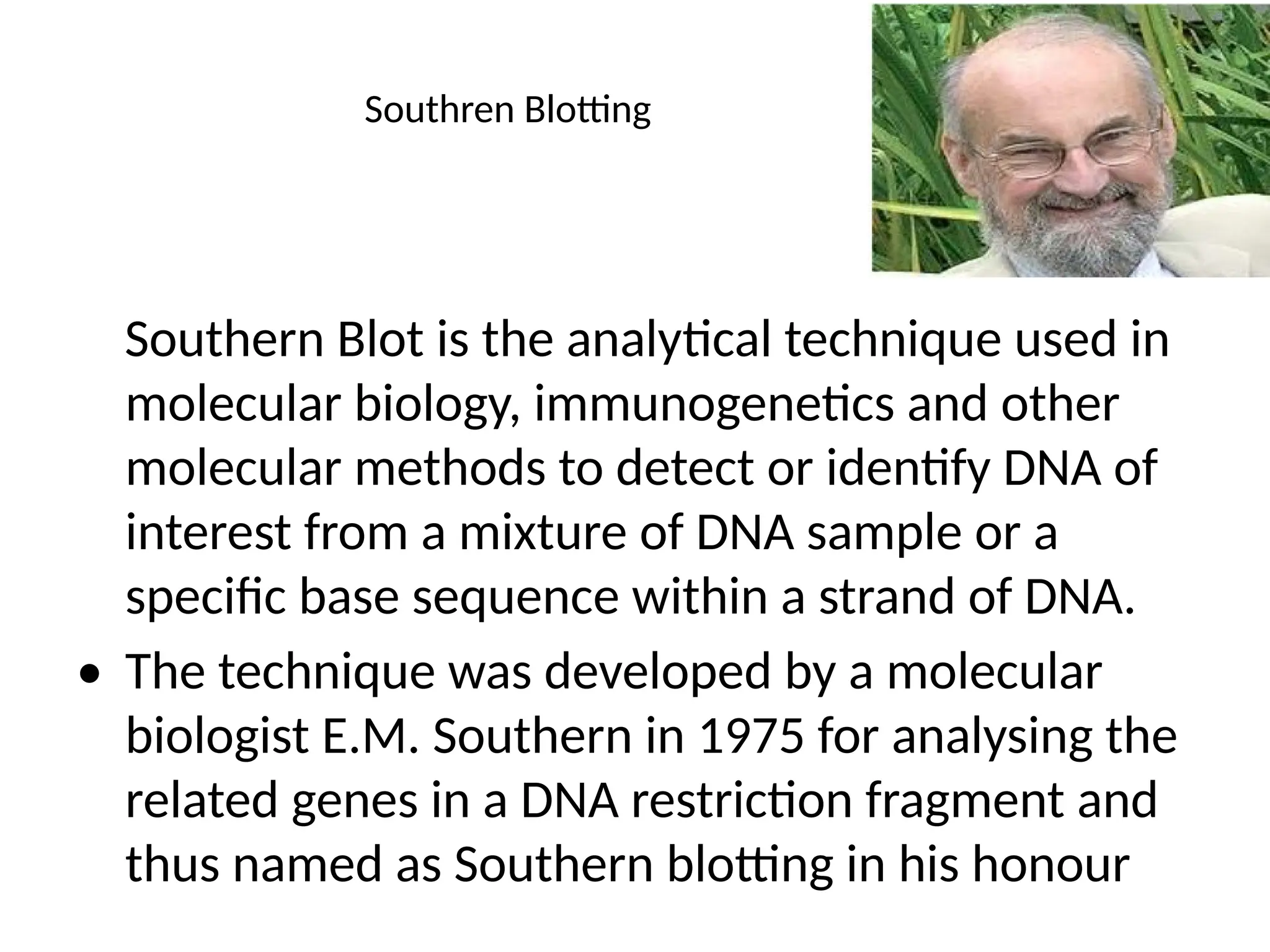
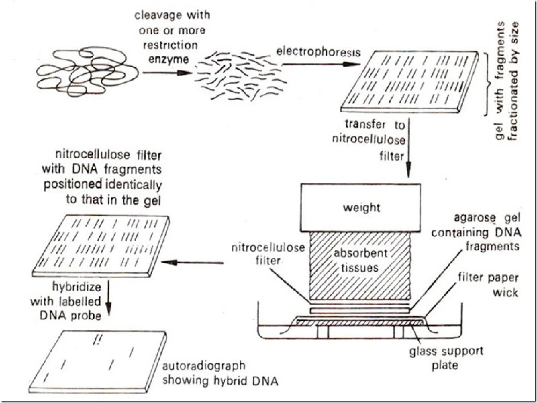
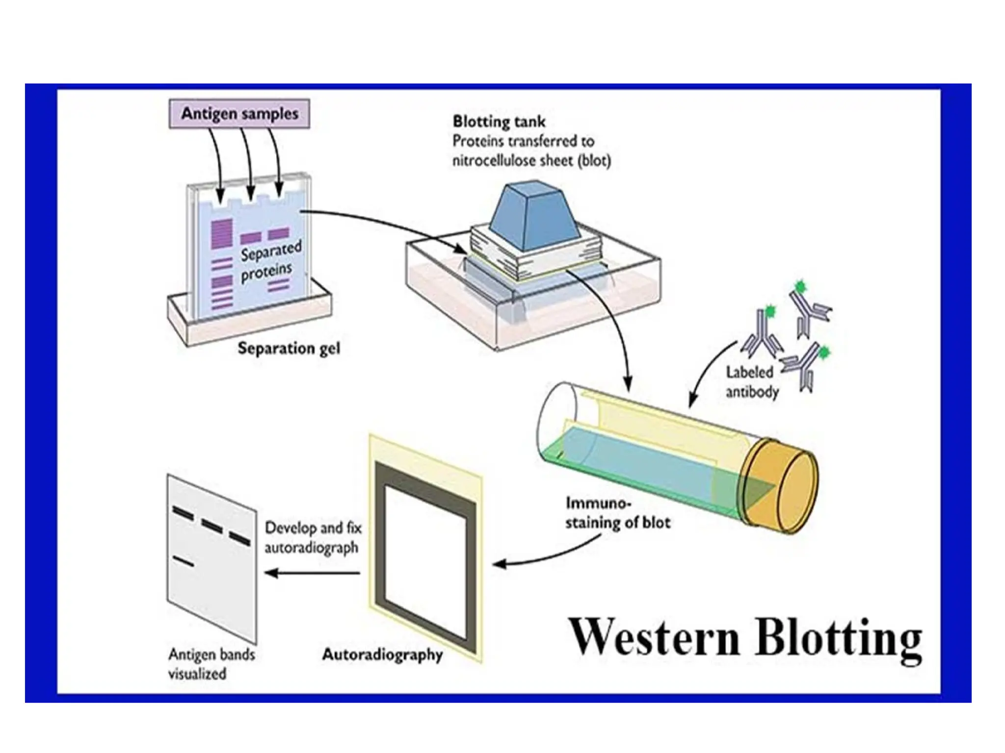
Blotting Technique
• What is blotting?
• Blots are techniques for transferring
DNA , RNA and proteins onto a carrier
so they can be separated, and often
follows the use of a gel electrophoresis.
The Southern blot is used for
transferring DNA, the Northern blot for
RNA and the western blot for PROTEIN.
• Eastern Blot for Carbohydrates
Eastern Blot
Southren Blotting
Southern Blot is the analytical technique used in
molecular biology, immunogenetics and other
molecular methods to detect or identify DNA of
interest from a mixture of DNA sample or a
specific base sequence within a strand of DNA.
• The technique was developed by a molecular
biologist E.M. Southern in 1975 for analysing the
related genes in a DNA restriction fragment and
thus named as Southern blotting in his honour
Principle
Hybridization
The process involves the transfer of electrophoresis-separated DNA
fragments to a carrier membrane which is usually nitrocellulose
and the subsequent detection of the target DNA fragment by
probe hybridization.
Hybridization refers to the process of forming a double-stranded
DNA molecule between a single-stranded DNA probe and a single-
stranded target DNA.
Since the probe and target DNA are complementary to each other,
the reaction is specific which aids in the detection of the specific
DNA fragment.
METHOD
• Restriction endonucleases are used to cut high-
molecular-weight DNA strands into smaller fragments.
• The DNA fragments are then electrophoresed on
an agarose gel to separate them by size.
• A sheet of nitrocellulose (or,
alternatively, nylone) membrane is placed on top of (or
below, depending on the direction of the transfer) the
gel.
METHOD
• Pressure is applied evenly to the gel (either
using suction, or by placing a stack of paper
towels and a weight on top of the membrane
and gel), to ensure good and even contact
between gel and membrane.
• The membrane is then baked in a vacuum or
regular oven at 80 °C for 2 hours (standard
conditions; nitrocellulose or nylon membrane)
or exposed to ultraviolet radiation.
METHOD
• The membrane is then exposed to a hybridization probe—a
single DNA fragment with a specific sequence whose presence
in the target DNA is to be determined.
• The probe DNA is labelled so that it can be detected, usually by
incorporating radioactivity or tagging the molecule with a
fluorescent or chromogenic dye.
• In some cases, the hybridization probe may be made from RNA,
rather than DNA. To ensure the specificity of the binding of the
probe to the sample DNA, most common hybridization methods
use salmon or herring sperm DNA for blocking of the membrane
surface and target DNA, deionized formamide, and detergents
such as SDS to reduce non-specific binding of the probe.
• After hybridization, excess probe is washed
from the membrane (typically using SSC buffer),
and the pattern of hybridization is visualized on
X-ray film by autoradiographyin the case of a
radioactive or fluorescent probe, or by
development of colour on the membrane if a
chromogenic detection method is used.
APPLICATIONS
• To identify specific DNA in a DNA sample
• To Isolate desired DNA for construction of rDNA
• Identify mutations, deletions, and gene
rearrangements
• Used in prognosis of cancer and in prenatal
diagnosis of genetic diseases
• In RFLP
• Used in phylogenetic analysis
• Diagnosis of HIV-1 and infectious disease
CONTINUED..
• In DNA fingerprinting:
1. Paternity and Maternity testing
2. Criminal identifications and Forensics
3. Personal Identification
Applications
• Identifying specific DNA in a DNA sample.
• Preparation of RFLP (Restriction Fragment Length Polymorphism)
maps
• Detection of mutations, deletions or gene rearrangements in DNA
• For criminal identification and DNA fingerprinting (VNTR)
• Detection and identification of trans gene in transgenic individual
• Mapping of restriction sites
• For diagnosis of infectious diseases
• Prognosis of cancer and prenatal diagnosis of genetic diseases
• Determination of the molecular weight of a restriction fragment
and to measure relative amounts in different samples.
Northern Blotting
• A northern blot is a laboratory method used to
detect specific RNA molecules among a mixture of
RNA. Northern blotting can be used to analyze a
sample of RNA from a particular tissue or cell type in
order to measure the RNA expression of particular
genes. This method was named for its similarity to
the technique known as a Southern blot
• BlotBase: a northern blot database
• Procedure:
• 1. The tissue or culture sample collected is first homogenized. The
samples may be representative of different types of culture for comparison
or it can be for the study of different stages of growth inside the culture.
• 2. The RNA sequence is separated in the electrophoresis unit an agarose
gel is used for the purpose of the nucleic acid separation.
• 3. Now the separated RNA sequence is transferred to the nylon
membrane. This is done by two mechanisms capillary action and the ionic
interaction.
• 4. The transfer operation is done by keeping the gel in the following
order. First, the agarose gel is placed on the bottom of the stack, followed by
the blotting membrane. On top of these paper towels a mild weight (glass
plate) is placed. The entire setup is kept in a beaker containing transfer
buffer.
• 5. RNA transferred to the nylon membrane is then fixed
using UV radiation.
• 6. The fixed nylon membrane is then mixed with probes.
The probes are specifically designed for the gene of interest,
so that they will hybridize with RNA sequences on the blot
corresponding to the sequence of interest.
• 7. The blot membrane is washed to remove unwanted
probe
• 8. Labeled probe is detected by chemiluminescence or
autoradiography. The result will be dark bands in x ray film.
• GEl and Probes
• The RNA samples are separated using agarose gels using formaldehyde as
denaturing agents but in small RNA or micro RNA sequences,
polyacrylamide sequences with urea as a denaturing agent also can be
used. Ethidium bromide can be used as a staining agent. Two types of
markers are for size marking. An RNA ladder and ribosomal subunit are
used for the identification of the size of the RNA sequences.
• Probes can be complementary to the whole or part of the RNA of interest.
They can be RNA, DNA or oligonucleotides of 25 complementary basepairs
to the target RNA. In case of RNA probes, invitro produced probes are used
as invivo probes can denature due to the rigorous washing. In case of
cDNA, the probes are labeled with radioactive isotopes, alkaline
phosphatase or horseradish peroxidase in case of chemiluminescence.
•
Western Blotting
• Western blot is the analytical technique used in
molecular biology, immunogenetics and other
molecular biology to detect specific proteins in a
sample of tissue homogenate or extract. Western
blotting is called so as the procedure is similar to
Southern blotting. While Southern blotting is done
to detect DNA, Western blotting is done for the
detection of proteins. Western blotting is also called
protein immunoblotting because an antibody is
used to specifically detect its antigen.
• Principle of western blotting:
• The technique consists of three major processes:
• 1. Separation of proteins by size (Electrophoresis).
• 2. Transfer to a solid support (Blotting)
• 3. Marking target protein using a proper primary and secondary antibody to
visualize (Detection).
• Electrophoresis used to separate proteins according to their electrophoretic
mobility which depends on charge, size of protein molecule and structure of the
proteins. Proteins are moved from within the gel onto a membrane made of
Nitrocellulose (NC) or Polyvinylidene difluoride (PVDF). Without pre-activation,
proteins combine with nitrocellulose membrane based on hydrophobic
interaction (Blotting). For detection of the proteins primary antibody and enzyme
conjugated secondary antibody are used. On addition of substrate, a substrate
reacts with the enzyme that is bound to the secondary antibody to generate
colored substance, namely, visible protein bands.
• In this technique a mixture of proteins is separated based on
molecular weight, and thus by type, through gel
electrophoresis. These results are then transferred to a
membrane producing a band for each protein. The membrane
is then incubated with labels antibodies specific to the protein
of interest. The unbound antibody is washed off leaving only
the bound antibody to the protein of interest. The bound
antibodies are then detected by developing the film. As the
antibodies only bind to the protein of interest, only one band
should be visible. The thickness of the band corresponds to the
amount of protein present; thus doing a standard can indicate
the amount of protein present.
• Western blotting is usually done on a tissue homogenate or
extract. It utilizes SDS-PAGE (Sodium dodecyl sulphate
polyacrylamide gel electrophoresis), a type of gel
electrophoresis to first separate various proteins in a mixture
on the basis of their shape and size. The protein bands thus
obtained are transferred onto a nitrocellulose or nylon
membrane where they are “probed” with antibodies specific to
the protein to be detected. The antigen–antibody complexes
that form on the band containing the protein recognized by the
antibody can be visualized in a variety of ways.
•
• If the protein of interest is bound by a radioactive antibody, its
position on the blot can be determined by exposing the
membrane to a sheet of X-ray film, a procedure called
autoradiography. However, the most generally used detection
procedures employ enzyme-linked antibodies against the
protein. After binding of the enzyme–antibody conjugate,
addition of a chromogenic substrate that produces a highly
colored and insoluble product causes the appearance of a
colored band at the site of the target antigen. The site of the
protein of interest can be determined with much higher
sensitivity if a chemiluminescent compound along with suitable
enhancing agents is used to produce light at the antigen site.
• Application of western blotting:
• 1. Identification of a specific protein in a complex mixture of proteins. In this method,
known antigens of well-defined molecular weight are separated by SDS-PAGE and
blotted onto nitrocellulose. The separated bands of known antigens are then probed
with the sample suspected of containing antibody specific for one or more of these
antigens. Reaction of an antibody with a band is detected by using either radiolabeled or
enzyme-linked secondary antibody that is specific for the species of the antibodies in the
test sample.
• 2. Estimation of the size of the protein as well as the amount of protein present in
the mixture.
• 3. It is most widely used as a confirmatory test for diagnosis of HIV, where this
procedure is used to determine whether the patient has antibodies that react with one
or more viral proteins or not.
• 4. Demonstration of specific antibodies in the serum for diagnosis of
neurocysticercosis and tubercular meningitis.
Blotting-Technique.pptx by taimoor khan 1

Blotting-Technique.pptx by taimoor khan 1

  • 1.
  • 2.
    • What isblotting? • Blots are techniques for transferring DNA , RNA and proteins onto a carrier so they can be separated, and often follows the use of a gel electrophoresis. The Southern blot is used for transferring DNA, the Northern blot for RNA and the western blot for PROTEIN. • Eastern Blot for Carbohydrates
  • 3.
  • 4.
    Southren Blotting Southern Blotis the analytical technique used in molecular biology, immunogenetics and other molecular methods to detect or identify DNA of interest from a mixture of DNA sample or a specific base sequence within a strand of DNA. • The technique was developed by a molecular biologist E.M. Southern in 1975 for analysing the related genes in a DNA restriction fragment and thus named as Southern blotting in his honour
  • 5.
    Principle Hybridization The process involvesthe transfer of electrophoresis-separated DNA fragments to a carrier membrane which is usually nitrocellulose and the subsequent detection of the target DNA fragment by probe hybridization. Hybridization refers to the process of forming a double-stranded DNA molecule between a single-stranded DNA probe and a single- stranded target DNA. Since the probe and target DNA are complementary to each other, the reaction is specific which aids in the detection of the specific DNA fragment.
  • 6.
    METHOD • Restriction endonucleasesare used to cut high- molecular-weight DNA strands into smaller fragments. • The DNA fragments are then electrophoresed on an agarose gel to separate them by size. • A sheet of nitrocellulose (or, alternatively, nylone) membrane is placed on top of (or below, depending on the direction of the transfer) the gel.
  • 7.
    METHOD • Pressure isapplied evenly to the gel (either using suction, or by placing a stack of paper towels and a weight on top of the membrane and gel), to ensure good and even contact between gel and membrane. • The membrane is then baked in a vacuum or regular oven at 80 °C for 2 hours (standard conditions; nitrocellulose or nylon membrane) or exposed to ultraviolet radiation.
  • 8.
    METHOD • The membraneis then exposed to a hybridization probe—a single DNA fragment with a specific sequence whose presence in the target DNA is to be determined. • The probe DNA is labelled so that it can be detected, usually by incorporating radioactivity or tagging the molecule with a fluorescent or chromogenic dye. • In some cases, the hybridization probe may be made from RNA, rather than DNA. To ensure the specificity of the binding of the probe to the sample DNA, most common hybridization methods use salmon or herring sperm DNA for blocking of the membrane surface and target DNA, deionized formamide, and detergents such as SDS to reduce non-specific binding of the probe.
  • 9.
    • After hybridization,excess probe is washed from the membrane (typically using SSC buffer), and the pattern of hybridization is visualized on X-ray film by autoradiographyin the case of a radioactive or fluorescent probe, or by development of colour on the membrane if a chromogenic detection method is used.
  • 11.
    APPLICATIONS • To identifyspecific DNA in a DNA sample • To Isolate desired DNA for construction of rDNA • Identify mutations, deletions, and gene rearrangements • Used in prognosis of cancer and in prenatal diagnosis of genetic diseases • In RFLP • Used in phylogenetic analysis • Diagnosis of HIV-1 and infectious disease
  • 12.
    CONTINUED.. • In DNAfingerprinting: 1. Paternity and Maternity testing 2. Criminal identifications and Forensics 3. Personal Identification
  • 13.
    Applications • Identifying specificDNA in a DNA sample. • Preparation of RFLP (Restriction Fragment Length Polymorphism) maps • Detection of mutations, deletions or gene rearrangements in DNA • For criminal identification and DNA fingerprinting (VNTR) • Detection and identification of trans gene in transgenic individual • Mapping of restriction sites • For diagnosis of infectious diseases • Prognosis of cancer and prenatal diagnosis of genetic diseases • Determination of the molecular weight of a restriction fragment and to measure relative amounts in different samples.
  • 14.
    Northern Blotting • Anorthern blot is a laboratory method used to detect specific RNA molecules among a mixture of RNA. Northern blotting can be used to analyze a sample of RNA from a particular tissue or cell type in order to measure the RNA expression of particular genes. This method was named for its similarity to the technique known as a Southern blot • BlotBase: a northern blot database
  • 15.
    • Procedure: • 1.The tissue or culture sample collected is first homogenized. The samples may be representative of different types of culture for comparison or it can be for the study of different stages of growth inside the culture. • 2. The RNA sequence is separated in the electrophoresis unit an agarose gel is used for the purpose of the nucleic acid separation. • 3. Now the separated RNA sequence is transferred to the nylon membrane. This is done by two mechanisms capillary action and the ionic interaction. • 4. The transfer operation is done by keeping the gel in the following order. First, the agarose gel is placed on the bottom of the stack, followed by the blotting membrane. On top of these paper towels a mild weight (glass plate) is placed. The entire setup is kept in a beaker containing transfer buffer.
  • 16.
    • 5. RNAtransferred to the nylon membrane is then fixed using UV radiation. • 6. The fixed nylon membrane is then mixed with probes. The probes are specifically designed for the gene of interest, so that they will hybridize with RNA sequences on the blot corresponding to the sequence of interest. • 7. The blot membrane is washed to remove unwanted probe • 8. Labeled probe is detected by chemiluminescence or autoradiography. The result will be dark bands in x ray film.
  • 17.
    • GEl andProbes • The RNA samples are separated using agarose gels using formaldehyde as denaturing agents but in small RNA or micro RNA sequences, polyacrylamide sequences with urea as a denaturing agent also can be used. Ethidium bromide can be used as a staining agent. Two types of markers are for size marking. An RNA ladder and ribosomal subunit are used for the identification of the size of the RNA sequences. • Probes can be complementary to the whole or part of the RNA of interest. They can be RNA, DNA or oligonucleotides of 25 complementary basepairs to the target RNA. In case of RNA probes, invitro produced probes are used as invivo probes can denature due to the rigorous washing. In case of cDNA, the probes are labeled with radioactive isotopes, alkaline phosphatase or horseradish peroxidase in case of chemiluminescence. •
  • 18.
    Western Blotting • Westernblot is the analytical technique used in molecular biology, immunogenetics and other molecular biology to detect specific proteins in a sample of tissue homogenate or extract. Western blotting is called so as the procedure is similar to Southern blotting. While Southern blotting is done to detect DNA, Western blotting is done for the detection of proteins. Western blotting is also called protein immunoblotting because an antibody is used to specifically detect its antigen.
  • 20.
    • Principle ofwestern blotting: • The technique consists of three major processes: • 1. Separation of proteins by size (Electrophoresis). • 2. Transfer to a solid support (Blotting) • 3. Marking target protein using a proper primary and secondary antibody to visualize (Detection). • Electrophoresis used to separate proteins according to their electrophoretic mobility which depends on charge, size of protein molecule and structure of the proteins. Proteins are moved from within the gel onto a membrane made of Nitrocellulose (NC) or Polyvinylidene difluoride (PVDF). Without pre-activation, proteins combine with nitrocellulose membrane based on hydrophobic interaction (Blotting). For detection of the proteins primary antibody and enzyme conjugated secondary antibody are used. On addition of substrate, a substrate reacts with the enzyme that is bound to the secondary antibody to generate colored substance, namely, visible protein bands.
  • 21.
    • In thistechnique a mixture of proteins is separated based on molecular weight, and thus by type, through gel electrophoresis. These results are then transferred to a membrane producing a band for each protein. The membrane is then incubated with labels antibodies specific to the protein of interest. The unbound antibody is washed off leaving only the bound antibody to the protein of interest. The bound antibodies are then detected by developing the film. As the antibodies only bind to the protein of interest, only one band should be visible. The thickness of the band corresponds to the amount of protein present; thus doing a standard can indicate the amount of protein present.
  • 22.
    • Western blottingis usually done on a tissue homogenate or extract. It utilizes SDS-PAGE (Sodium dodecyl sulphate polyacrylamide gel electrophoresis), a type of gel electrophoresis to first separate various proteins in a mixture on the basis of their shape and size. The protein bands thus obtained are transferred onto a nitrocellulose or nylon membrane where they are “probed” with antibodies specific to the protein to be detected. The antigen–antibody complexes that form on the band containing the protein recognized by the antibody can be visualized in a variety of ways. •
  • 23.
    • If theprotein of interest is bound by a radioactive antibody, its position on the blot can be determined by exposing the membrane to a sheet of X-ray film, a procedure called autoradiography. However, the most generally used detection procedures employ enzyme-linked antibodies against the protein. After binding of the enzyme–antibody conjugate, addition of a chromogenic substrate that produces a highly colored and insoluble product causes the appearance of a colored band at the site of the target antigen. The site of the protein of interest can be determined with much higher sensitivity if a chemiluminescent compound along with suitable enhancing agents is used to produce light at the antigen site.
  • 24.
    • Application ofwestern blotting: • 1. Identification of a specific protein in a complex mixture of proteins. In this method, known antigens of well-defined molecular weight are separated by SDS-PAGE and blotted onto nitrocellulose. The separated bands of known antigens are then probed with the sample suspected of containing antibody specific for one or more of these antigens. Reaction of an antibody with a band is detected by using either radiolabeled or enzyme-linked secondary antibody that is specific for the species of the antibodies in the test sample. • 2. Estimation of the size of the protein as well as the amount of protein present in the mixture. • 3. It is most widely used as a confirmatory test for diagnosis of HIV, where this procedure is used to determine whether the patient has antibodies that react with one or more viral proteins or not. • 4. Demonstration of specific antibodies in the serum for diagnosis of neurocysticercosis and tubercular meningitis.