
Download to read offline
![SSR Inst. Int. J. Life Sci. ISSN (O): 2581-8740 | ISSN (P): 2581-8732
Misra and Chandravati, 2019
DOI:10.21276/SSR-IIJLS.2019.5.5.4
Copyright © 2015 - 2019| SSR-IIJLS by Society for Scientific Research under a CC BY-NC 4.0 International License Volume 05 | Issue 05 | Page 2393
A Study on Hysteroscopy as a Primary Diagnostic Modality in
Patients of Abnormal Uterine Bleeding
Malvika Misra
1
*, DR Chandravati
2
1
Assistant Professor, Department of Obstetrics and Gynecology, Dr. Ram Manohar Lohia Institute of Medical Sciences,
Lucknow, India
2
Emeritus Professor, Department of Obstetrics and Gynecology, Krishna Medical Centre, Lucknow, India
*Address for Correspondence: Dr. Malvika Misra, Assistant Professor, Department of Obstetrics and Gynecology, Dr.
Ram Manohar Lohia Institute of Medical Sciences, Lucknow, India
E-mail: drmalvikam@gmail.com
Received: 12 Apr 2019/ Revised: 19 Jul 2019/ Accepted: 22 Aug 2019
ABSTRACT
Background: Among women, abnormal uterine bleeding and infertility rates are rapidly increasing. The clinician generally prefers
physical examination, methodical histopathology report to diagnose the pregnancy related disorders, systemic conditions and
disease related to the genital tract. Diagnostic hysteroscopy is a precious contrivance to diagnose widespread disorders and
diseases related to female genital tract and uterus, such as abnormal uterine bleeding since hysteroscopy assists to understand
the mechanism for the regulation of normal cycle.
Methods: The study had been carried out in the Department of Obstetrics and Gynecology, Krishna Medical Center, Lucknow from
September 2012 to July 2017. Total of 400 patients were included in this randomized study. The study allocated histopathologic
analysis, diagnostic hysteroscopy, intervention with or without anesthesia and postoperative diagnosis of patients.
Results: Of all the cases, who underwent diagnostic hysteroscopy, about 44% of cases were normal and 56% of cases were found
with various abnormalities. 22% of cases had endometrial hyperplasia, 11% of cases had an endometrial polyp, 10% of cases had
submucous myoma and 9% of cases had atrophy. On observing the clinical presentation, 42% of cases had menorrhagia, 20% of
cases had polymenorrhoea, 16% of cases had metorrhagia and 22% of cases had reported with postmenopausal bleeding. About
60% of diagnostic hysteroscopies were performed without anesthesia, however, an ascending trend was reported for general
anesthesia. Primary infertility was reported in 44% of cases and 56% of cases were reported with secondary infertility.
Conclusion: Our study concluded that the patients with pregnancy-related disorders and disease related to the genital tract and
uterus should first undergo diagnostic hysteroscopy to provide a diagnosis with high accuracy and specificity.
Key-words: Abnormal Uterine bleeding, Anesthesia, Diagnostic hysteroscopy, Infertility, Tamoxifen therapy
INTRODUCTION
Abnormal uterine bleeding (AUB) becomes a common
gynecological problem encountered in the family
practice setting, with complicated clinical presentations.
A regular history, physical examination, and laboratory
evaluation possibly enables a clinician to find out the
causes of disorders related to fertility such as genital
tract pathology, iatrogenic causes, as well as medications
in women under childbearing age [1]
.
How to cite this article
Misra M, Chandravati DR. A Study on Hysteroscopy as a Primary
Diagnostic Modality in Patients of Abnormal Uterine Bleeding. SSR
Inst. Int. J. Life. Sci., 2019; 5(5): 2393-2401.
Access this article online
https://iijls.com/
A national study on mechanism of abnormal uterine
bleeding reported those over a period of 10 years,
menstrual disorder were observed in 19% of 20 million
patient visits to a physician’s clinic for gynecologic
condition [2]
. Besides this, 25% surgeries at gynecologist
clinic were done to treat abnormal uterine bleeding.
Women experienced several changes in frequency of the
menstrual period, duration of menstrual period, or
quantity of flow, as well as bleeding among two cycles
[2,3]
. The abnormal uterine bleeding in postmenopausal
women, vaginal bleeding after 12 months or more of
cessation of menses, and/or irregular bleeding in
postmenopausal women who have been getting
12month or more then 12 month hormone therapy [4,5]
.
The first contemplation is pregnancy for women of
childbearing age facing AUB [6]
. The most possible
Research Article](https://image.slidesharecdn.com/studyhysteroscopyprimarydiagnosticpatientsabnormaluterinebleeding-230718111956-a037e437/85/Study_Hysteroscopy_Primary_Diagnostic_Patients_Abnormal_Uterine_Bleeding-pdf-1-320.jpg)
![SSR Inst. Int. J. Life Sci. ISSN (O): 2581-8740 | ISSN (P): 2581-8732
Misra and Chandravati, 2019
DOI:10.21276/SSR-IIJLS.2019.5.5.4
Copyright © 2015 - 2019| SSR-IIJLS by Society for Scientific Research under a CC BY-NC 4.0 International License Volume 05 | Issue 05 | Page 2393
A Study on Hysteroscopy as a Primary Diagnostic Modality in
Patients of Abnormal Uterine Bleeding
Malvika Misra
1
*, DR Chandravati
2
1
Assistant Professor, Department of Obstetrics and Gynecology, Dr. Ram Manohar Lohia Institute of Medical Sciences,
Lucknow, India
2
Emeritus Professor, Department of Obstetrics and Gynecology, Krishna Medical Centre, Lucknow, India
*Address for Correspondence: Dr. Malvika Misra, Assistant Professor, Department of Obstetrics and Gynecology, Dr.
Ram Manohar Lohia Institute of Medical Sciences, Lucknow, India
E-mail: drmalvikam@gmail.com
Received: 12 Apr 2019/ Revised: 19 Jul 2019/ Accepted: 22 Aug 2019
ABSTRACT
Background: Among women, abnormal uterine bleeding and infertility rates are rapidly increasing. The clinician generally prefers
physical examination, methodical histopathology report to diagnose the pregnancy related disorders, systemic conditions and
disease related to the genital tract. Diagnostic hysteroscopy is a precious contrivance to diagnose widespread disorders and
diseases related to female genital tract and uterus, such as abnormal uterine bleeding since hysteroscopy assists to understand
the mechanism for the regulation of normal cycle.
Methods: The study had been carried out in the Department of Obstetrics and Gynecology, Krishna Medical Center, Lucknow from
September 2012 to July 2017. Total of 400 patients were included in this randomized study. The study allocated histopathologic
analysis, diagnostic hysteroscopy, intervention with or without anesthesia and postoperative diagnosis of patients.
Results: Of all the cases, who underwent diagnostic hysteroscopy, about 44% of cases were normal and 56% of cases were found
with various abnormalities. 22% of cases had endometrial hyperplasia, 11% of cases had an endometrial polyp, 10% of cases had
submucous myoma and 9% of cases had atrophy. On observing the clinical presentation, 42% of cases had menorrhagia, 20% of
cases had polymenorrhoea, 16% of cases had metorrhagia and 22% of cases had reported with postmenopausal bleeding. About
60% of diagnostic hysteroscopies were performed without anesthesia, however, an ascending trend was reported for general
anesthesia. Primary infertility was reported in 44% of cases and 56% of cases were reported with secondary infertility.
Conclusion: Our study concluded that the patients with pregnancy-related disorders and disease related to the genital tract and
uterus should first undergo diagnostic hysteroscopy to provide a diagnosis with high accuracy and specificity.
Key-words: Abnormal Uterine bleeding, Anesthesia, Diagnostic hysteroscopy, Infertility, Tamoxifen therapy
INTRODUCTION
Abnormal uterine bleeding (AUB) becomes a common
gynecological problem encountered in the family
practice setting, with complicated clinical presentations.
A regular history, physical examination, and laboratory
evaluation possibly enables a clinician to find out the
causes of disorders related to fertility such as genital
tract pathology, iatrogenic causes, as well as medications
in women under childbearing age [1]
.
How to cite this article
Misra M, Chandravati DR. A Study on Hysteroscopy as a Primary
Diagnostic Modality in Patients of Abnormal Uterine Bleeding. SSR
Inst. Int. J. Life. Sci., 2019; 5(5): 2393-2401.
Access this article online
https://iijls.com/
A national study on mechanism of abnormal uterine
bleeding reported those over a period of 10 years,
menstrual disorder were observed in 19% of 20 million
patient visits to a physician’s clinic for gynecologic
condition [2]
. Besides this, 25% surgeries at gynecologist
clinic were done to treat abnormal uterine bleeding.
Women experienced several changes in frequency of the
menstrual period, duration of menstrual period, or
quantity of flow, as well as bleeding among two cycles
[2,3]
. The abnormal uterine bleeding in postmenopausal
women, vaginal bleeding after 12 months or more of
cessation of menses, and/or irregular bleeding in
postmenopausal women who have been getting
12month or more then 12 month hormone therapy [4,5]
.
The first contemplation is pregnancy for women of
childbearing age facing AUB [6]
. The most possible
Research Article](https://image.slidesharecdn.com/studyhysteroscopyprimarydiagnosticpatientsabnormaluterinebleeding-230718111956-a037e437/75/Study_Hysteroscopy_Primary_Diagnostic_Patients_Abnormal_Uterine_Bleeding-pdf-1-2048.jpg)
![SSR Inst. Int. J. Life Sci. ISSN (O): 2581-8740 | ISSN (P): 2581-8732
Misra and Chandravati, 2019
DOI:10.21276/SSR-IIJLS.2019.5.5.4
Copyright © 2015 - 2019| SSR-IIJLS by Society for Scientific Research under a CC BY-NC 4.0 International License Volume 05 | Issue 05 | Page 2394
reasons behind pregnancy-related bleeding comprise of
spontaneous pregnancy loss, ectopic pregnancy,
placenta previa, abruptio placentae, and trophoblastic
disease [3,7]
. Patients are supposed to have queries
regarding patterns, contraception, and activity related to
reproductive life. In establishing pregnancy-related
disorders, a number of examinations are done, including
bimanual pelvic examination, a beta-subunit human
chorionic gonadotropin test as well as transvaginal
sonography [8]
. Subsequently, iatrogenic reasons of
abnormal uterine bleeding should be investigated.
Medication might be the reason behind the onset of
bleeding, which includes anticoagulants, reuptake
inhibitors of selective serotonin, antipsychotics,
corticosteroids, hormonal medications, and tamoxifen.
Ginseng, ginkgo, and soy supplements are few herbal
substances that alter the estrogen levels and parameters
of clotting, and therefore they may cause menstrual
irregularities [5]
. The evaluation of abnormal uterine
bleeding significantly depends on the age of the patient,
risk factors associated with endometrial cancer includes
high body mass index (BMI), anovulatory cycles,
nulliparity, age exceeding 35 years and tamoxifen
therapy [8]
. Medical management is recommended for
patients at low risk for endometrial carcinoma in
premenopausal women who are diagnosed with
presumed dysfunctional uterine bleeding. Endometrial
evaluation of subtle genital tract pathology is suggested
for patients having high risk of endometrial cancer as
well as at low risk, who keep abnormally bleeding
continuously despite of medical management [9]
.
Recently, hysteroscopy is the most accepted procedure
for the analysis and/or for treatment of various
pathologies of the endometrial cavity, tubal ostia and/or
endocervical canal for diagnosis alone or for diagnostic
and operating procedure in the same sitting [10,11]
. A
review article published in 2011, introduces the
diagnostic and operative indications for hysteroscopy. It
includes abnormal uterine bleeding, which might be
premenopausal or postmenopausal, sub-mucosal,
endometrial polyps, intramural, intrauterine adhesions,
fibroids, and Mullerian anomalies retained products of
conception and intrauterine devices or other foreign
bodies and endocervical polyps [5,10]
. Several conditions
such as viable intrauterine pregnancy, active pelvic
infection, uterine cancer or cervical cancer are
contraindications to hysteroscopy [11]
. The probabilities
to carry out hysteroscopy without anesthesia or with
local anesthesia allocate outpatient settings and speedy
recovery. The vaginoscopic technique is carried out, that
is devoid of tenaculum or speculum, and anesthesia [12]
.
Mostly the diagnosis and the minor operations can be
done without anesthetic or can be performed with a
local anesthetic [13]
. General or regional anesthesia is
ideal for patients undergoing extensive operative
procedures, or patients with comorbidities that
necessitate intensive monitoring [14,15]
. Dilation and
Curettage (D&C) are traditionally used to evaluate
abnormal uterine bleeding. It is a blind procedure and
the endometrium is transported to pathologist for the
analyses of histological patterns and reporting [1,16]
. The
procedure completely depends on co-operation of the
pathologist to get outcomes; therefore the procedure is
believed to be less accurate.
The study has been undertaken to evaluate the
indication for diagnostic hysteroscopy, type of
anesthesia used during the procedure, the role of
hysteroscopy for evaluation of abnormal uterine
bleeding and finding accuracy among hysteroscopy and
histopathology via comparative analysis. The study also
focused towards the adverse condition affecting the
fertility of patients. Furthermore, the study also
evaluated the ratio and percentage of cases facing
abnormal uterine bleeding, subsequent to the
completion of procedure or treatment.
MATERIALS AND METHODS
The present “randomized study” was an approach to
overcome the failure of abnormal uterine bleeding
treatment. The study had been carried out in the
Department of Obstetrics and Gynecology, Krishna
Medical Center, Lucknow, India from September 2012 to
July 2017. The subject included 400 patients that had
been attended OPD and underwent hysteroscopy and
D&C subsequent to the exclusion of comorbid illness.
Inclusion criteria
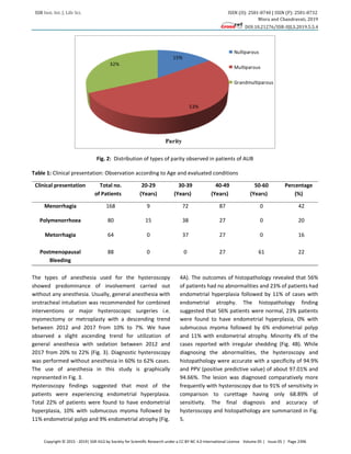
1. Patients had age between 20 to 60 years with AUB
2. Multiparous, nulliparous and grand multiparous
3. Patients did not require emergency treatment [16]
Exclusion criteria
1. Patients with profuse bleeding and large fibroids or
multiple fibroids.
2. Cases having genital tract infection and genital tract
malignancies [16]](https://image.slidesharecdn.com/studyhysteroscopyprimarydiagnosticpatientsabnormaluterinebleeding-230718111956-a037e437/85/Study_Hysteroscopy_Primary_Diagnostic_Patients_Abnormal_Uterine_Bleeding-pdf-2-320.jpg)
![SSR Inst. Int. J. Life Sci. ISSN (O): 2581-8740 | ISSN (P): 2581-8732
Misra and Chandravati, 2019
DOI:10.21276/SSR-IIJLS.2019.5.5.4
Copyright © 2015 - 2019| SSR-IIJLS by Society for Scientific Research under a CC BY-NC 4.0 International License Volume 05 | Issue 05 | Page 2395
Before proceeding to hysteroscopy, the investigations
were done, including PAP smear, vaginal bacteriologic
tests, haemograms [17]
. The patients were subjected to
detailed history taking, physical examination as well as
specific examination in the form of per speculum and per
vaginal examination. Blood investigations and urine
investigations include hemoglobin (Hb%), ABO and
Rhesus (Rh), blood sugar level, time of bleeding, time is
taken while blood clotting, aPTT, INR urine routine and
microscopy were ordered for all patients [18]
. TVS of all
the patients were done. Detailed information of all the
patients was obtained before taking up for any
procedure [15]
. Hysteroscopy and diagnostic D&C were
done for each of these patients. Hysteroscopic-guided
curetting were taken and sent for histopathological
analysis. The findings at USG, D&C reports, hysteroscopy
were compared with each other. The procedures were
done under anesthesia in the operation theater.
Generally, the antibiotics were not routinely
administered during hysteroscopy for avoiding surgical
site infection or endocarditis as post hysteroscopy
infection occurred in less than 1% of women [12]
.
Antibiotics were not regularly prescribed during the
procedure [15]
.
RESULTS
Hysteroscopy was carried out in this study by means of 5
mm hysteroscope with 30 degrees oblique lens in
patients of abnormal uterine bleeding and was
performed from September 2012 to July 2017. The study
was conducted with a group of 400 patients who were
presented with abnormal uterine bleeding. The
endometrium was transported to pathology for
histopathological investigations.
In the present study, maximum numbers of patients
were between 40–49 years of age. The oldest patient
was 52 years old and youngest one was about 22 years
old. 17.5% patient’s age incidences between 20–29
years, 16% patient’s age incidences between 30–39
years, 0.75% patient’s age incidences between 50–60
years, however the maximum number of patients
incidence between 40–49 years that were 65.75% of all
the patients.
The 16 patients of age between 20–29 years had less
than 6 months of symptoms, however, 72 patients of age
between 40–49 years had symptoms for 6 months to 1
year and 64 patients of the same age group had
symptoms for 1 year. Only 2 patients of the age group
between 50–60 years had 6 months to 1 year of
symptoms and only single case was reported symptoms
for 1 year (Fig. 1). About 53% of patients were
multiparous and belong to the age group between 40–49
years. Nulliparity was observed in 15% in patients
between 20–29 years of age (Fig. 2).
Polymenorrhoea was a frequent clinical presentation in
patient of age between 40–49 years and menorrhagia
were mostly observed in patients of age between 30–39
years. The clinical presentations for patients of the age
group between 20–29 years were not specified shown in
Table 1.
˂ 6 Month
1 year
0
20
40
60
80
20-29 30-39 40-49 50-60
No.
of
Patients
Age of Patients
Age incidance with respect to the duration of
symptoms
˂ 6 Month
6 Month to
1year
Fig. 1: Distribution of Patients according to age, having Symptoms of Abnormal menstrual cycle and uterine bleeding](https://image.slidesharecdn.com/studyhysteroscopyprimarydiagnosticpatientsabnormaluterinebleeding-230718111956-a037e437/85/Study_Hysteroscopy_Primary_Diagnostic_Patients_Abnormal_Uterine_Bleeding-pdf-3-320.jpg)
![SSR Inst. Int. J. Life Sci. ISSN (O): 2581-8740 | ISSN (P): 2581-8732
Misra and Chandravati, 2019
DOI:10.21276/SSR-IIJLS.2019.5.5.4
Copyright © 2015 - 2019| SSR-IIJLS by Society for Scientific Research under a CC BY-NC 4.0 International License Volume 05 | Issue 05 | Page 2399
0
5
10
15
20
25
30
%
of
cases
identified
TypesofPathology
Abnormal uterine bleeding
Percentage
Fig. 6: Data of different Pathology for abnormal uterine bleeding
Whereas- Hys biopsy- Hysteroscopic biopsy; IUD- Intrauterine devices
DISCUSSION
This study was done with the patients of the age group
between 20–60 years. However the maximum
abnormalities were observed in cases of age between
35–45 years. The study given by Panda et al.[19]
identified
that the maximum age incidence between 35–45 yrs and
the range of age in Gianninoto’s series was 38–40 years
and consequently most common incidence was between
30–45 yrs, however, Trotsenburg acknowledged the
maximum age incidence between 41–50 yrs [18,19]
.
The most common clinical symptoms presented in this
study was menorrhagia in 42% of cases.
Postmenopausal bleeding was present in 22% cases and
20% cases shown Polymenorrhoea [20]
. The series given
by Panda et al. [19]
recognized menorrhagia in 60% of
cases followed by Polymenorrhagia and Metrorrhagia
[15,21]
. In our study, 46% were found normal on
hysteroscopy whereas 54% of patients were identified
with abnormal symptoms related to reproductive health.
Since 1999, the specialists of reproductive gynecology
have disputes regarding the inclusion of hysteroscopy in
infertility investigation. Hysteroscopy detected
endometrial polyp, submucous fibroid and all cases of
endometrial hyperplasia accurately [15,20]
. European
society of human reproduction and embryology
conducted a study on hysteroscopy with endometrial
biopsy and thus concluded that the hysteroscopy with
endometrial biopsy is the “Gold standard” to investigate
abnormal uterine bleeding [19-22]
. Dilatation and
curettage was considered as obsolete because it is a
blind procedure with a complication rate of 6% to 8%
and low sensitivity for local and pedunculated intra-
cavitary lesions [9,16]
. The Lesions were more accurately
visualized through hysteroscopy therefore biopsy can be
done simultaneously. A study carried out at the
University of Wisconsin, Madison evaluated that the
hysteroscopy with biopsy allocates the visualization of
the endometrial cavity and is considered as the gold
standard for endometrial assessment [23]
.
The series provided by specialists of reproductive
gynecology concluded that hysteroscopy is necessary for
infertility investigated by Revel and Shushan [22]
.
However, the World Health Organization recommends
hysterosalpingography (HSG) for the management of
infertile women because HSG provides information on
tubal potency or blockage [15,23]
. World Health
Organization recommended hysteroscopy when an
intra-uterine abnormality was diagnosed via clinical or
complementary examination such as ultrasound and/or
HSG or after the failure of in vitro fertilization [24,25]
. Even
so, most of the specialists experience that hysteroscopy
is a more accurate tool because of their low false
positive and false-negative rates of intrauterine
abnormality and as a result, most of the specialist of
infertility utilizes hysteroscopy as “first-line routine
exam” for infertility patients [26-29]
.](https://image.slidesharecdn.com/studyhysteroscopyprimarydiagnosticpatientsabnormaluterinebleeding-230718111956-a037e437/85/Study_Hysteroscopy_Primary_Diagnostic_Patients_Abnormal_Uterine_Bleeding-pdf-7-320.jpg)
![SSR Inst. Int. J. Life Sci. ISSN (O): 2581-8740 | ISSN (P): 2581-8732
Misra and Chandravati, 2019
DOI:10.21276/SSR-IIJLS.2019.5.5.4
Copyright © 2015 - 2019| SSR-IIJLS by Society for Scientific Research under a CC BY-NC 4.0 International License Volume 05 | Issue 05 | Page 2400
CONCLUSIONS
The study revealed that the hysteroscopy is safe and
relatively more accurate in comparison to another
procedure to diagnose the patients, who had abnormal
uterine bleeding. Therefore, the procedure can be
considered as an advantage for women facing infertility
problems. The abnormalities and clinical presentations of
AUB and other genital tract malignancies have been
easily diagnosed by using hysteroscopy as well as it can
be operated and treated in the same sitting. The
gynecologists prefer hysteroscopy over D&C USG and/or
HSG because these procedures failed to detect cervical
or uterine pathologies and cancer as well as the results
obtained through this procedure illustrates the high rate
of false positive. Therefore, it possibly will lead to the
wrong diagnosis and therapeutic decision. Moreover, the
outcomes reveled through this procedure are less
specific and have had low sensitivity compared to
hysteroscopy, therefore fails to identify the lesion.
Our study provided information on the effectiveness and
accuracy of hysteroscopy. We concluded that
hysteroscopy should be the first and foremost
examination to be done for patients experiencing
abnormal menstrual and/or abnormal uterine bleeding.
As well as it can provide a decisive outline for further
improvement in management and follow-up of patient
with abnormal uterine bleeding and infertility, thus,
benefitting in terms of time loss in reaching to an
endpoint for the final accurate diagnosis for the patient.
This study is a strong data source that may facilitate
innovative ideas among researchers to improve
technology.
CONTRIBUTION OF AUTHORS
Research concept- Dr. Chandravati
Research design- Dr. Malvika Misra
Supervision- Dr. Chandravati
Materials- Dr. Chandravati
Data collection- Dr. Malvika Misra
Data analysis and Interpretation- Dr. Malvika Misra
Literature search- Dr. Malvika Misra
Writing article- Dr. Malvika Misra
Critical review- Dr. Chandravati
Article editing- Dr. Malvika Misra
Final approval- Dr. Chandravati
REFERENCES
[1] Nicholson WK, Ellison SA, Grason H, Powe NR.
Patterns of ambulatory care use for gynecologic
conditions: a national study. Am. J. Obstet. Gynecol.,
2001; 184: 523–30.
[2] Livingstone M, Fraser IS. Mechanisms of abnormal
uterine bleeding. Hum. Reprod. Update, 2002; 8: 60–
67.
[3] Janet RA, Sharon KH, Robert MW. Abnormal uterine
bleeding. Am. Fam. Physician, 2004; 69(8): 1915–26.
[4] Lethaby A, Farquhar C, Sarkis A, Roberts H, Jepson R,
et al. Hormone replacement therapy in
postmenopausal women: endometrial hyperplasia
and irregular bleeding. Cochrane Database Syst. Rev.,
2003; 4: CD000402.
[5] ACOG practice bulletin. Clinical management
guidelines for obstetrician-gynecologists. Use of
botanicals for management of menopausal
symptoms. Obstet. Gynecol., 2001; 96(6 suppl): 01–
11.
[6] Shwayder JM. Pathophysiology of abnormal uterine
bleeding. Obstet. Gynecol. Clin. North Am., 2000; 27:
219–34.
[7] Oriel KA, Schrager S. Abnormal uterine bleeding. Am.
Fam. Physician, 1999; 60: 1371–80.
[8] Baggish MS. Operative Hysteroscopy. In: Rock JA,
Jones HW III, editors. TeLinde’s Operative
Gynecology 9th
ed. Philadelphia: Lippincott Williams
& Wilkins; 2003: 379–411.
[9] Sheth SS, Nerukar NM, Mangeshkar PS. Hysteroscopy
in abnormal uterine bleeding. J. Obstet. Gynecol.
India, 1990; 40: 451.
[10]Hargreave T, Mellows H. WHO Manual for the
Standardized Investigation and Diagnosis of the
Infertile Couple, The Press Syndicate of the
University of Cambridge, Cambridge, UK, 1993.
[11]Koskas M, Mergui JL, Yazbeck C. Office hysteroscopy
for infertility: a series of 557 consecutive cases.
Obstet. Gynecol. Int., 2010: 168096.
[12]Price TM, Harris JB. Fulminant hepatic failure due to
herpes simplex after hysteroscopy. Obstet. Gynecol.,
2001; 98: 954.
[13]Garbin O, Kutnahorsky R, Gollner JL. Vaginoscopic
versus conventional approaches to outpatient
diagnostic hysteroscopy: a two-centre randomized
prospective study. Hum. Reprod., 2006; 21: 2996–
300.](https://image.slidesharecdn.com/studyhysteroscopyprimarydiagnosticpatientsabnormaluterinebleeding-230718111956-a037e437/85/Study_Hysteroscopy_Primary_Diagnostic_Patients_Abnormal_Uterine_Bleeding-pdf-8-320.jpg)
![SSR Inst. Int. J. Life Sci. ISSN (O): 2581-8740 | ISSN (P): 2581-8732
Misra and Chandravati, 2019
DOI:10.21276/SSR-IIJLS.2019.5.5.4
Copyright © 2015 - 2019| SSR-IIJLS by Society for Scientific Research under a CC BY-NC 4.0 International License Volume 05 | Issue 05 | Page 2401
[14]Bettocchi S, Ceci O, Di Venere R. Advanced Rowe
operative office hysteroscopy without anesthesia:
analysis of 501 cases treated with a 5 Fr bipolar
electrode. Hum. Reprod., 2002; 17: 2435–38.
[15]Stefanescu A, Marinescu B. Diagnostic hysteroscopy-
A retrosoective study of 1545 cases. Medica., 2012;
7(4): 309–14.
[16]Kumaresan S, Perumal D. A clinical study of
diagnostic hysteroscopy in abnormal uterine
bleeding and its histopathological correlation. Int. J.
Reprod. Contracept. Obstet. Gynecol., 2018; 7: 396–
400.
[17]De Sa Rosa e de Silva AC, Rosa e Silva JC, Candido dos
Reis FJ. Routine office hysteroscopy in the
investigation of infertile couples before assisted
reproduction. J. Reprod. Med., 2005; 50(7): 501-06.
[18]Krampl E, Bourne T, Hurlen-Solbakken H, Istre O.
Transvaginal ultrasonography, sonohysterography
and operative hysteroscopy for the evaluation of
abnormal uterine bleeding. Acta. Obstet. Gynecol.
Scand., 2001; 80: 616-22.
[19]Panda A, Parulekar SV, Gupta A. Diagnostic
hysteroscopy in abnormal uterine bleeding and its
histopathological correlation. J. Obstet. Gynecol.
India, 1999; 175: 74-76.
[20]Gianninoto A, Morana C, Campione C. Diagnostic
hysteroscopy in abnormal uterine bleeding. Five
years experience. Minerva. Ginecol., 2003; 55: 57–
61.
[21]VanTrotsenburg M, Wieser F, Naegle F. Diagnostic
hysteroscopy for the investigation of abnormal
uterine bleeding in premenopausal patients. Contrib.
Gynecol. Obstet., 2000; 20: 21–26.
[22]Revel A, Shushan A. Investigation of infertile
couple/Hysteroscopy with Endometrial biopsy is the
goal standard investigation for AUB. Human Reprod.
Hum. Reprod., 2002; 17(8): 1947–49.
[23]Oriel KA, Schrager S. Abnormal uterine bleeding. Am.
Fam. Physician, 1999; 60(5): 1371–80.
[24]Golan A, Eilat E, Ron-El R. Hysteroscopy is superior to
hysterosalpingography in infertility investigation.
Acta. Obstet. Gynecol. Scand., 1996; 75: 654–56. doi:
10.3109/00016349609054692.
[25]Wang CW, Lee CL, Lay YM. Comparison of
hysterosalpingography and hysteroscopy in female
infertility. J. Am. Assoc. Gynecol. Laparosc., 1996;
3:581-84.
[26]Prevedourakis C, Loutradis D, Kalianidis C, et al.
Hysterosalpingogra phy and hysteroscopy in female
infertility. Hum. Reprod., 1994; 9: 2353-55. doi:
10.1093/oxfordjournals.humrep.a138451.
[27]Goldstein SR, Zeltser I, Horan CK, Snyder JR, Schwartz
LB.Ultrasonography-based triage for perimenopausal
patients with abnormal uterine bleeding. Am. J.
Obstet. Gynecol., 1997; 177: 102-08.
[28]Dijkhuizen FP, Mol BW, Brolmann HA, Heintz AP. The
accuracy of endometrial sampling in the diagnosis of
patients with endometrial carcinoma and
hyperplasia: a meta-analysis. Cancer, 2000; 8: 1765-
72. doi: 10.1002/1097-0142(20001015)89:8<1765::AI
D-CNCR17>3.0.CO;2-F.
[29]Rosenthal AN, Panoskaltsis T, Smith T, Soutter WP.
The frequency of significant pathology in women
attending a general gynaecological service for
postcoital bleeding. BJOG, 2001; 108: 103-06.
Open Access Policy:
Authors/Contributors are responsible for originality, contents, correct references, and ethical issues. SSR-IIJLS publishes all articles under Creative
Commons Attribution- Non-Commercial 4.0 International License (CC BY-NC). https://creativecommons.org/licenses/by-nc/4.0/legalcode](https://image.slidesharecdn.com/studyhysteroscopyprimarydiagnosticpatientsabnormaluterinebleeding-230718111956-a037e437/85/Study_Hysteroscopy_Primary_Diagnostic_Patients_Abnormal_Uterine_Bleeding-pdf-9-320.jpg)
This study evaluated diagnostic hysteroscopy as a primary diagnostic modality in 400 patients with abnormal uterine bleeding seen between 2012-2017. Most patients were between 40-49 years old. Hysteroscopy found endometrial abnormalities in 56% of patients, most commonly endometrial hyperplasia (22%), followed by endometrial polyps (11%) and submucous myomas (10%). Histopathological examination agreed with hysteroscopy findings in most cases. The study concluded that hysteroscopy provides accurate diagnosis for abnormal uterine bleeding and should be the first line investigation for evaluating causes of abnormal bleeding and infertility.


![Polymer [ बहुलक ] Chemistry Notes PDF - Irfanullah Mehar - JJ Sir Chemistry.pdf](https://cdn.slidesharecdn.com/ss_thumbnails/polymerchemistrynotespdf-irfanullahmehar-jjsirchemistry-260210172118-3f9b37f7-thumbnail.jpg?width=640&height=640&fit=bounds)


