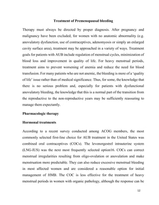
1
MINISTRY OF EDUCATION AND SCIENCE
OF THE RUSSIAN FEDERATION
“PENZA STATE UNIVERSITY”
MEDICAL INSTITUTE
DEPARTMENT OF OBSTETRICS AND GYNECOLOGY
Abstract
In the discipline obstetrics and Gynecology
« Abnormal uterine bleeding in premenopausal age»
Course 31.05.01. –General Medicine
COMPLETED: Patel Rushil Nileshbhai
GROUP:19LL5(a)
CHECKED: Dr. A.F.Shtakh
Associate Professor of Obstetrics
and Gynecology
Penza 2023
2
MINISTRY OF EDUCATION AND SCIENCE OF THE RUSSIAN
FEDERATION
PENZA STATE UNIVERSITY
"APPROVED":
Head Department of Obstetrics and Gynecology
Candidate of Medical Sciences, Associate Professor
______________ A.F. Shtakh
EXERCISE
for term paper on the discipline "Obstetrics and Gynecology"
Theme: Abnormal uterine bleeding in premenopausal age
Student: Patel Rushil Nileshbhai
group:19LL5(a)
Necessary:
a) conduct an analytical and synthetic review of information resources on a
given topic;
b) highlight the main problematic issues;
c) provide information in the form of an academic medical history;
d) execute a report on the work, formulate the main conclusions;
e) design - printed on A4 paper, font Times New Roman, font size 14, spacing
1.5; stapled in a binder.
Completion date - no later than 2 days before the end of the cycle
Supervisor _____________________________________
3
Introduction
Premenopausal is defined as ‘the period around the onset of menopause that
is often marked by various physical signs such as hot flushes and menstrual
irregularities'. Perhaps another way to think of this is that premenopause is the mirror
image of adolescence, which is the coming on to the reproductive years, while
premenopause is the coming off from the reproductive years. Although the median
age of menopause, at least in North America, is 51 years, the premenopause is often
highly variable in its age of onset, duration and bleeding patterns. Regular menstrual
cycles are associated with ovulation and production of progesterone in the luteal
phase. Anovulatory cycles can be highly variable in their bleeding patterns.
The definition of abnormal uterine bleeding (AUB) is ‘flow outside of normal
volume, duration, regularity or frequency’. One-third of patient visits to the
gynecologist are for AUB and it accounts for more than 70% of all gynecological
consults in the premenopausal and postmenopausal years.
Most patients with chronic AUB require medical attention but can be managed
in an outpatient setting. Occasionally, an exacerbation of chronic AUB is severe
enough to necessitate emergency medical care. A thorough evaluation of patients is
important for two main reasons:
(1) to exclude serious pathology such as carcinoma or complex atypical hyperplasia,
(2) to identify the cause of bleeding so that proper therapy (which, in some cases,
may be expectant management) can be embarked upon.
Prevalence and causes
4
Premenopausal bleeding is challenging to define because distinguishing
normal from abnormal uterine bleeding (AUB) during a time in life when a woman’s
menstrual cycles are dynamically changing requires an extra degree of clinical
acumen and attention. The American College of Obstetricians and Gynecologists
(ACOG) defines AUB as: ‘bleeding from the uterine corpus that is abnormal in its
regularity, volume, frequency or duration and is not associated with pregnancy.’1
This entity is further separated into acute AUB, which requires immediate
assessment of the patient’s hemodynamic stability and consideration of acute
management modalities, and chronic AUB, which is defined as AUB that is present
for most of the past 6 months. Regardless of acute or chronic nature, the differential
diagnosis is the same and the PALM-COEIN diagnostic. The ‘PALM’ refers to
uterine structural abnormalities and the ‘COEIN’ refers to conditions unrelated to
structural abnormalities of the uterus.
Fig:1 Classification
Although menstrual irregularity is normal during perimenopause, some
unusual bleeding can be a sign of a problem that needs medical attention (see
5
"Possible causes of abnormal uterine bleeding in Premenopausal women"). See your
clinician if you experience any of the following:
● very heavy monthly bleeding, especially with clots (for example, soaking
through a sanitary product every hour for more than a day)
● bleeding after sexual intercourse
● spotting or bleeding between menstrual periods
● several menstrual cycles that are shorter than 21 days
● several periods that last three days longer than usual
● more than three months without a period.
Possible causes of abnormal uterine bleeding in Premenopausal women
Hormonal imbalance: May cause an absence of periods (amenorrhea).
Chronic failure to ovulate can result in an overgrowth of cells lining the uterus,
which is a risk factor for abnormal bleeding and for endometrial cancer.
Fibroid : May cause heavy bleeding (menorrhagia). These benign tumors
often grow larger during perimenopause and tend to subside after menopause.
Endometrial polyps : May cause heavy, prolonged, or irregular bleeding
(often spotting). These benign growths of the uterine lining may or may not need to
be removed.
Contraceptives: Oral contraceptives may cause various irregular bleeding
patterns (skipped pills can cause bleeding between periods). Unmedicated IUDs may
increase menstrual flow; hormone-treated IUDs may reduce flow.
Thyroid problems: Hypothyroidism can cause heavy bleeding. Both hypo-
and hyperthyroidism are associated with the absence of periods
6
Clotting problems: Irregular bleeding may be caused by inherited clotting
disorders, such as von Willebrand disease (a rare hereditary bleeding disorder that
impairs the blood's ability to clot).
Pregnancy: Ectopic pregnancy, miscarriage, placenta previa, and other
problem pregnancies can cause irregular bleeding.
Polycystic ovary syndrome: An endocrine disorder characterized by lack of
ovulation and few or absent periods. Periods that do occur may be heavy due to
abnormal buildup of the uterine lining.
Possibly due to a change in weight, chronic stress, heavy exercise, illness,
perimenopause, or psychotropic medications.
Diagnosis in women presenting with abnormal uterine bleeding
AUB is an umbrella term that encompasses heavy menstrual bleeding (HMB,
previously referred to as menorrhagia) and intermenstrual bleeding (IMB, previously
referred to as metrorrhagia). The goal of diagnosis is to distinguish women with
anatomic causes (cancer, hyperplasia, polyps, leiomyomas) from women with
normal anatomy where the cause may be ovulatory dysfunction, adenomyosis
without endometrial abnormalities, and, less likely, coagulopathic or iatrogenic
reasons. As with all good medical practice, diagnosis begins with a thorough history
and physical examination followed by appropriate laboratory and imaging tests as
indicated.
History should include a relevant family history including underlying
bleeding disorders, and use of medications or herbal preparations that might affect
7
bleeding such as ginseng, ginkgo, motherwort, contraceptives, non-steroidal anti-
inflammatory drugs (NSAIDs), and warfarin or heparin or their derivatives.
Physical examination should include assessment of body mass index, thyroid
examination, a pelvic examination including speculum findings to rule out cervical
or vaginal causes, and bimanual pelvic assessment including size and contour of the
uterus.
Laboratory assessment should include a full blood count and iron studies as
well as tests for bleeding disorders if suspected or indicated. Pregnancy testing and
thyroid screening may also be appropriate. Although many women may be unsure
of how often or how long they bleed, a careful history of bleeding patterns, frequency
and heaviness is critical and will often assist with diagnosis. For instance, cyclic
HMB without IMB would be unlikely to be carcinoma or even hyperplasia. The
commonest cause of irregular bleeding is anovulatory uterine bleeding. Most often,
anovulatory bleeding is not associated with anatomical abnormalities.
Shortcomings of endometrial biopsy
After a single study by Stovall and colleagues, blind endometrial sampling
with disposable suction piston devices became the standard approach to patients with
AUB. Stovall performed such an outpatient biopsy on 40 patients with known
carcinoma in the week prior to their hysterectomy and obtained endometrial
carcinoma in 39 of the 40 samples, thus reporting 97.5% accuracy. This was widely
publicized, marketed and promoted and was rapidly accepted as ‘standard of care’.
In a similar study, Guido and colleagues performed blind endometrial sampling in
65 patients with known carcinoma in the operating room just prior to their
hysterectomy. They missed 11/65 cancers (sensitivity only 83%) but, upon opening
8
all those uteri, they reported that, when the cancers occupied 50% or more of the
endometrial surface, the biopsy was 100% accurate. Others did similar studies. In
women with known carcinomas, the sensitivity of blind sampling was only 84% and
68%, yielding a false negative rate of 16% and 32%, respectively. These studies
involved blind biopsies done on women with known carcinoma.
Imaging techniques
The primary imaging test of the uterus for the evaluation of AUB is
transvaginal ultrasound. However, not all uteri lend themselves to a meaningful
ultrasound examination such as in cases of co-existing myomas, previous surgery,
marked obesity, axial uterus or adenomyosis. Hysteroscopy as a diagnostic tool may
also be employed, although it is more expensive, requires more anesthesia and, if
performed, is preferably done in an office setting. Newer disposable hysteroscopes
make this recommendation easier to follow.
Transvaginal ultrasonography
The vaginal probe provides a degree of image magnification as if we were
doing ultrasound through a low-power microscope and can be considered a form of
‘sonomicroscopy’. We see things with the vaginal probe that we could not see with
our naked eye, if we could hold the structure in our hand at arm's length and squint
at it. Early observational studies and subsequent large multicenter trials, mostly from
Western Europe, clearly established the fact that, in postmenopausal women with
bleeding, a thin distinct endometrial echo is indicative of a lack of significant tissue
and has higher negative predictive value for endometrial cancer than blind
endometrial sampling. Premenopausal women have cycling of the endometrium
based on the erratic estrogen production of Premenopausal ovaries. Thus, the use of
9
transvaginal ultrasound in such patients must be timed to the end of a bleeding
episode when the endometrial echo will be as thin as one would expect throughout
the whole month. This protocol prevents misinterpretation of endometrial moguls,
which can occur because of the heterogeneity of the topography of the functionalis
as it proliferates.
Clinical Management
Evaluation and Workup : Discussion of patient history should include review of
the patient’s bleeding pattern, contraceptive use, risk factors for endometrial cancer,
medical problems, personal or family history of underlying bleeding disorders, and
current medications.
Physical exam should include speculum and bimanual exams and thyroid
exam. Laboratory assessment should include a full blood count and iron studies as
well as tests for bleeding disorders if suspected or indicated. Pregnancy testing and
thyroid screening may also be appropriate. Although many women may be unsure
of how often or how long they bleed, a careful history of bleeding patterns, frequency
and heaviness is critical and will often assist with diagnosis. The commonest cause
of irregular bleeding is anovulatory uterine bleeding. Most often, anovulatory
bleeding is not associated with anatomical abnormalities.
10
Fig 2. Approach to management of abnormal uterine bleeding in
Premenopausal patients. After ruling out underlying medical disorders and
pathology requiring surgical intervention, management should incorporate need for
contraception and patient preferences.
11
Structural causes of bleeding are best assessed with ultrasound, which may
also guide the need for endometrial sampling to evaluate for hyperplasia or
malignancy. If possible, ultrasound performed 4-6 days after onset of bleeding will
yield maximum information about the endometrium. If the endometrial stripe is
irregular or not clearly identified, sonohysterography can help distinguish between
global and focal changes.
Generally, endometrial sampling should be performed in women over 45
years old with AUB, and those younger than 45 with a history of unopposed estrogen
exposure, risk factors for endometrial cancer, or failed medical management.
Significant focal pathology, such as polyps or submucosal myomas, will likely be
missed by a blind endometrial biopsy. Hysteroscopy may be indicated in selected
cases when focal disease is strongly suspected or visualized on ultrasound; however,
it is more expensive and invasive.
Evaluation of the likely time until menopause can be helpful when discussing
treatment options. Women who are close to menopause may prefer temporizing
medical management to avoid surgery, while those for whom menopause is more
distant may prioritize efficacy over limiting invasive procedures.
An anti-Mullerian hormone (AMH) level >200 ng/mL makes menopause
highly unlikely within the next five years, regardless of a woman’s age, whereas a
very low AMH level predicts menopause in next year with a positive predictive value
ranging from 51% at <48 years of age to 79% at ³51 years of age.
12
Treatment of Premenopausal bleeding
Therapy must always be directed by proper diagnosis. After pregnancy and
malignancy have been excluded, for women with no anatomic abnormality (e.g.
anovulatory dysfunction, use of contraceptives, adenomyosis or simply an enlarged
cavity surface area), treatment may be approached in a variety of ways. Treatment
goals for patients with AUB include regulation of menstrual cycles, minimization of
blood loss and improvement in quality of life. For heavy menstrual periods,
treatment aims to prevent worsening of anemia and reduce the need for blood
transfusion. For many patients who are not anemic, the bleeding is more of a ‘quality
of life’ issue rather than of medical significance. Thus, for some, the knowledge that
there is no serious problem and, especially for patients with dysfunctional
anovulatory bleeding, the knowledge that this is a normal part of the transition from
the reproductive to the non-reproductive years may be sufficiently reassuring to
manage them expectantly.
Pharmacologic therapy
Hormonal treatments
According to a recent survey conducted among ACOG members, the most
commonly selected first-line choice for AUB treatment in the United States was
combined oral contraceptives (COCs). The levonorgestrel intrauterine system
(LNG-IUS) was the next most frequently selected option16. COCs can correct
menstrual irregularities resulting from oligo-ovulation or anovulation and make
menstruation more predictable. They can also reduce excessive menstrual bleeding
in most affected women and are considered a reasonable option for initial
management of HMB. The COC is less effective for the treatment of heavy
menstrual periods in women with organic pathology, although the response can be
13
variable. Menstrual blood loss is reduced by about 50% in women using COCs, and
the reduction is most apparent during the first 2 days of the menstrual flow.
Extended-cycle or continuous COCs with the 20 lg ethinylestradio l/100 lg
levonorgestrel formulation, with either 84/7 regimens that shorten the hormone-free
interval and decrease the number of days of bleeding per year, or the 365-day
regimen designed to eliminate bleeding altogether, are associated with significant
reduced bleeding when compared with cyclic COCs. Shortening of the hormone-
free interval from 7 to 4 days significantly decreases the number of withdrawal
bleeding days in each cycle and increases the amenorrhea rate. Many patients who
are already on COCs experience what we refer to as ‘breakthrough bleeding’.
Usually, adjustment of the dose or type of pill can overcome this, although persistent
bleeding of such a nature should trigger further diagnostic evaluation in case some
co-existing organic pathology is also present. For those women with a
contraindication to estrogen therapy, progesterone therapy or the LNG-IUS can be
an alternative. Because of the effect of levonorgestrel on the endometrium, the
duration and amount of menstrual bleeding are reduced. This effect may begin
during the first menstrual cycle after the placement of the device, and bleeding
becomes progressively less over time. Many of the progesterone-only, long-acting,
reversible contraceptive methods have been used for the treatment of HMB. Patients
should be counseled about the inconvenience of breakthrough bleeding observed
during the initial months of treatment. The timing of complete suppression of the
menstrual cycle is variable depending on the method. Amenorrhea occurs in
approximately 30–40% and 20–80% in 12 months after the single-rod progestin
contraceptive implant and the LNG-IUS, respectively. Use of depot
medroxyprogesterone acetate results in relatively high rates of amenorrhea by the
fourth dose (approximately as high as 90% in some studies) and has traditionally
been used extensively to suppress menses.
14
Non-steroidal anti-inflammatory drugs
NSAIDs reduce prostaglandin synthesis. Prostaglandins may have a role in aberrant
neovascularization leading to dysfunctional uterine bleeding. Oral NSAIDs are an
excellent treatment to reduce HMB. When compared with placebo, they decrease
menstrual cramping and reduce menstrual blood loss by 33%. There is no evidence
of a difference between individual NSAIDs in reducing HMB. Mefenamic acid 500
mg three times a day for 5 days and ibuprofen 600 mg every 6 h or 800 mg every 8
h are commonly prescribed during the first 3 days of the menstrual cycle to reduce
blood loss and menstrual cramping.
Tranexamic acid
Tranexamic acid reversibly blocks lysine-binding sites on plasminogen, preventing
plasmin and fibrin polymer interaction and resulting in fibrin degradation, the
stabilization of clots, and reduction of bleeding. It is contraindicated in women with
increased risk for thromboembolism. It has been routinely used for many years to
reduce blood loss and the need for blood transfusion during and after surgical
procedures. It has been used for the treatment of HMB outside of the United States
for several decades. Its therapeutic effect is superior to placebo, and results in
significant reduction in objective measurement of idiopathic HMB. Tranexamic acid
has no effect on the duration of bleeding or in relieving unscheduled bleeding. In all
studies analyzed, only mild to moderate side-effects were reported, mostly
gastrointestinal, and there were no reports of thromboembolic events. The FDA has
approved tranexamic acid at a per oral dosage of 1300 mg (two 650-mg tablets) three
times daily for 5 days each menstrual cycle. The oral bioavailability of tranexamic
acid is only about 35%, which makes frequent administrations necessary. The
disadvantages of frequent administrations include decreased patient compliance and
15
increased risk of gastrointestinal side-effects, most commonly nausea, vomiting and
diarrhea.
16
Non-medical approaches to abnormal uterine bleeding
Endometrial ablation
Endometrial ablation is a minimally invasive alternative for the treatment of
HMB and is associated with a high level of patient satisfaction. It is usually reserved
for patients refractory to medical treatment and who wish to avoid definitive
treatment with hysterectomy. Although endometrial ablation is a less invasive
surgical alternative to hysterectomy, it does not eliminate surgical risk and is
followed by further surgery within 4 years in up to 38% of the women who undergo
this treatment. Endometrial ablation should be considered as an alternative to
hysterectomy, especially in older women who have opted to retain their uterus and
not in women with a desire for future fertility. Patient age is an important predictor
of treatment success. In contrast to the original resectoscopic endometrial ablation,
several new ablation devices offer the advantage of being performed in an office
setting. A discussion of the various devices is beyond the scope of this White Paper;
however, a word about patient selection is crucial. Careful patient selection seems
to be the key to decrease the occurrence of endometrial cancer after ablation. In a
recent systematic review where 22 post-ablation endometrial cancer cases were
reviewed, time to diagnosis of endometrial cancer ranged from 2 weeks to 10 years
following endometrial ablation.. Most patients had symptoms of persistent bleeding
or pain after the procedure. Eighty-six percent of the cancer patients presented with
risk factors for endometrial cancer such as obesity, complex atypical endometrial
hyperplasia, diabetes, hypertension and postmenopausal status. Thus, endometrial
ablation should be restricted to premenopausal women who have low risk factors for
endometrial cancer and who have documented normal endometrial histopathologic
features at pre-ablation evaluation.
17
Uterine fibroids
Uterine fibroids occur in 25% of women of reproductive age and are often
associated with abnormal uterine bleeding, particularly irregular and/or heavy
bleeding. Although hysterectomy remains a common treatment for uterine fibroids,
other treatments should be discussed in order that women are able to make an
informed choice. A combination of presenting clinical symptoms, fibroid size and
location, and the patient’s desire for fertility will influence the choice of therapeutic
modality offered and accepted by a woman to treat her symptomatic fibroids.
Treatments for fibroids Non-surgical treatment
There is no medical treatment that can cure fibroids but several treatments are
available that provide symptom relief. In Premenopausal women, such therapies are
employed with the goal of satisfactorily alleviating symptoms until the menopause.
Tranexamic acid and mefenamic acid. Tranexamic acid (discussed above) has been
used as first-line treatment for HMB and is frequently used for those with small
fibroids despite little evidence of efficacy. However, the safety and efficacy of the
modified-release variant have been proven in women with fibroids when compared
to placebo.
Mefenamic acid is a NSAID that is commonly used for dysmenorrhea and
leads to a modest reduction in HMB in women without fibroids, although it is less
effective than tranexamic acid. There have been no trials to date to show the benefits
of NSAIDs in women with fibroids.
Levonorgestrel-releasing intrauterine system.
18
The LNG-IUS is used for treatment of HMB. Studies on women with fibroids
have demonstrated relief of menstrual symptoms and a non-significant effect on
volume. The overall incidence of spontaneous expulsion of the LNG-IUS is 9.6%
over a 3- year period. This is increased to 15.8% in the presence of fibroids.
Gonadotropin-releasing hormone agonists.
Gonadotropinreleasing hormone (GnRH) agonists may be used prior to
fibroid surgery since they reduce both uterine volume and fibroid size. They correct
pre-operative iron deficiency anemia and reduce intra-operative blood loss. If uterine
size is such that a mid-line incision is planned, this can be avoided in many women
with the use of GnRH agonists. However, some argue that they render myomectomy
more difficult because they destroy tissue planes, increase the risk of recurrence, and
are associated with side-effects in situations where they confer no benefit, or where
alternative cheaper drugs with fewer side-effects are available. Add-back therapy
may be initiated at the same time as the treatment with a GnRH agonist to reduce the
hypoestrogenic sideeffects such as vasomotor symptoms and loss of bone mineral
density, if use for more than 6 months is anticipated in those who do not desire
surgery.
Selective progesterone receptor modulators.
Ulipristal acetate (UPA) is currently the only selective progesterone receptor
modulator (SPRM) clinically available in some countries. UPA induced amenorrhea
in 63.1% of women and controlled menstrual bleeding in 91% in a dose of 5 mg/day
and in 71.3% and 92%, respectively, in a dose of 10 mg/day, although irregular
bleeding often occurs in those with submucous fibroids. UPA also has some effect
on fibroid size, although less than GnRH agonists.
19
The potential long-term effects of UPA on the endometrium are under study
due to an unusual histological pattern of benign, ‘non-physiologic’ endometrial
change which occurred in many UPA-treated women. These are referred to as PAEC
(progesterone modulator-associated endometrial changes). Common side-effects of
its use are headaches, nasopharyngitis, abdominal pain and hot flushes. The anti-
progesterone properties of mifepristone have also been utilized for the treatment of
fibroids and have been shown to be effective in reducing fibroid size and improving
the quality of life at low doses.
Other medical treatments
Other medical treatments can be used, although frequently they are less
effective in the presence of fibroids. Those that induce amenorrhea such as the oral
contraceptive pill and norethisterone acetate may be valuable, and studies have been
undertaken with aromatase inhibitors.
Radiological treatment:
uterine artery embolization Uterine artery embolization (UAE), performed by
an appropriately trained interventional radiologist, is a minimally invasive treatment
option for uterine fibroids. A catheter is inserted through the femoral artery in the
groin under local anesthetic and directed towards the uterine arteries using
fluoroscopy. The uterine artery is blocked on each side using an appropriate embolic
agent. The objective of UAE is completely to infarct all the fibroid tissue while
preserving the uterus, ovaries and surrounding pelvic structures.
UAE was used initially for massive obstetric hemorrhage. It is indicated for
symptomatic fibroids and is an alternative to myomectomy, since it allows
conservation of the uterus and only involves a short hospital stay. The most common
20
problem associated with UAE is post-procedure pain which can usually be
controlled by analgesics. Expulsion of a necrotic fibroid, chronic vaginal discharge
and premature ovarian failure are less common effects associated with UAE. A
recently published Cochrane review concluded that UAE is a safe and effective
treatment for fibroid-related menstrual disorders. Major complications were rare and
reported not to be more common than after surgery. Since the effects on fertility and
pregnancy are still unclear, it is a good option for most Premenopausal women. UAE
is also associated with some decrease in ovarian function in women older than 45
years which may lead to menopause.
Surgical treatment
Hysterectomy: Hysterectomy is the established surgical treatment for women
who no longer plan to give birth and have completed their family. Although
hysterectomy is a major surgical procedure for women with fibroids, it results in
resolution of most symptoms, particularly when related to menstrual disorders, and
has a high satisfaction rate.
Myomectomy
Myomectomy is a surgical procedure to remove uterine fibroids and
reconstruct the uterus. It is used as a fertility-sparing option. Myomectomy can be
associated with considerable bleeding, risk of hysterectomy, prolonged
postoperative stay, postoperative adhesion formation and recurrence of the fibroids.
Myomectomy can be performed as an open procedure and by laparoscopy or
hysteroscopy, depending on the position of the fibroids and the skill of the surgeon.
It may also depend on the size and number of fibroids that can be treated
laparoscopically, and the skills required are often only available in specialist units.
21
These constraints also apply to vaginal myomectomy. Hysteroscopic myomectomy
is most suitable for fibroids under 5 cm in diameter and where a majority of the
fibroid is within the uterine cavity. Submucosal or pedunculated fibroids distort the
endometrial cavity and may be covered with vessels that break down, thus leading
to irregular bleeding. Those less than about 5 cm in diameter can be removed
hysteroscopically, a procedure now facilitated by the development of new
instrumentation that has increased the safety and ease of the procedure. If larger than
5 cm in diameter, then a two-step procedure might be utilized and/or with prior
administration of a drug such as a GnRH agonist or a SPRM that will shrink the
fibroid.
Malignant and premalignant disease
Endometrial polyps: Endometrial polyps are usually removed as their malignant
potential is uncertain and they may cause irregular bleeding. Removal can often be
carried out under a local anesthetic. Although a wide range of benign gynecological
problems present with abnormal bleeding, it is also a common presenting symptom
of endometrial cancer, endometrial hyperplasia, cervical cancer and, less commonly,
cancer of the vagina or even vulva.
Treatments for endometrial hyperplasia
Endometrial hyperplasia of low malignant potential Often cases of endometrial
hyperplasia of low malignant potential, previously referred to as simple or complex
hyperplasia without atypia, may be handled with conservative management and
appropriately timed endometrial resampling to check for regression. More often,
however, progestogens, administered either orally or by LNG-IUS, are employed. A
systematic review of progestogens for treatment of hyperplasia concluded that the
22
LNG-IUS is a good treatment for non-atypical hyperplasia, with both high success
and high compliance rates. Definitive therapy in selected cases may utilize
hysterectomy
Atypical hyperplasia
Cases of atypical hyperplasia are usually treated with total hysterectomy and
bilateral salpingo-oophorectomy. In selected cases, especially fertility-sparing, high-
dose progestogens are utilized.
Gynecologic malignancies resulting in abnormal uterine bleeding
A detailed account of the treatment of gynecological malignancies is beyond
the scope of this article. Most often, this will involve referral to appropriate
specialists consultants.
Conclusion
In our study, uterine fibroid was the leading cause of AUB for which
hysterectomy was done. Radiological and pathological evaluation correlated well to
23
diagnose fibroids. USG has good specificity to diagnose adenomyosis, however,
because of low sensitivity it is important not to rule out adenomyosis based on USG
findings alone. USG alone is not sufficient to diagnose endometrial hyperplasia.
However, USG detected thick endometrium in postmenopausal woman is a good
indicator of endometrial pathology. Regarding endometrial finding, histopathology
revealed hyperplasia in majority of cases of which simple typical type was the
predominant.
Multiple factors should be considered when choosing among treatment
options for abnormal uterine bleeding including the cause and acuity of the
bleeding, fertility and contraceptive preferences, medical comorbidities, adverse
effects, cost, and relative effectiveness. If the underlying cause of bleeding can be
identified and treated, symptoms may resolve without the need for additional
intervention. Anemia is an indication for treatment, as is bleeding that negatively
affects the patient's quality of life. Because exposure to unopposed estrogen
increases the risk of endometrial cancer, treatment of anovulatory abnormal uterine
bleeding involves inducing ovulatory cycles or administering supplemental
progesterone to antagonize estrogen's proliferative effect on the endometrium.
24
References
1. Jacobs P, Cumming DC. Use of health services associated with increased
menstrual loss in the United States. Am J Obstet Gynecol. 2003;188(2):343–
348
2. Committee on Practice Bulletins—Gynecology. diagnosis of abnormal
uterine bleeding in reproductive-aged women. Obstet Gynecol.
2012;120(1):197–206.
3. Fraser IS, Critchley HO, Broder M, Munro MG. The recommendations on
terminologies and abnormal uterine bleeding. Semin Reprod Med. 2011
4. Ahuja SP, Hertweck SP. Overview of bleeding disorders in adolescent
females with menorrhagia. J Pediatr Adolesc Gynecol. 2010;23(6 suppl):S15–
S21.
5. Hoffman BL, Schorge JO, Bradshaw KD, Halvorson LM, Schaffer JI, Corton
MM. Williams Gynecology. 3rd ed. New York, NY: 2016.
6. Mahajan N, Aggarwal M, Bagga A. Health issues of menopausal women in
North India. J Midlife Health.
7. Kumar P, Malhotra N. Clinical types of abnormal uterine bleeding. In: Kumar
P, editor. Jeffcoate’s Principle of Gynecology. 7th ed.
8. Munro MG, Critchley HO, Fraser IS. The FIGO systems for nomenclature
and classification of causes of abnormal uterine bleeding in the reproductive
years.
9. Telner DE, Jakubovicz D. Approach to diagnosis and management of
abnormal uterine bleeding. Can Fam Physician.
10.Jetley S, Rana S, Jairajpuri ZS. Morphological spectrum of endometrial
pathology in middle-aged women with atypical uterine bleeding.

Abnormal uterine bleeding in premenopausal age.docx

  • 1.
    1 MINISTRY OF EDUCATIONAND SCIENCE OF THE RUSSIAN FEDERATION “PENZA STATE UNIVERSITY” MEDICAL INSTITUTE DEPARTMENT OF OBSTETRICS AND GYNECOLOGY Abstract In the discipline obstetrics and Gynecology « Abnormal uterine bleeding in premenopausal age» Course 31.05.01. –General Medicine COMPLETED: Patel Rushil Nileshbhai GROUP:19LL5(a) CHECKED: Dr. A.F.Shtakh Associate Professor of Obstetrics and Gynecology Penza 2023
  • 2.
    2 MINISTRY OF EDUCATIONAND SCIENCE OF THE RUSSIAN FEDERATION PENZA STATE UNIVERSITY "APPROVED": Head Department of Obstetrics and Gynecology Candidate of Medical Sciences, Associate Professor ______________ A.F. Shtakh EXERCISE for term paper on the discipline "Obstetrics and Gynecology" Theme: Abnormal uterine bleeding in premenopausal age Student: Patel Rushil Nileshbhai group:19LL5(a) Necessary: a) conduct an analytical and synthetic review of information resources on a given topic; b) highlight the main problematic issues; c) provide information in the form of an academic medical history; d) execute a report on the work, formulate the main conclusions; e) design - printed on A4 paper, font Times New Roman, font size 14, spacing 1.5; stapled in a binder. Completion date - no later than 2 days before the end of the cycle Supervisor _____________________________________
  • 3.
    3 Introduction Premenopausal is definedas ‘the period around the onset of menopause that is often marked by various physical signs such as hot flushes and menstrual irregularities'. Perhaps another way to think of this is that premenopause is the mirror image of adolescence, which is the coming on to the reproductive years, while premenopause is the coming off from the reproductive years. Although the median age of menopause, at least in North America, is 51 years, the premenopause is often highly variable in its age of onset, duration and bleeding patterns. Regular menstrual cycles are associated with ovulation and production of progesterone in the luteal phase. Anovulatory cycles can be highly variable in their bleeding patterns. The definition of abnormal uterine bleeding (AUB) is ‘flow outside of normal volume, duration, regularity or frequency’. One-third of patient visits to the gynecologist are for AUB and it accounts for more than 70% of all gynecological consults in the premenopausal and postmenopausal years. Most patients with chronic AUB require medical attention but can be managed in an outpatient setting. Occasionally, an exacerbation of chronic AUB is severe enough to necessitate emergency medical care. A thorough evaluation of patients is important for two main reasons: (1) to exclude serious pathology such as carcinoma or complex atypical hyperplasia, (2) to identify the cause of bleeding so that proper therapy (which, in some cases, may be expectant management) can be embarked upon. Prevalence and causes
  • 4.
    4 Premenopausal bleeding ischallenging to define because distinguishing normal from abnormal uterine bleeding (AUB) during a time in life when a woman’s menstrual cycles are dynamically changing requires an extra degree of clinical acumen and attention. The American College of Obstetricians and Gynecologists (ACOG) defines AUB as: ‘bleeding from the uterine corpus that is abnormal in its regularity, volume, frequency or duration and is not associated with pregnancy.’1 This entity is further separated into acute AUB, which requires immediate assessment of the patient’s hemodynamic stability and consideration of acute management modalities, and chronic AUB, which is defined as AUB that is present for most of the past 6 months. Regardless of acute or chronic nature, the differential diagnosis is the same and the PALM-COEIN diagnostic. The ‘PALM’ refers to uterine structural abnormalities and the ‘COEIN’ refers to conditions unrelated to structural abnormalities of the uterus. Fig:1 Classification Although menstrual irregularity is normal during perimenopause, some unusual bleeding can be a sign of a problem that needs medical attention (see
  • 5.
    5 "Possible causes ofabnormal uterine bleeding in Premenopausal women"). See your clinician if you experience any of the following: ● very heavy monthly bleeding, especially with clots (for example, soaking through a sanitary product every hour for more than a day) ● bleeding after sexual intercourse ● spotting or bleeding between menstrual periods ● several menstrual cycles that are shorter than 21 days ● several periods that last three days longer than usual ● more than three months without a period. Possible causes of abnormal uterine bleeding in Premenopausal women Hormonal imbalance: May cause an absence of periods (amenorrhea). Chronic failure to ovulate can result in an overgrowth of cells lining the uterus, which is a risk factor for abnormal bleeding and for endometrial cancer. Fibroid : May cause heavy bleeding (menorrhagia). These benign tumors often grow larger during perimenopause and tend to subside after menopause. Endometrial polyps : May cause heavy, prolonged, or irregular bleeding (often spotting). These benign growths of the uterine lining may or may not need to be removed. Contraceptives: Oral contraceptives may cause various irregular bleeding patterns (skipped pills can cause bleeding between periods). Unmedicated IUDs may increase menstrual flow; hormone-treated IUDs may reduce flow. Thyroid problems: Hypothyroidism can cause heavy bleeding. Both hypo- and hyperthyroidism are associated with the absence of periods
  • 6.
    6 Clotting problems: Irregularbleeding may be caused by inherited clotting disorders, such as von Willebrand disease (a rare hereditary bleeding disorder that impairs the blood's ability to clot). Pregnancy: Ectopic pregnancy, miscarriage, placenta previa, and other problem pregnancies can cause irregular bleeding. Polycystic ovary syndrome: An endocrine disorder characterized by lack of ovulation and few or absent periods. Periods that do occur may be heavy due to abnormal buildup of the uterine lining. Possibly due to a change in weight, chronic stress, heavy exercise, illness, perimenopause, or psychotropic medications. Diagnosis in women presenting with abnormal uterine bleeding AUB is an umbrella term that encompasses heavy menstrual bleeding (HMB, previously referred to as menorrhagia) and intermenstrual bleeding (IMB, previously referred to as metrorrhagia). The goal of diagnosis is to distinguish women with anatomic causes (cancer, hyperplasia, polyps, leiomyomas) from women with normal anatomy where the cause may be ovulatory dysfunction, adenomyosis without endometrial abnormalities, and, less likely, coagulopathic or iatrogenic reasons. As with all good medical practice, diagnosis begins with a thorough history and physical examination followed by appropriate laboratory and imaging tests as indicated. History should include a relevant family history including underlying bleeding disorders, and use of medications or herbal preparations that might affect
  • 7.
    7 bleeding such asginseng, ginkgo, motherwort, contraceptives, non-steroidal anti- inflammatory drugs (NSAIDs), and warfarin or heparin or their derivatives. Physical examination should include assessment of body mass index, thyroid examination, a pelvic examination including speculum findings to rule out cervical or vaginal causes, and bimanual pelvic assessment including size and contour of the uterus. Laboratory assessment should include a full blood count and iron studies as well as tests for bleeding disorders if suspected or indicated. Pregnancy testing and thyroid screening may also be appropriate. Although many women may be unsure of how often or how long they bleed, a careful history of bleeding patterns, frequency and heaviness is critical and will often assist with diagnosis. For instance, cyclic HMB without IMB would be unlikely to be carcinoma or even hyperplasia. The commonest cause of irregular bleeding is anovulatory uterine bleeding. Most often, anovulatory bleeding is not associated with anatomical abnormalities. Shortcomings of endometrial biopsy After a single study by Stovall and colleagues, blind endometrial sampling with disposable suction piston devices became the standard approach to patients with AUB. Stovall performed such an outpatient biopsy on 40 patients with known carcinoma in the week prior to their hysterectomy and obtained endometrial carcinoma in 39 of the 40 samples, thus reporting 97.5% accuracy. This was widely publicized, marketed and promoted and was rapidly accepted as ‘standard of care’. In a similar study, Guido and colleagues performed blind endometrial sampling in 65 patients with known carcinoma in the operating room just prior to their hysterectomy. They missed 11/65 cancers (sensitivity only 83%) but, upon opening
  • 8.
    8 all those uteri,they reported that, when the cancers occupied 50% or more of the endometrial surface, the biopsy was 100% accurate. Others did similar studies. In women with known carcinomas, the sensitivity of blind sampling was only 84% and 68%, yielding a false negative rate of 16% and 32%, respectively. These studies involved blind biopsies done on women with known carcinoma. Imaging techniques The primary imaging test of the uterus for the evaluation of AUB is transvaginal ultrasound. However, not all uteri lend themselves to a meaningful ultrasound examination such as in cases of co-existing myomas, previous surgery, marked obesity, axial uterus or adenomyosis. Hysteroscopy as a diagnostic tool may also be employed, although it is more expensive, requires more anesthesia and, if performed, is preferably done in an office setting. Newer disposable hysteroscopes make this recommendation easier to follow. Transvaginal ultrasonography The vaginal probe provides a degree of image magnification as if we were doing ultrasound through a low-power microscope and can be considered a form of ‘sonomicroscopy’. We see things with the vaginal probe that we could not see with our naked eye, if we could hold the structure in our hand at arm's length and squint at it. Early observational studies and subsequent large multicenter trials, mostly from Western Europe, clearly established the fact that, in postmenopausal women with bleeding, a thin distinct endometrial echo is indicative of a lack of significant tissue and has higher negative predictive value for endometrial cancer than blind endometrial sampling. Premenopausal women have cycling of the endometrium based on the erratic estrogen production of Premenopausal ovaries. Thus, the use of
  • 9.
    9 transvaginal ultrasound insuch patients must be timed to the end of a bleeding episode when the endometrial echo will be as thin as one would expect throughout the whole month. This protocol prevents misinterpretation of endometrial moguls, which can occur because of the heterogeneity of the topography of the functionalis as it proliferates. Clinical Management Evaluation and Workup : Discussion of patient history should include review of the patient’s bleeding pattern, contraceptive use, risk factors for endometrial cancer, medical problems, personal or family history of underlying bleeding disorders, and current medications. Physical exam should include speculum and bimanual exams and thyroid exam. Laboratory assessment should include a full blood count and iron studies as well as tests for bleeding disorders if suspected or indicated. Pregnancy testing and thyroid screening may also be appropriate. Although many women may be unsure of how often or how long they bleed, a careful history of bleeding patterns, frequency and heaviness is critical and will often assist with diagnosis. The commonest cause of irregular bleeding is anovulatory uterine bleeding. Most often, anovulatory bleeding is not associated with anatomical abnormalities.
  • 10.
    10 Fig 2. Approachto management of abnormal uterine bleeding in Premenopausal patients. After ruling out underlying medical disorders and pathology requiring surgical intervention, management should incorporate need for contraception and patient preferences.
  • 11.
    11 Structural causes ofbleeding are best assessed with ultrasound, which may also guide the need for endometrial sampling to evaluate for hyperplasia or malignancy. If possible, ultrasound performed 4-6 days after onset of bleeding will yield maximum information about the endometrium. If the endometrial stripe is irregular or not clearly identified, sonohysterography can help distinguish between global and focal changes. Generally, endometrial sampling should be performed in women over 45 years old with AUB, and those younger than 45 with a history of unopposed estrogen exposure, risk factors for endometrial cancer, or failed medical management. Significant focal pathology, such as polyps or submucosal myomas, will likely be missed by a blind endometrial biopsy. Hysteroscopy may be indicated in selected cases when focal disease is strongly suspected or visualized on ultrasound; however, it is more expensive and invasive. Evaluation of the likely time until menopause can be helpful when discussing treatment options. Women who are close to menopause may prefer temporizing medical management to avoid surgery, while those for whom menopause is more distant may prioritize efficacy over limiting invasive procedures. An anti-Mullerian hormone (AMH) level >200 ng/mL makes menopause highly unlikely within the next five years, regardless of a woman’s age, whereas a very low AMH level predicts menopause in next year with a positive predictive value ranging from 51% at <48 years of age to 79% at ³51 years of age.
  • 12.
    12 Treatment of Premenopausalbleeding Therapy must always be directed by proper diagnosis. After pregnancy and malignancy have been excluded, for women with no anatomic abnormality (e.g. anovulatory dysfunction, use of contraceptives, adenomyosis or simply an enlarged cavity surface area), treatment may be approached in a variety of ways. Treatment goals for patients with AUB include regulation of menstrual cycles, minimization of blood loss and improvement in quality of life. For heavy menstrual periods, treatment aims to prevent worsening of anemia and reduce the need for blood transfusion. For many patients who are not anemic, the bleeding is more of a ‘quality of life’ issue rather than of medical significance. Thus, for some, the knowledge that there is no serious problem and, especially for patients with dysfunctional anovulatory bleeding, the knowledge that this is a normal part of the transition from the reproductive to the non-reproductive years may be sufficiently reassuring to manage them expectantly. Pharmacologic therapy Hormonal treatments According to a recent survey conducted among ACOG members, the most commonly selected first-line choice for AUB treatment in the United States was combined oral contraceptives (COCs). The levonorgestrel intrauterine system (LNG-IUS) was the next most frequently selected option16. COCs can correct menstrual irregularities resulting from oligo-ovulation or anovulation and make menstruation more predictable. They can also reduce excessive menstrual bleeding in most affected women and are considered a reasonable option for initial management of HMB. The COC is less effective for the treatment of heavy menstrual periods in women with organic pathology, although the response can be
  • 13.
    13 variable. Menstrual bloodloss is reduced by about 50% in women using COCs, and the reduction is most apparent during the first 2 days of the menstrual flow. Extended-cycle or continuous COCs with the 20 lg ethinylestradio l/100 lg levonorgestrel formulation, with either 84/7 regimens that shorten the hormone-free interval and decrease the number of days of bleeding per year, or the 365-day regimen designed to eliminate bleeding altogether, are associated with significant reduced bleeding when compared with cyclic COCs. Shortening of the hormone- free interval from 7 to 4 days significantly decreases the number of withdrawal bleeding days in each cycle and increases the amenorrhea rate. Many patients who are already on COCs experience what we refer to as ‘breakthrough bleeding’. Usually, adjustment of the dose or type of pill can overcome this, although persistent bleeding of such a nature should trigger further diagnostic evaluation in case some co-existing organic pathology is also present. For those women with a contraindication to estrogen therapy, progesterone therapy or the LNG-IUS can be an alternative. Because of the effect of levonorgestrel on the endometrium, the duration and amount of menstrual bleeding are reduced. This effect may begin during the first menstrual cycle after the placement of the device, and bleeding becomes progressively less over time. Many of the progesterone-only, long-acting, reversible contraceptive methods have been used for the treatment of HMB. Patients should be counseled about the inconvenience of breakthrough bleeding observed during the initial months of treatment. The timing of complete suppression of the menstrual cycle is variable depending on the method. Amenorrhea occurs in approximately 30–40% and 20–80% in 12 months after the single-rod progestin contraceptive implant and the LNG-IUS, respectively. Use of depot medroxyprogesterone acetate results in relatively high rates of amenorrhea by the fourth dose (approximately as high as 90% in some studies) and has traditionally been used extensively to suppress menses.
  • 14.
    14 Non-steroidal anti-inflammatory drugs NSAIDsreduce prostaglandin synthesis. Prostaglandins may have a role in aberrant neovascularization leading to dysfunctional uterine bleeding. Oral NSAIDs are an excellent treatment to reduce HMB. When compared with placebo, they decrease menstrual cramping and reduce menstrual blood loss by 33%. There is no evidence of a difference between individual NSAIDs in reducing HMB. Mefenamic acid 500 mg three times a day for 5 days and ibuprofen 600 mg every 6 h or 800 mg every 8 h are commonly prescribed during the first 3 days of the menstrual cycle to reduce blood loss and menstrual cramping. Tranexamic acid Tranexamic acid reversibly blocks lysine-binding sites on plasminogen, preventing plasmin and fibrin polymer interaction and resulting in fibrin degradation, the stabilization of clots, and reduction of bleeding. It is contraindicated in women with increased risk for thromboembolism. It has been routinely used for many years to reduce blood loss and the need for blood transfusion during and after surgical procedures. It has been used for the treatment of HMB outside of the United States for several decades. Its therapeutic effect is superior to placebo, and results in significant reduction in objective measurement of idiopathic HMB. Tranexamic acid has no effect on the duration of bleeding or in relieving unscheduled bleeding. In all studies analyzed, only mild to moderate side-effects were reported, mostly gastrointestinal, and there were no reports of thromboembolic events. The FDA has approved tranexamic acid at a per oral dosage of 1300 mg (two 650-mg tablets) three times daily for 5 days each menstrual cycle. The oral bioavailability of tranexamic acid is only about 35%, which makes frequent administrations necessary. The disadvantages of frequent administrations include decreased patient compliance and
  • 15.
    15 increased risk ofgastrointestinal side-effects, most commonly nausea, vomiting and diarrhea.
  • 16.
    16 Non-medical approaches toabnormal uterine bleeding Endometrial ablation Endometrial ablation is a minimally invasive alternative for the treatment of HMB and is associated with a high level of patient satisfaction. It is usually reserved for patients refractory to medical treatment and who wish to avoid definitive treatment with hysterectomy. Although endometrial ablation is a less invasive surgical alternative to hysterectomy, it does not eliminate surgical risk and is followed by further surgery within 4 years in up to 38% of the women who undergo this treatment. Endometrial ablation should be considered as an alternative to hysterectomy, especially in older women who have opted to retain their uterus and not in women with a desire for future fertility. Patient age is an important predictor of treatment success. In contrast to the original resectoscopic endometrial ablation, several new ablation devices offer the advantage of being performed in an office setting. A discussion of the various devices is beyond the scope of this White Paper; however, a word about patient selection is crucial. Careful patient selection seems to be the key to decrease the occurrence of endometrial cancer after ablation. In a recent systematic review where 22 post-ablation endometrial cancer cases were reviewed, time to diagnosis of endometrial cancer ranged from 2 weeks to 10 years following endometrial ablation.. Most patients had symptoms of persistent bleeding or pain after the procedure. Eighty-six percent of the cancer patients presented with risk factors for endometrial cancer such as obesity, complex atypical endometrial hyperplasia, diabetes, hypertension and postmenopausal status. Thus, endometrial ablation should be restricted to premenopausal women who have low risk factors for endometrial cancer and who have documented normal endometrial histopathologic features at pre-ablation evaluation.
  • 17.
    17 Uterine fibroids Uterine fibroidsoccur in 25% of women of reproductive age and are often associated with abnormal uterine bleeding, particularly irregular and/or heavy bleeding. Although hysterectomy remains a common treatment for uterine fibroids, other treatments should be discussed in order that women are able to make an informed choice. A combination of presenting clinical symptoms, fibroid size and location, and the patient’s desire for fertility will influence the choice of therapeutic modality offered and accepted by a woman to treat her symptomatic fibroids. Treatments for fibroids Non-surgical treatment There is no medical treatment that can cure fibroids but several treatments are available that provide symptom relief. In Premenopausal women, such therapies are employed with the goal of satisfactorily alleviating symptoms until the menopause. Tranexamic acid and mefenamic acid. Tranexamic acid (discussed above) has been used as first-line treatment for HMB and is frequently used for those with small fibroids despite little evidence of efficacy. However, the safety and efficacy of the modified-release variant have been proven in women with fibroids when compared to placebo. Mefenamic acid is a NSAID that is commonly used for dysmenorrhea and leads to a modest reduction in HMB in women without fibroids, although it is less effective than tranexamic acid. There have been no trials to date to show the benefits of NSAIDs in women with fibroids. Levonorgestrel-releasing intrauterine system.
  • 18.
    18 The LNG-IUS isused for treatment of HMB. Studies on women with fibroids have demonstrated relief of menstrual symptoms and a non-significant effect on volume. The overall incidence of spontaneous expulsion of the LNG-IUS is 9.6% over a 3- year period. This is increased to 15.8% in the presence of fibroids. Gonadotropin-releasing hormone agonists. Gonadotropinreleasing hormone (GnRH) agonists may be used prior to fibroid surgery since they reduce both uterine volume and fibroid size. They correct pre-operative iron deficiency anemia and reduce intra-operative blood loss. If uterine size is such that a mid-line incision is planned, this can be avoided in many women with the use of GnRH agonists. However, some argue that they render myomectomy more difficult because they destroy tissue planes, increase the risk of recurrence, and are associated with side-effects in situations where they confer no benefit, or where alternative cheaper drugs with fewer side-effects are available. Add-back therapy may be initiated at the same time as the treatment with a GnRH agonist to reduce the hypoestrogenic sideeffects such as vasomotor symptoms and loss of bone mineral density, if use for more than 6 months is anticipated in those who do not desire surgery. Selective progesterone receptor modulators. Ulipristal acetate (UPA) is currently the only selective progesterone receptor modulator (SPRM) clinically available in some countries. UPA induced amenorrhea in 63.1% of women and controlled menstrual bleeding in 91% in a dose of 5 mg/day and in 71.3% and 92%, respectively, in a dose of 10 mg/day, although irregular bleeding often occurs in those with submucous fibroids. UPA also has some effect on fibroid size, although less than GnRH agonists.
  • 19.
    19 The potential long-termeffects of UPA on the endometrium are under study due to an unusual histological pattern of benign, ‘non-physiologic’ endometrial change which occurred in many UPA-treated women. These are referred to as PAEC (progesterone modulator-associated endometrial changes). Common side-effects of its use are headaches, nasopharyngitis, abdominal pain and hot flushes. The anti- progesterone properties of mifepristone have also been utilized for the treatment of fibroids and have been shown to be effective in reducing fibroid size and improving the quality of life at low doses. Other medical treatments Other medical treatments can be used, although frequently they are less effective in the presence of fibroids. Those that induce amenorrhea such as the oral contraceptive pill and norethisterone acetate may be valuable, and studies have been undertaken with aromatase inhibitors. Radiological treatment: uterine artery embolization Uterine artery embolization (UAE), performed by an appropriately trained interventional radiologist, is a minimally invasive treatment option for uterine fibroids. A catheter is inserted through the femoral artery in the groin under local anesthetic and directed towards the uterine arteries using fluoroscopy. The uterine artery is blocked on each side using an appropriate embolic agent. The objective of UAE is completely to infarct all the fibroid tissue while preserving the uterus, ovaries and surrounding pelvic structures. UAE was used initially for massive obstetric hemorrhage. It is indicated for symptomatic fibroids and is an alternative to myomectomy, since it allows conservation of the uterus and only involves a short hospital stay. The most common
  • 20.
    20 problem associated withUAE is post-procedure pain which can usually be controlled by analgesics. Expulsion of a necrotic fibroid, chronic vaginal discharge and premature ovarian failure are less common effects associated with UAE. A recently published Cochrane review concluded that UAE is a safe and effective treatment for fibroid-related menstrual disorders. Major complications were rare and reported not to be more common than after surgery. Since the effects on fertility and pregnancy are still unclear, it is a good option for most Premenopausal women. UAE is also associated with some decrease in ovarian function in women older than 45 years which may lead to menopause. Surgical treatment Hysterectomy: Hysterectomy is the established surgical treatment for women who no longer plan to give birth and have completed their family. Although hysterectomy is a major surgical procedure for women with fibroids, it results in resolution of most symptoms, particularly when related to menstrual disorders, and has a high satisfaction rate. Myomectomy Myomectomy is a surgical procedure to remove uterine fibroids and reconstruct the uterus. It is used as a fertility-sparing option. Myomectomy can be associated with considerable bleeding, risk of hysterectomy, prolonged postoperative stay, postoperative adhesion formation and recurrence of the fibroids. Myomectomy can be performed as an open procedure and by laparoscopy or hysteroscopy, depending on the position of the fibroids and the skill of the surgeon. It may also depend on the size and number of fibroids that can be treated laparoscopically, and the skills required are often only available in specialist units.
  • 21.
    21 These constraints alsoapply to vaginal myomectomy. Hysteroscopic myomectomy is most suitable for fibroids under 5 cm in diameter and where a majority of the fibroid is within the uterine cavity. Submucosal or pedunculated fibroids distort the endometrial cavity and may be covered with vessels that break down, thus leading to irregular bleeding. Those less than about 5 cm in diameter can be removed hysteroscopically, a procedure now facilitated by the development of new instrumentation that has increased the safety and ease of the procedure. If larger than 5 cm in diameter, then a two-step procedure might be utilized and/or with prior administration of a drug such as a GnRH agonist or a SPRM that will shrink the fibroid. Malignant and premalignant disease Endometrial polyps: Endometrial polyps are usually removed as their malignant potential is uncertain and they may cause irregular bleeding. Removal can often be carried out under a local anesthetic. Although a wide range of benign gynecological problems present with abnormal bleeding, it is also a common presenting symptom of endometrial cancer, endometrial hyperplasia, cervical cancer and, less commonly, cancer of the vagina or even vulva. Treatments for endometrial hyperplasia Endometrial hyperplasia of low malignant potential Often cases of endometrial hyperplasia of low malignant potential, previously referred to as simple or complex hyperplasia without atypia, may be handled with conservative management and appropriately timed endometrial resampling to check for regression. More often, however, progestogens, administered either orally or by LNG-IUS, are employed. A systematic review of progestogens for treatment of hyperplasia concluded that the
  • 22.
    22 LNG-IUS is agood treatment for non-atypical hyperplasia, with both high success and high compliance rates. Definitive therapy in selected cases may utilize hysterectomy Atypical hyperplasia Cases of atypical hyperplasia are usually treated with total hysterectomy and bilateral salpingo-oophorectomy. In selected cases, especially fertility-sparing, high- dose progestogens are utilized. Gynecologic malignancies resulting in abnormal uterine bleeding A detailed account of the treatment of gynecological malignancies is beyond the scope of this article. Most often, this will involve referral to appropriate specialists consultants. Conclusion In our study, uterine fibroid was the leading cause of AUB for which hysterectomy was done. Radiological and pathological evaluation correlated well to
  • 23.
    23 diagnose fibroids. USGhas good specificity to diagnose adenomyosis, however, because of low sensitivity it is important not to rule out adenomyosis based on USG findings alone. USG alone is not sufficient to diagnose endometrial hyperplasia. However, USG detected thick endometrium in postmenopausal woman is a good indicator of endometrial pathology. Regarding endometrial finding, histopathology revealed hyperplasia in majority of cases of which simple typical type was the predominant. Multiple factors should be considered when choosing among treatment options for abnormal uterine bleeding including the cause and acuity of the bleeding, fertility and contraceptive preferences, medical comorbidities, adverse effects, cost, and relative effectiveness. If the underlying cause of bleeding can be identified and treated, symptoms may resolve without the need for additional intervention. Anemia is an indication for treatment, as is bleeding that negatively affects the patient's quality of life. Because exposure to unopposed estrogen increases the risk of endometrial cancer, treatment of anovulatory abnormal uterine bleeding involves inducing ovulatory cycles or administering supplemental progesterone to antagonize estrogen's proliferative effect on the endometrium.
  • 24.
    24 References 1. Jacobs P,Cumming DC. Use of health services associated with increased menstrual loss in the United States. Am J Obstet Gynecol. 2003;188(2):343– 348 2. Committee on Practice Bulletins—Gynecology. diagnosis of abnormal uterine bleeding in reproductive-aged women. Obstet Gynecol. 2012;120(1):197–206. 3. Fraser IS, Critchley HO, Broder M, Munro MG. The recommendations on terminologies and abnormal uterine bleeding. Semin Reprod Med. 2011 4. Ahuja SP, Hertweck SP. Overview of bleeding disorders in adolescent females with menorrhagia. J Pediatr Adolesc Gynecol. 2010;23(6 suppl):S15– S21. 5. Hoffman BL, Schorge JO, Bradshaw KD, Halvorson LM, Schaffer JI, Corton MM. Williams Gynecology. 3rd ed. New York, NY: 2016. 6. Mahajan N, Aggarwal M, Bagga A. Health issues of menopausal women in North India. J Midlife Health. 7. Kumar P, Malhotra N. Clinical types of abnormal uterine bleeding. In: Kumar P, editor. Jeffcoate’s Principle of Gynecology. 7th ed. 8. Munro MG, Critchley HO, Fraser IS. The FIGO systems for nomenclature and classification of causes of abnormal uterine bleeding in the reproductive years. 9. Telner DE, Jakubovicz D. Approach to diagnosis and management of abnormal uterine bleeding. Can Fam Physician. 10.Jetley S, Rana S, Jairajpuri ZS. Morphological spectrum of endometrial pathology in middle-aged women with atypical uterine bleeding.