Download to read offline
![SSR Institute of International Journal of Life Sciences
ISSN (O): 2581-8740 | ISSN (P): 2581-8732
Dash et al., 2023
DOI: 10.21276/SSR-IIJLS.2023.9.6.10
Copyright © 2015–2023| SSR-IIJLS by Society for Scientific Research under a CC BY-NC 4.0 International License Volume 09 | Issue 06 | Page 3428
Assessment of Acromion Morphology in Association with Shoulder
Impingement Syndrome using MRI
Abhisek Dash1
, Chandra Sekhar Pradhan2
, Gopal Chandra Patro3*
, Jyotibhanja Das4
1
Senior Resident, Department of Orthopaedic Surgery, SLN Medical College and Hospital, Koraput, Odisha, India
2
Associate Professor, Department of Orthopaedic Surgery, MKCG Medical College and Hospital, Berhampur, Odisha,
India
3
Senior Resident, Department of Orthopaedic Surgery, MKCG Medical College and Hospital, Berhampur, Odisha, India
4
Junior Resident, Department of Orthopaedic Surgery, MKCG Medical College and Hospital, Berhampur, Odisha, India
*Address for Correspondence: Dr. Gopal Chandra Patro, Senior Resident, Department of Orthopaedic Surgery, MKCG
Medical College and Hospital, Berhampur, Odisha, India
E-mail: gopal8694patro@gmail.com
Received: 07 Jun 2023/ Revised: 14 Aug 2023/ Accepted: 11 Oct 2023
ABSTRACT
Background: The third most common musculoskeletal symptom in orthopaedic clinical practice is a sore shoulder, which can cause
significant morbidity. It has been reported that 7–27% of the general population has it, and 36–66% of overhead arm athletes have it.
Pathophysiology includes functional, degenerative, and mechanical factors. Most shoulder pain is subacromial pain syndrome (SAPS),
often known as ‘shoulder impingement syndrome’. Impingement hypothesis: shoulder joint structures mechanically clash. SAPS accounts
for 36–48% of shoulder discomfort.
Methods: This observational study was conducted in the Department of Orthopaedics, MKCG Medical College and
Hospital, Berhampur, among Eastern Indian outpatients. The study included adult patients (ages 18–75) of both sexes
who presented to MKCG Medical College and Hospital's OPD with shoulder pain from December 2020 to November
2022 and were diagnosed with Shoulder Impingement Syndrome (SIS). Thorough histories and clinical exams were done.
The Department of Radiology, MKCG Medical College and Hospital, Berhampur, performed conventional shoulder MRIs
on the selected participants.
Results: Most cases and controls were Type-II (43.3%), followed by Type-I (28.3% and 30%, 29.2% of the total group). The study's
least common acromial shape was type-IV, seen in 5% of cases and 10% of controls (7.5% of the sample). Fisher's exact test
showed no significant connection between subacromial impingement and acromial shape (p=0.65). With a p-value of 0.045, cases
had a significantly greater acromial width (8.12±2.16 mm) than controls (7.51±0.81 mm).
Conclusion: Sub-acromial impingement was unrelated to acromion morphology. There was no correlation between acromial
morphology and rotator cuff injuries.
Key-words: Shoulder Impingement Syndrome, Acromion Morphology, MRI
INTRODUCTION
A painful shoulder is the third most encountered
musculoskeletal complaint in day-to-day
orthopaedic clinical practice that can lead to
considerable morbidity [1]
.
How to cite this article
Dash A, Pradhan CS, Patro GC, Das J. Assessment of Acromion
Morphology in Association with Shoulder Impingement Syndrome
using MRI. SSR Inst. Int. J. Life Sci., 2023; 9(6): 3428-3438.
Access this article online
http://iijls.com/
Its prevalence ranges between 7% and 27% in the
general population, as reported [2,3]
. It is even more
frequent among athletes who regularly perform
overhead arm activity, which can be as high as 36 to
66% [4-6]
. Considering the pathophysiology, its causes
can be numerous: functional, degenerative, and
mechanical.
The most common diagnosis of shoulder pain is
subacromial pain syndrome (SAPS) [7]
, routinely
referred to as ‘shoulder impingement syndrome’
(SIS) [8]
. The impingement hypothesis supposes a
Research Article](https://image.slidesharecdn.com/assessmentacromionmorphologyassociationshoulderimpingementsyndromemri-240119141145-d89c3c77/75/Assessment_Acromion_Morphology_Association_Shoulder_Impingement_Syndrome_MRI-pdf-1-2048.jpg)
![SSR Institute of International Journal of Life Sciences
ISSN (O): 2581-8740 | ISSN (P): 2581-8732
Dash et al., 2023
DOI: 10.21276/SSR-IIJLS.2023.9.6.10
Copyright © 2015–2023| SSR-IIJLS by Society for Scientific Research under a CC BY-NC 4.0 International License Volume 09 | Issue 06 | Page 3429
pathophysiological mechanism in which there is
mechanical conflict among the various structures of
the shoulder joint. Out of all types of shoulder pain,
the prevalence of SAPS is 36 to 48% [7,9]
.
SAPS is a clinical syndrome with painful entrapment
of specific soft tissues in an area of the shoulder
joint. Patients affected are generally over 40 years of
age, suffering from chronic pain without any known
history of preceding trauma. SIS is most commonly
diagnosed in the sixth decade [2,7-11]
. This syndrome
usually causes pain on elevating the arm, thus
limiting the shoulder range of motion [12,13]
. Patients
complain of pain on elevating the arm between 70°
and 120° (the “painful arc”), on attempting forced
movement above the head, and also while lying on the
affected side [14]
.
This syndrome, if left untreated, could result in
rotator cuff disruption which then persists to cause
secondary osteoarthritis of the shoulder, severely
restricting shoulder movement in the end [12]
.
The pathophysiologic cause of shoulder
impingement syndrome is multifactorial, and the
relative importance of each component remains
debated. Nowadays, the mechanisms contributing to
rotator cuff disease are mainly classified into
intrinsic and extrinsic factors. The intrinsic factors
include abnormalities within the rotator cuff itself:
alteration in the collagen fibre microstructure,
tensile overload, aging, decrease in microvascular
supply, and traumatism, which usually results in
degeneration of the tendon itself [15]
. The extrinsic
factors are mainly some anatomic variables such as
acromial morphology or abnormal scapular
biomechanics, acromial spurs, morphology of the
coracoacromial ligament and acromioclavicular joint,
which would narrow the subacromial space and
increase pressure on tendons by impingement from
bony structures or surrounding soft tissues [15,16]
.
There still exist debates on which mechanism is
primary or secondary, but in most patients, it seems
to be an interaction between both.
Some have suggested that extrinsic osseous
impingement is the primary etiologic mechanism of
rotator cuff disease and that osseous impingement
is related to several causes, such as acquired and
often degenerative bone production, congenital and
developmental variants in bone shape and os
acromiale [17-20]
. Osteophytes, hypertrophic changes,
and bony spurs in the acromion have been
considered major extrinsic causes of SIS.
Subacromial or acromioclavicular spurs were
reported in almost half of SIS cases [21]
.
In some cases, the symptoms are relieved by
conservative treatment alone. However, some
patients with specific structural pathology, such as
bone spur of acromion or compression of acromial
undersurface, might require surgical procedures,
such as acromioplasty, for definitive treatment
[2,17,22-24]
. Subacromial decompression and
acromioplasty are regular surgical methods
performed over a long period to treat SIS. However, it
is still controversial as to precisely which part of the
acromion needs to be resected or decompressed.
A significant component of osseous impingement is
the morphologic characteristics of the acromion.
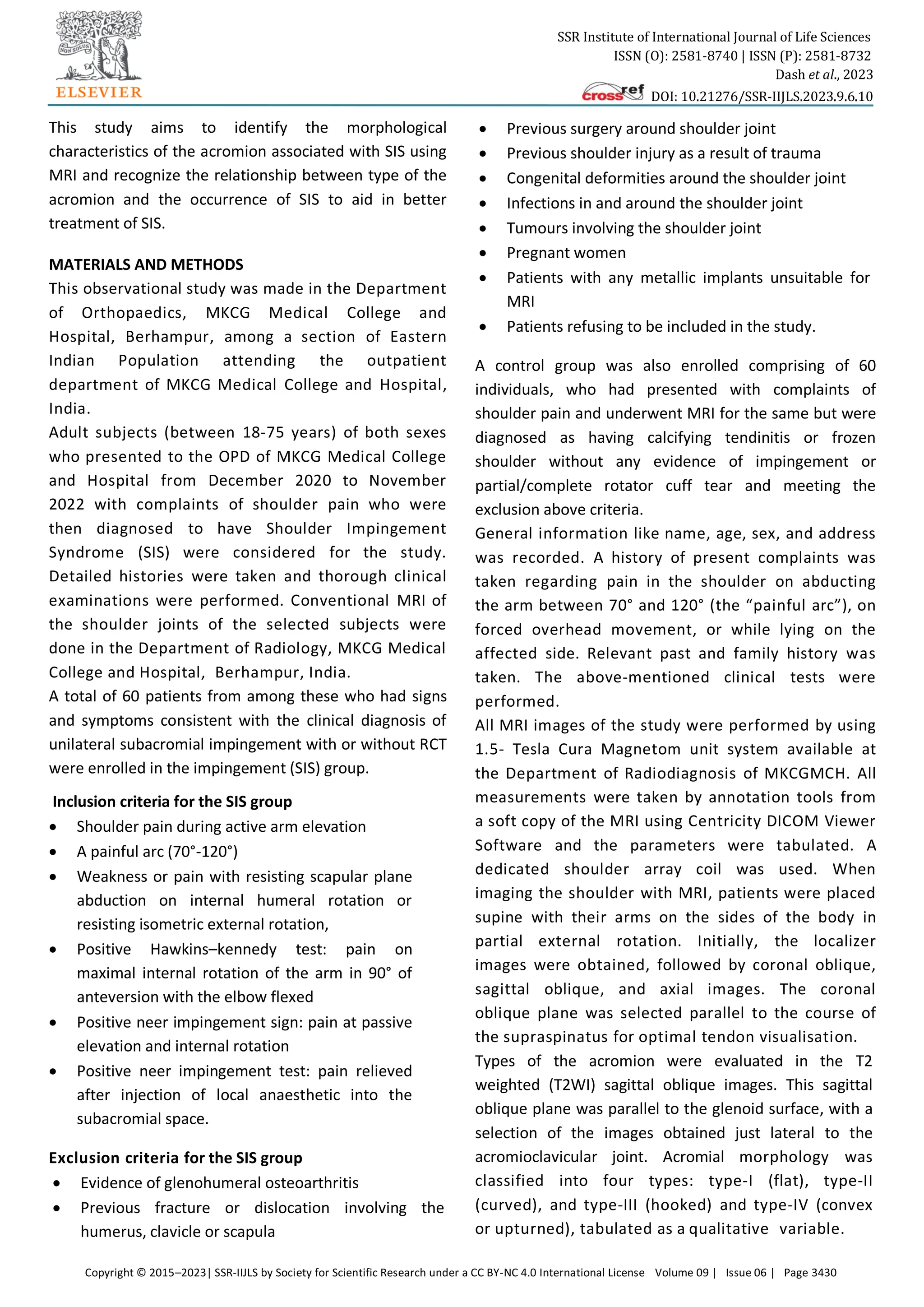
Consequently, numerous attempts have been made
to grade acromial morphologic features. The most
notable is the flat (type I), curved (type II), and
hooked (type III) classification by Bigliani and
colleagues [25]
, who originally described the
acromion by using anatomic specimens. This
classification has subsequently been applied to
acromia by using multiple imaging modalities. All
analyses have been subjective to variable degrees,
and significant intraobserver and interobserver
variability has been demonstrated [26-28]
.
The pathogenesis of SIS seems to be related to the
morphology of the acromion, which is usually
assessed through the commonly used parameters on
standard plain radiographs including the acromial
type, acromial slope, acromial tilt, lateral acromial
angle, critical shoulder angle, acromial index,
acromiohumeral distance, etc [29]
. However, with
only a plain radiograph of the acromion in the
supraspinatus outlet view, it is notoriously difficult
to image the acromion and distinguish the hooked
from the non-hooked acromion with anterior spurs
[30,31]
.
Magnetic resonance imaging (MRI) makes it possible to
depict the shape of acromion in its longitudinal axis with
better evaluation of these acromial morphological
factors, which have been suggested to influence the
rotator cuff status [32,33]
.](https://image.slidesharecdn.com/assessmentacromionmorphologyassociationshoulderimpingementsyndromemri-240119141145-d89c3c77/75/Assessment_Acromion_Morphology_Association_Shoulder_Impingement_Syndrome_MRI-pdf-2-2048.jpg)

![SSR Institute of International Journal of Life Sciences
ISSN (O): 2581-8740 | ISSN (P): 2581-8732
Dash et al., 2023
DOI: 10.21276/SSR-IIJLS.2023.9.6.10
Copyright © 2015–2023| SSR-IIJLS by Society for Scientific Research under a CC BY-NC 4.0 International License Volume 09 | Issue 06 | Page 3435
Table 8: Distribution of rotator cuff tears among different acromial types
Acromion shape Full thickness (%)
(N=6)
Partial thickness (%)
(N=7)
Total (%)
(N=13)
Type-I 1 (16.7) 2 (28.6) 3 (23.1)
Type-II 1 (16.7) 3 (42.9) 4 (30.8)
Type-III 4 (66.7) 2 (28.6) 6 (46.2)
Type-IV 0 (0) 0 (0) 0 (0.0)
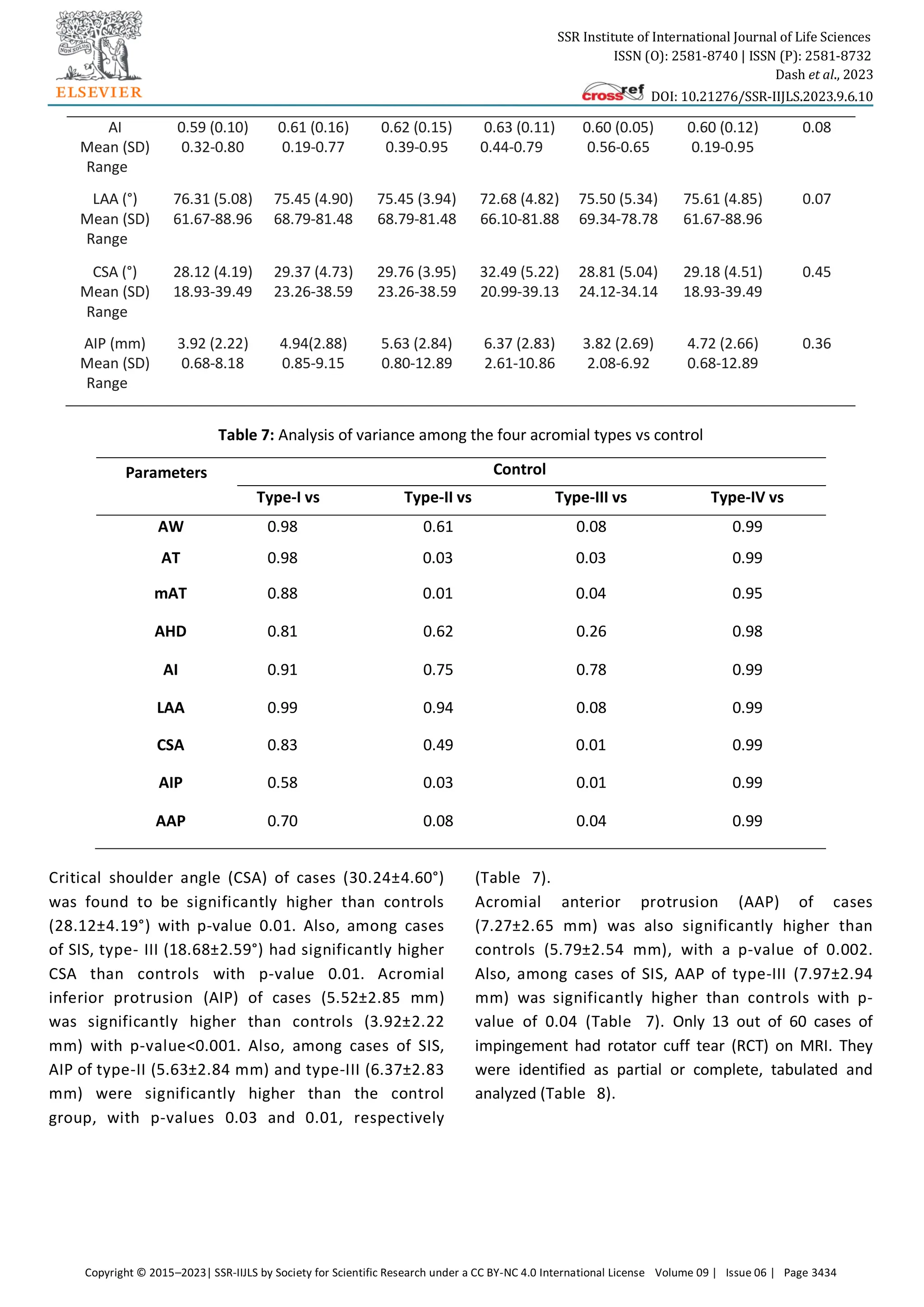
DISCUSSION
This study was therefore designed to analyse the
morphologic characteristics of different acromia in
patients with subacromial impingement with or without
partial or complete thickness RCT by using MRI as the
diagnostic modality of choice to elucidate the
relationship between acromial shapes and SIS
clearly, which could, in turn, prove a helpful guide in
the diagnosis and management of impingement and
rotator cuff tendinopathy.
Similar to previous studies [34]
, the current study also
observed that type-II acromial shape is the most
commonly encountered type among patients and
control groups (43.33% in both groups). Conversely,
type-IV acromion shape was the least prevalent
among case and control groups (5% and 10%,
respectively). In both groups, there was no
significant demographic difference (p>0.05) in age
and sex regarding the occurrence of impingement.
This is contrary to a previous study by Paraskevas et
al. [34], which noted that type-I acromion was
significantly more common in females (13 or 56.5%
vs. 10 or 43.4%). In contrast, type-III was
significantly more common in males (9 or 56.2% vs.
7 or 43.7% in females). The present study noted
almost equal sex distribution among all four
acromion types. With p-value of 0.65 using the
Fisher exact test, the present study found no
statistically significant relationship between
acromion shape and subacromial impingement. In
this study, the acromial morphometric differences
between impingement and control patients indicate
significant anterior and inferior prominence of the
impinged acromia. These results were consistent
with previous reports [35]
.
Nevertheless, this data could only support the
correlation, but not the causal relationship between
subacromial impingement and acromion
morphology.
Previously, a side-to-side comparison by Li et al. [36]
in
2017 demonstrated significant morphological
differences between the affected and non-affected
shoulders on CT imaging in patients of SIS. On the other
hand, the differences were negligible in controls. These
findings in this previous study may provide added
credibility to the intrinsic factor theory in the
development of subacromial impingement: initially,
rotator cuff tendon degeneration occurs, further
leading to unbalanced force couples around the
shoulder with resultant antero-superior
glenohumeral instability; subsequently, as a
mechanism of compensation and to restrict this
instability, bone spurs gradually grow along the
coracoacromial arch, creating a deformed acromion.
In this case, it is quite rational to expect a
morphological difference between the affected and
non-affected acromia in impingement patients.
In the present study, it was impossible to compare
side-to-side with the unaffected normal shoulder of
the impingement patients due to cost and time
constraints. Hence, the conclusions of the previous
study by Li et al. [36]
, though they could not be verified,
must be borne in mind while evaluating the cause and
planning out optimal treatment in patients with
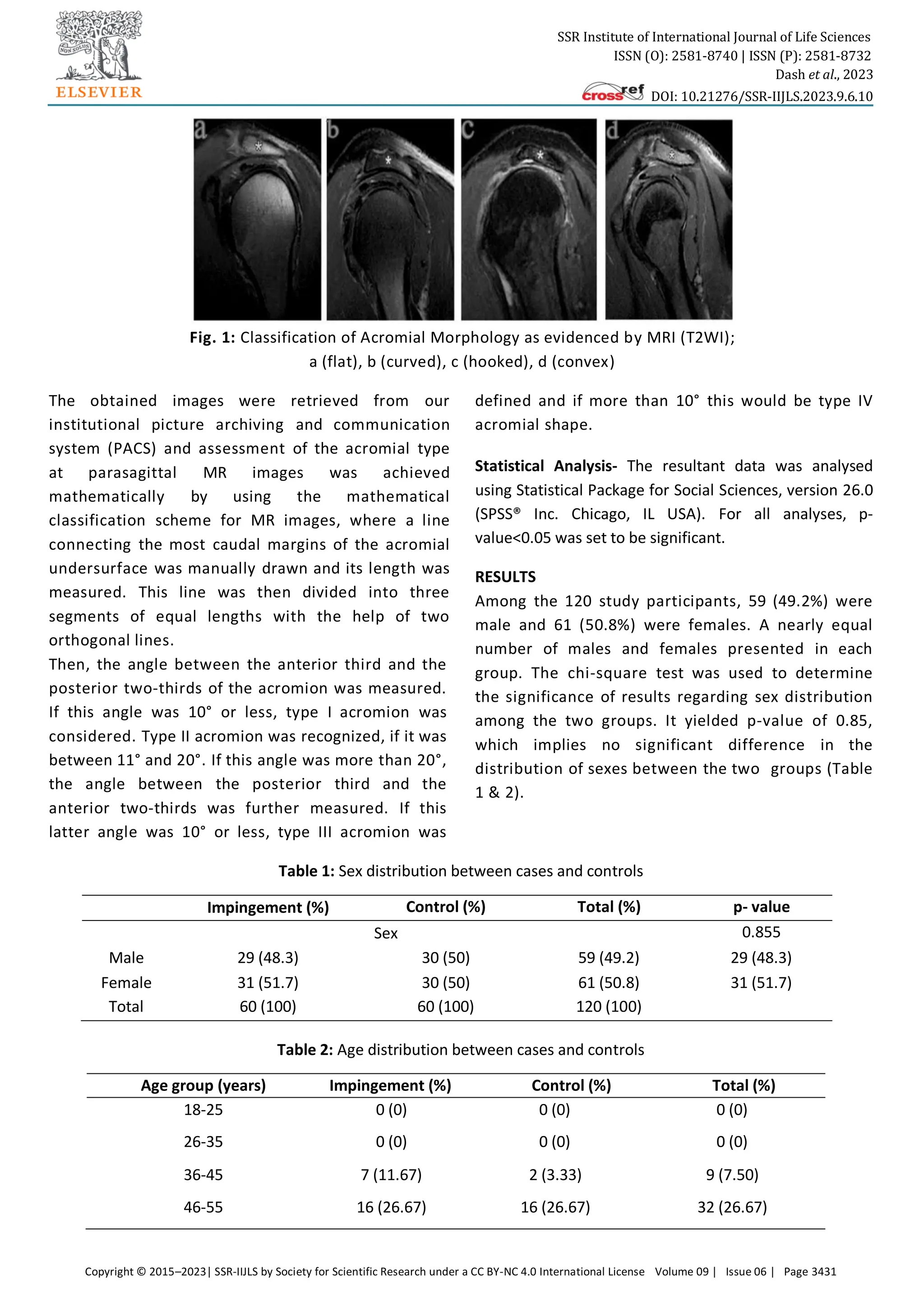
subacromial impingement. The mean acromial width
(AW) in patients with impingement (8.12 mm) was
significantly thicker than in the control group (7.51 mm),
with a p-value of 0.04. This is by the conclusion of
previous studies [37]
. The AW of Type-III (8.75 mm),
though markedly thicker than the other three types and](https://image.slidesharecdn.com/assessmentacromionmorphologyassociationshoulderimpingementsyndromemri-240119141145-d89c3c77/75/Assessment_Acromion_Morphology_Association_Shoulder_Impingement_Syndrome_MRI-pdf-8-2048.jpg)
![SSR Institute of International Journal of Life Sciences
ISSN (O): 2581-8740 | ISSN (P): 2581-8732
Dash et al., 2023
DOI: 10.21276/SSR-IIJLS.2023.9.6.10
Copyright © 2015–2023| SSR-IIJLS by Society for Scientific Research under a CC BY-NC 4.0 International License Volume 09 | Issue 06 | Page 3436
the control, was statistically insignificant. Therefore,
revealed significant inferior protrusion of impinged
acromia, with significantly lower AT, mAT and
significantly higher AIP. These findings agree with the
previous CT-based study by Li et al. [36]
In this study, the impingement group (7.27 mm)
exhibited significantly higher AAP than the control group
(5.79 mm), with a p-value of 0.01, implying significant
anterior prominence of the impinged acromia. The study
also reveals significantly higher AAP of type-III acromia
(7.97 mm), compared with other types and controls.
These findings align with Li et al. [36]
. The present study
reveals significantly lower AHD among impinged
acromia (7.85 mm) than controls (8.64 mm) with a p-
value of 0.03. However, no significant variation was
found in the AHD among the four acromial types in
SIS patients. These findings align with previous
studies [38]
and signify the presence of marked
superior migration of the humeral head in impinged
acromia independent of the acromial shape.
In the present study, the AI of impinged acromia
(0.62) was higher than controls (0.59), signifying a
more excellent coverage of the subacromial tissues by
the acromion. However, this difference in AI between
cases and controls was non-significant (p=0.13), nor
did it display any significant variation among the four
acromial types. This is in line with the previous
similar study conducted by Li et al. [36]
. In this study,
the LAA of impinged acromia (74.90°), though relatively
lesser than controls (76.31°) again, the difference was
found to be statistically insignificant (p=0.11), neither
was there any significant difference in LAA among the
four acromial types. However, type-III acromia (72.68°)
displayed a trend towards lower LAA compared with the
other types.
The present study tries to establish the relationship
between CSA and subacromial impingement. The CSA
of impinged acromia (30.24°) was significantly
greater than controls (28.12°) with a p-value of 0.01,
implying increased acromial cover laterally over the
glenoid. This finding aligns with Li et al. [36]
. Among the
cases, type-III acromia (32.49°) demonstrated
significantly higher CSA than other acromial types and
controls.
CONCLUSIONS
There was non-significant relationship between
acromion shape and subacromial impingement.
There was no significant association established
between acromial shape and rotator cuff tears.
Compared to controls, the impinged acromia are
significantly thicker (higher AW), more prominent
inferiorly (lower AT, mAT, & higher AIP) and
anteriorly (higher AAP), with increased lateral
acromial cover over the glenoid (greater CSA),
superior migration of the humeral head (lesser AHD)
and narrowing of the subacromial space. This study,
therefore, concludes that shoulder impingement is
associated with significant acromial morphological
variations.
Genetic factors influencing acromial differences and
personalized therapeutic strategies could be the subject
of future research. Understanding the causes of SIS can
potentially improve patient outcomes by implementing
more effective treatments and safeguards in the future.
Beyond aiding in the diagnostic process, this study paves
the way for improved clinical practices and a broader
comprehension of shoulder-related conditions.
ACKNOWLEDGMENTS
The authors are highly grateful to the respective
Universities and Principals of relevant Institutions to
carry out the present investigations.
CONTRIBUTION OF AUTHORS
Research concept- Abhisek Dash
Research design- Chandra Sekhar Pradhan
Supervision- Gopal Chandra Patro
Materials- Abhisek Dash
Data collection- Abhisek Dash
Data analysis and Interpretation- Abhisek Dash
Literature search- Gopal Chandra Patro
Writing article- Chandra Sekhar Pradhan
Critical review- Gopal Chandra Patro
Article editing- Jyotibhanja Das
Final approval- Gopal Chandra Patro
REFERENCES
[1] Urwin M, Symmons D, Allison T, et al. Estimating the
burden of musculoskeletal disorders in the
community: the comparative prevalence of
symptoms at different anatomical sites, and the
relation to social deprivation. Ann Rheum Dis.,](https://image.slidesharecdn.com/assessmentacromionmorphologyassociationshoulderimpingementsyndromemri-240119141145-d89c3c77/75/Assessment_Acromion_Morphology_Association_Shoulder_Impingement_Syndrome_MRI-pdf-9-2048.jpg)
![SSR Institute of International Journal of Life Sciences
ISSN (O): 2581-8740 | ISSN (P): 2581-8732
Dash et al., 2023
DOI: 10.21276/SSR-IIJLS.2023.9.6.10
Copyright © 2015–2023| SSR-IIJLS by Society for Scientific Research under a CC BY-NC 4.0 International License Volume 09 | Issue 06 | Page 3437
1998; 57: 649–55.
[2] Garving C, Jakob S, Bauer I, Nadjar R, et al.
Impingement syndrome of the shoulder. Dtsch
Arztebl Int., 2017; 114: 765-76.
[3] De-Yang TJ, Tan AHC. Shoulder impingement
syndrome, a common affliction of the shoulder:
a comprehensive review. Proc Singapore
Healthc., 2014; 23: 297-305.
[4] Myklebust G, Hasslan L, Bahr R, Steffen K. High
prevalence of shoulder pain among elite
Norwegian female handball players. Scand. J
Med Sci Sports, 2013; 23: 288–94.
[5] Oliveira, Mod V, et al. Shoulder pain in
adolescent athletes: prevalence, associated
factors and its influence on upper limb function.
Braz J Phys Ther., 2017; 21: 107–13.
[6] Rodeo SA, Nguyen JT, Cavanaugh JT, Patel Y, Adler
RS. Clinical and ultrasonographic evaluations of
the shoulders of elite swimmers. Am J Sports
Med., 2016; 44: 3214–21.
[7] Juel NG, Natvig B. Shoulder diagnoses in
secondary care, a one year cohort. BMC
Musculoskelet Disord., 2014; 15: 89.
[8] Neer CS, Welsh RP. The shoulder in sports.
Orthop Clin North Am., 1997; 8: 583–91.
[9] Van der Windt DA, Koes BW, de Jong, BA, Bouter
LM. Shoulder disorders in general practice:
incidence, patient characteristics, and
management. Ann Rheum Dis., 1995; 54: 959–64.
[10]Makela M, Heliovaara M, Sainio P, Knekt P,
Impivaara O, et al. Shoulder joint impairment
among Finns aged 30 years or over: prevalence,
risk factors and co- morbidity. Rheumatol., 1999;
38: 656–62.
[11]Yamaguchi K, Ditsios K, Middleton WD, Hildebolt
CF, Galatz LM, et al. The demographic and
morphological features of rotator cuff disease. A
comparison of asymptomatic and symptomatic
shoulders. J Bone Joint Surg Am., 2006; 88:
1699–704.
[12]Blom AW, Warwick D, Whitehouse MR. Apley and
Solomon's system of orthopaedics and trauma.
10th
ed Boca Raton: Taylor & Francis, 2018.
[13]Lazaro R. Shoulder impingement syndromes:
implications on physical therapy examination and
intervention. J Jpn Phys Ther Assoc., 2005; 8: 1-7.
[14]Habermeyer P. Schulterchirurgie. Munchen:
Elsevier, Urban & Fischer 2010; 4th
edition.
[15]Almekinders LC, Weinhold PS, Maffulli N.
Compression etiology in tendinopathy. Clin
Sports Med., 2003; 22: 703–10.
[16]Matthews TJ, Hand GC, Rees JL, Athanasou NA, et
al. Pathology of the torn rotator cuff tendon.
Reduction in potential for repair as tear size
increases. J Bone Joint Surg Br., 2006; 88: 489–
495.
[17]Neer CS II. Anterior acromioplasty for the chronic
impingement syndrome in the shoulder: a
preliminary report. J Bone Joint Surg Am., 1992;
54: 41–50.
[18]Yazici M, Kopuz C, Gulman B. Morphologic
variants of acromion in neonatal cadavers. J
Pediatr Orthop., 1995; 15: 644–47.
[19]Nicholson GP, Goodman DA, Flatow EL, Bigliani
LU. The acromion: morphologic condition and
age-related changes-a study of 420 scapulas. J
Shoulder Elbow Surg., 1996; 5: 1–11.
[20]Hutchinson MR, et al. Arthroscopic
decompression of shoulder impingement
secondary to os acromiale. Arthroscopy, 1993; 9:
28–32.
[21]Seeger LL, Gold RH, Bassett LW, Ellman H.
Shoulder impingement syndrome: MR findings in
53 shoulders. AJR Am J Roentgenol., 1998; 150:
343-47.
[22]Dhillon KS. Subacromial impingement syndrome
of the shoulder: a musculoskeletal disorder or a
medical myth? Malays Orthop J., 2019; 13: 1-7.
[23]Edwards SL, Bell JE, Bigliani LU. Subacromial
impingement. In: Wilk KE, Reinold MM, Andrews
JR, editors. The Athlete's Shoulder. 2nd
ed.
Philadelphia: Churchill Livingstone, 2009; pp.
115-22.
[24]Morrison DS, Frogameni AD, Woodworth P. Non-
operative treatment of subacromial impingement
syndrome. J Bone Joint Surg Am., 1997; 79: 732-
37.
[25]Bigliani LU, Morrison DS, et al. The morphology of
the acromion and its relationship to rotator cuff
tears. Orthop Trans., 1996; 10: 216.
[26]Zuckerman JD, Kummer FJ, Cuomo F, Greller M.
Interobserver reliability of acromial morphology
classification: an anatomic study. J Shoulder
Elbow Surg., 1997; 6: 286–87.](https://image.slidesharecdn.com/assessmentacromionmorphologyassociationshoulderimpingementsyndromemri-240119141145-d89c3c77/75/Assessment_Acromion_Morphology_Association_Shoulder_Impingement_Syndrome_MRI-pdf-10-2048.jpg)
![SSR Institute of International Journal of Life Sciences
ISSN (O): 2581-8740 | ISSN (P): 2581-8732
Dash et al., 2023
DOI: 10.21276/SSR-IIJLS.2023.9.6.10
Copyright © 2015–2023| SSR-IIJLS by Society for Scientific Research under a CC BY-NC 4.0 International License Volume 09 | Issue 06 | Page 3438
[27]Jacobson SR, Speer KP, Moor JT, et al. Reliability
of radiographic assessment of acromial
morphology. J Shoulder Elbow Surg., 1995; 4:
449–53.
[28]Peh WC, Farmer TH, Totty WG. Acromial arch
shape: assessment with MR imaging. Radiol.,
1995; 195: 501–05.
[29]Balke M, Schmidt C, Dedy N, et al. Correlation of
acromial morphology with impingement
syndrome and rotator cuff tears. Acta
Orthopaedica., 2013; 84(2): 178– 83.
[30]Bigliani LU, Morrison DS, Ticker JB, et al.
Relationship of acromial architecture and
diseases of the rotator cuff (in German).
Orthopade, 1991; 20: 302-09.
[31]Nho SJ, Yadav H, Shindle MK, et al. Rotator cuff
degeneration: etiology and pathogenesis. Am J
Sports Med., 2008; 36(5): 987–93.
[32]Morag Y, Jacobson A, Miller B, et al. MR imaging
of rotator cuff injury: what the clinician needs to
know? Radio Graphics, 2006; 26: 1045-65.
[33]Hirano M, Ide J, Takagi K. Acromial shapes and
extension of rotator cuff tears: magnetic
resonance imaging evaluation. J Shoulder Elbow
Surg., 2002, 576-78.
[34]Paraskevas G, Tzaveas A, Papaziogas B, et al.
Morphological parameters of the acromion. Folia
Morphol., 2008; 67(4): 255–60.
[35]Nagerl H, Kubein-Meesenburg D, Cotta H,
Fanghanel J, Kirsch S: Biomechanical principles in
diarthroses and synarthroses. II: The humerus
articulation as a ball-and- socket joint. Z Orthop
Ihre Grenzgeb., 1993; 131: 293–301.
[36]Li X, Xu W, Hu N, Liang X, Huang W, et al.
Relationship between acromial morphological
variation and subacromial impingement: a three-
dimensional analysis. PLoS One, 2017; 12:
e0176193.
[37]Habermeyer PS. München: Elsevier, Urban &
Fischer 2010; 4th
edition.
[38]Harrison AK, Flatow EL: Subacromial
impingement syndrome. J Am Acad Orthop Surg.,
2011; 19: 701–08.
Open Access Policy:
Authors/Contributors are responsible for originality, contents, correct references, and ethical issues. SSR-IIJLS publishes all articles under Creative
Commons Attribution- Non-Commercial 4.0 International License (CC BY-NC). https://creativecommons.org/licenses/by-nc/4.0/legalcode](https://image.slidesharecdn.com/assessmentacromionmorphologyassociationshoulderimpingementsyndromemri-240119141145-d89c3c77/75/Assessment_Acromion_Morphology_Association_Shoulder_Impingement_Syndrome_MRI-pdf-11-2048.jpg)
This study assesses acromion morphology's relationship with shoulder impingement syndrome (SIS) using MRI among patients at MKCG Medical College, India. Findings indicate that while subacromial impingement was not significantly linked to acromion shape, cases presented with a larger acromial width compared to controls. The study concludes that acromion morphology does not correlate with SIS or rotator cuff injuries.