The document provides a comprehensive overview of viral structure and classification, detailing components such as nucleic acids, capsids, and envelopes. It discusses various virus morphologies, including helical, polyhedral, and complex structures, while also classifying viruses based on their genetic material (DNA or RNA). Additionally, the document outlines specific virus families and their characteristics, including examples like herpesviruses, adenoviruses, and retroviruses.


![Herpes viruses1
Herpesviruses are divided into three groups:
• α-herpesviruses are fast-growing cytolytic viruses that
establish latent infections in neurones (e.g. herpes simplex
and varicella zoster);
• β-herpesviruses are slow-growing viruses that become
latent in secretory glands and kidneys (e.g. cytomegalovirus
[CMV], HHV6 and 7);
• γ-herpesviruses are latent in lymphoid tissues (e.g. Epstein–
Barr virus [EBV], HHV-8).
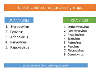
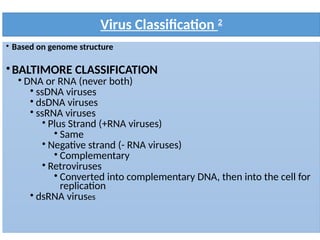
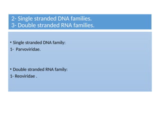
Classification
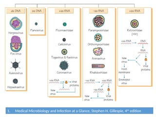
Medical Microbiology and Infection at a Glance, Stephen H. Gillespie, 4th
edition](https://image.slidesharecdn.com/virusclassification-250101094302-06bbb5c1/85/Structures-and-classification-of-viruses-22-320.jpg)