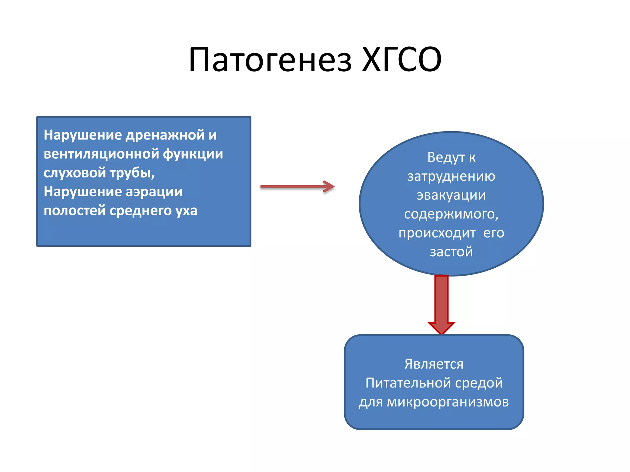
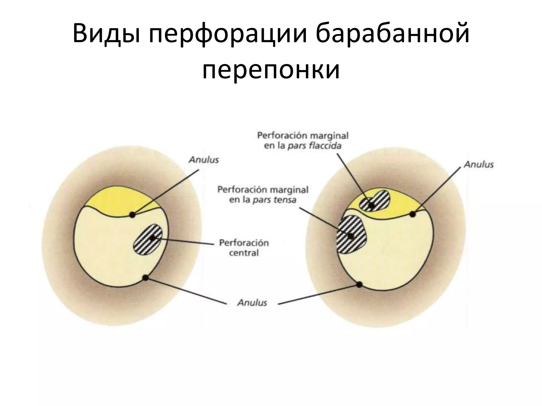
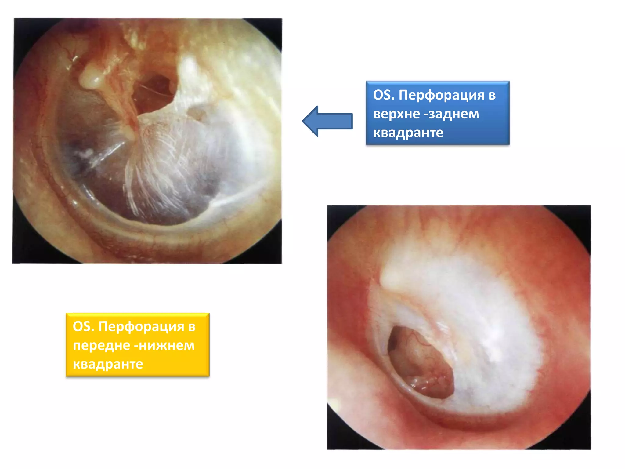
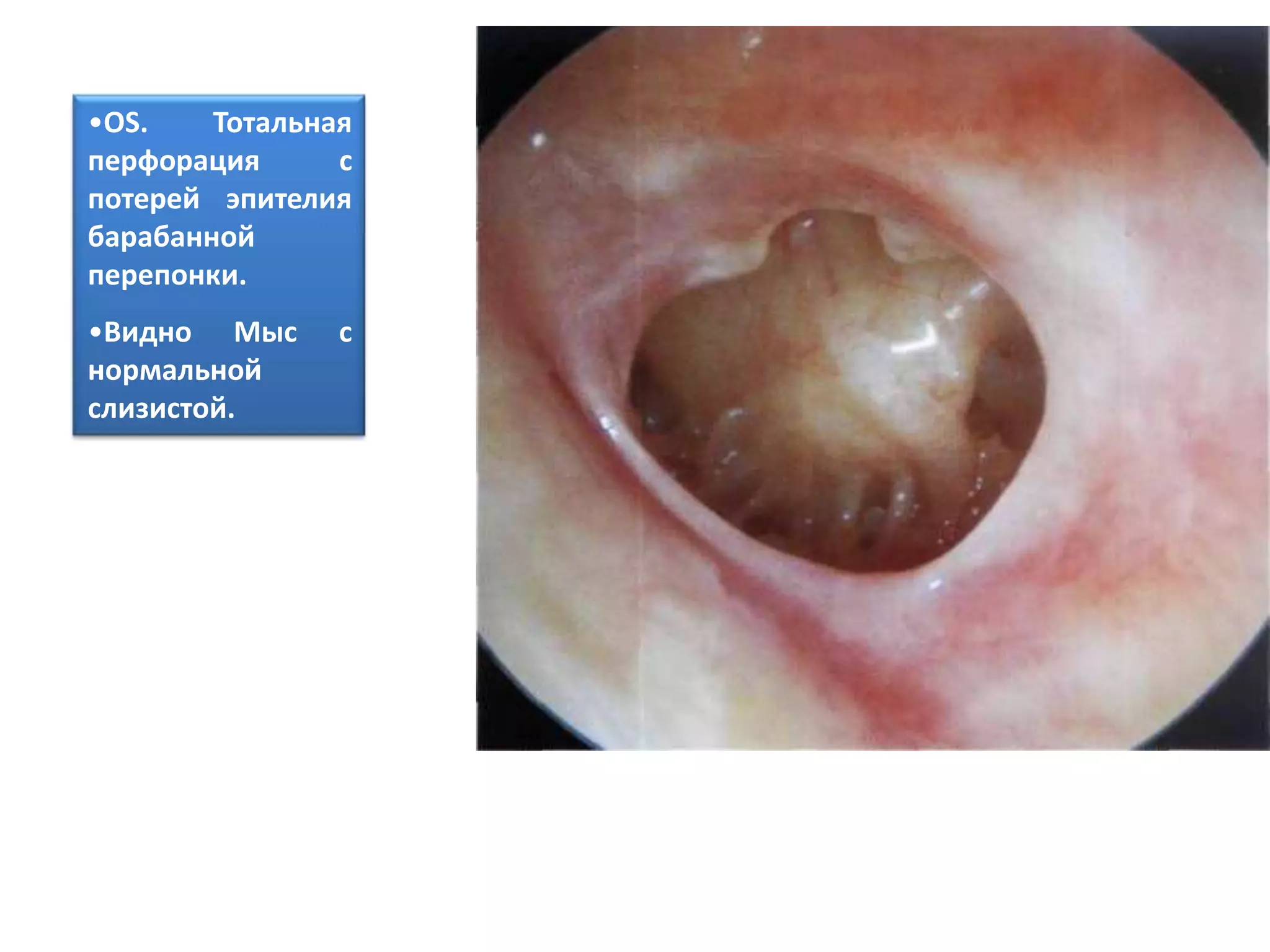
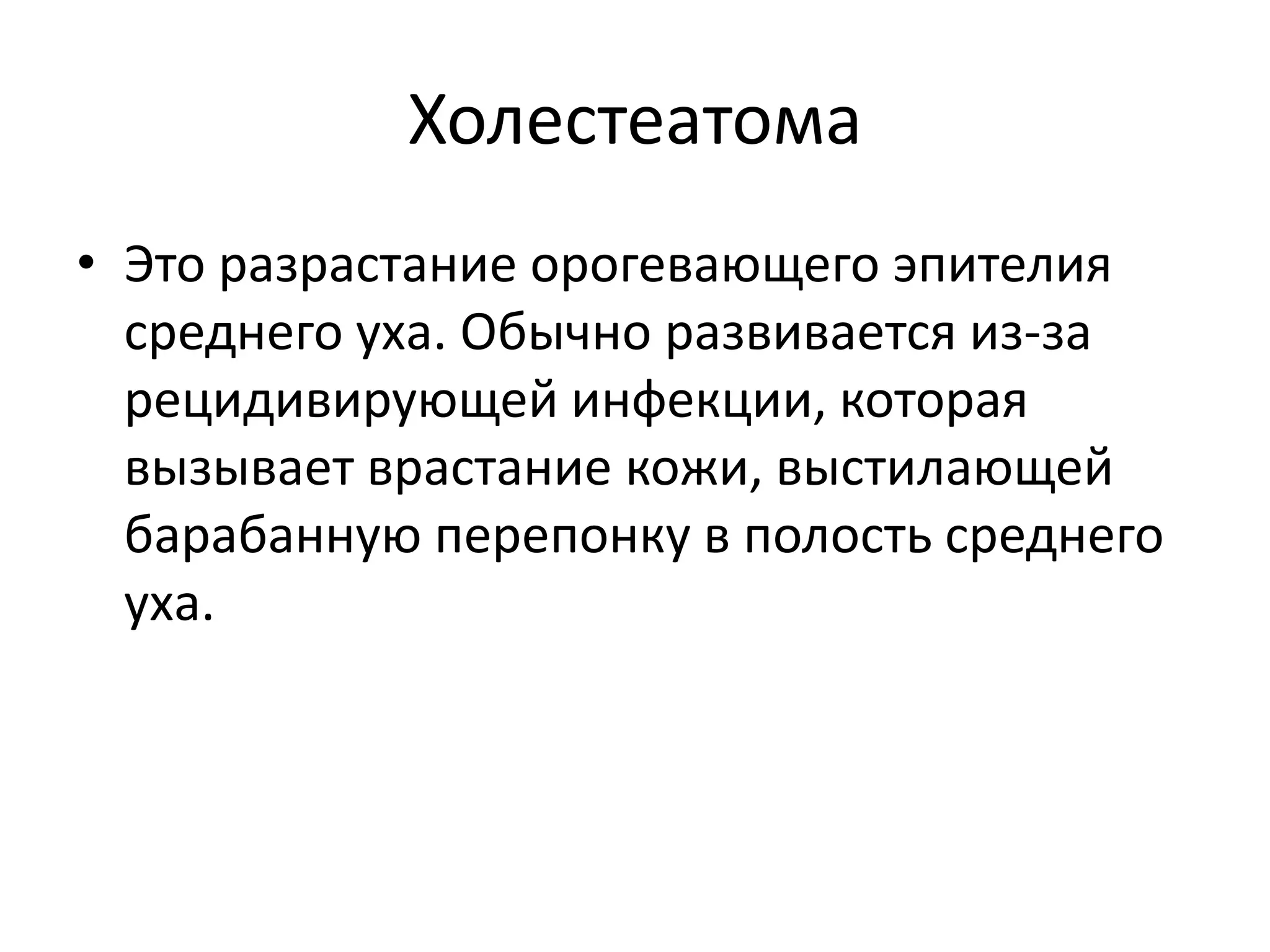
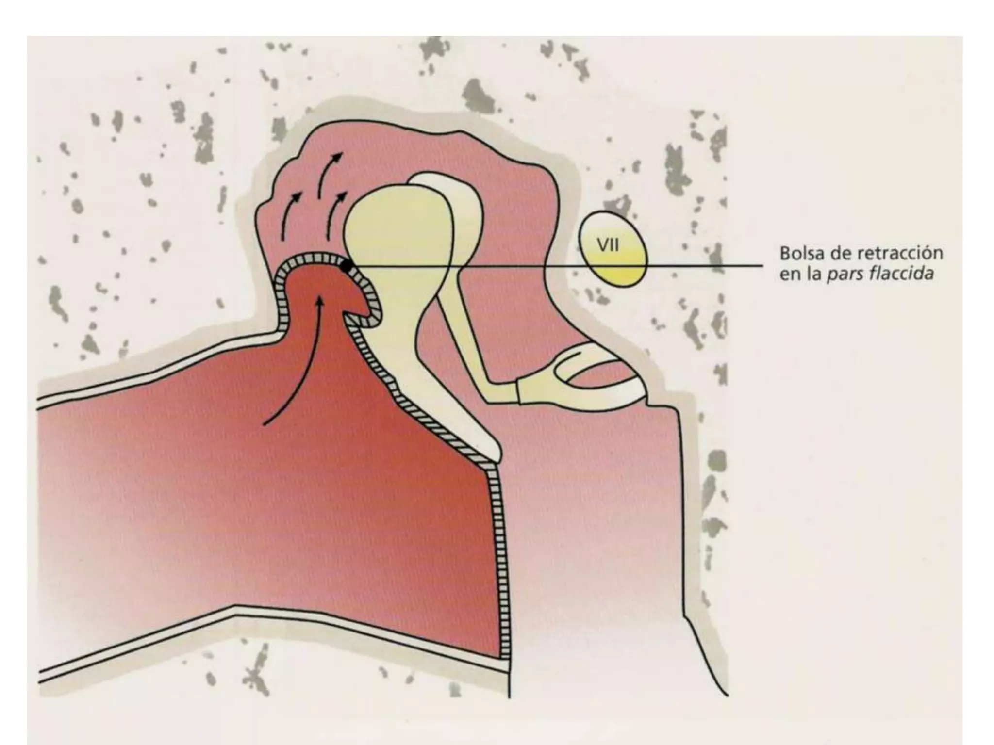
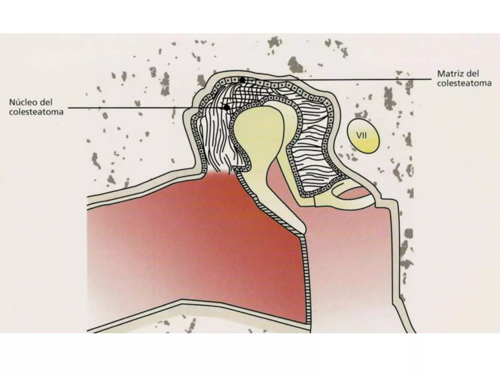
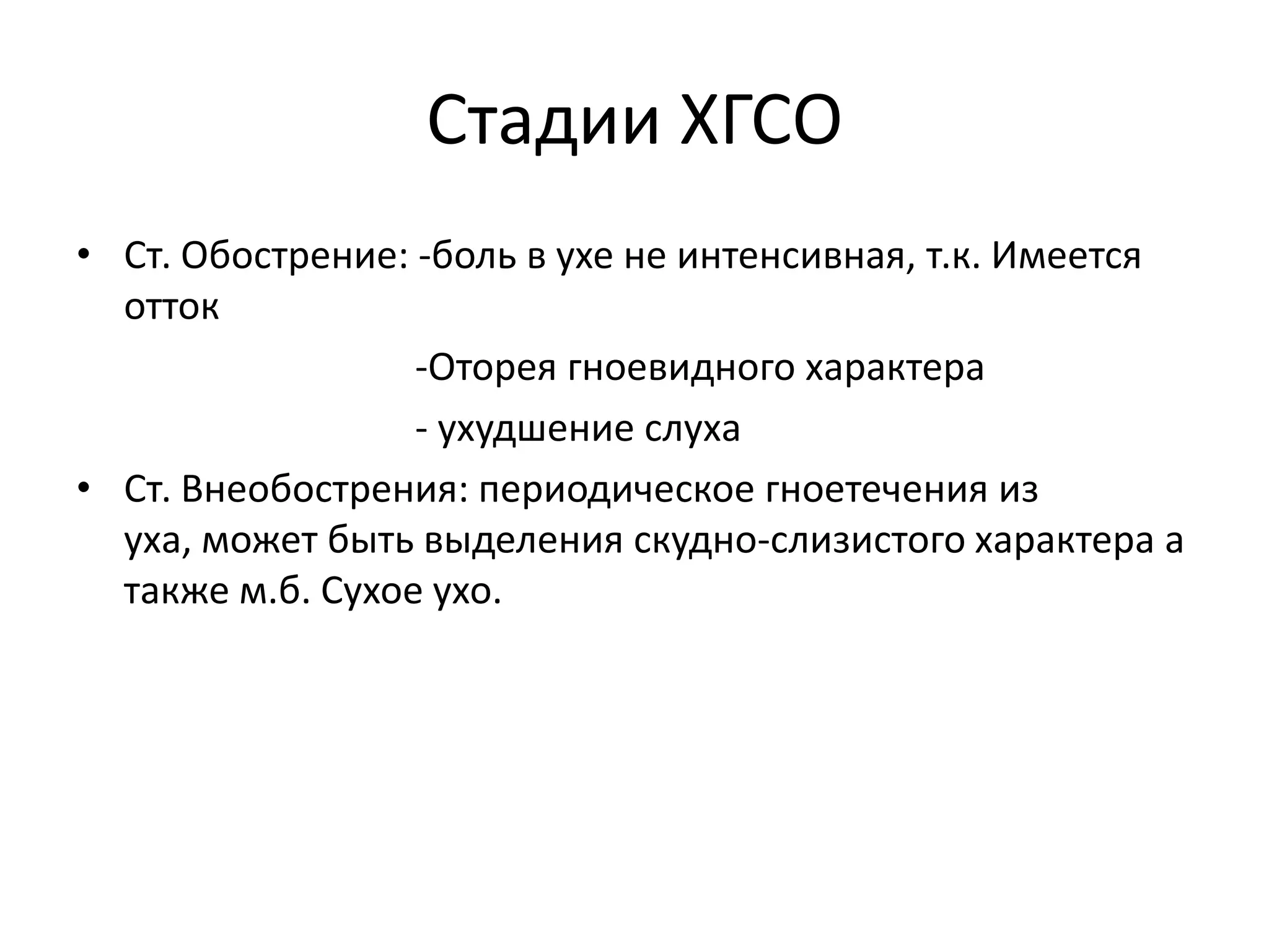
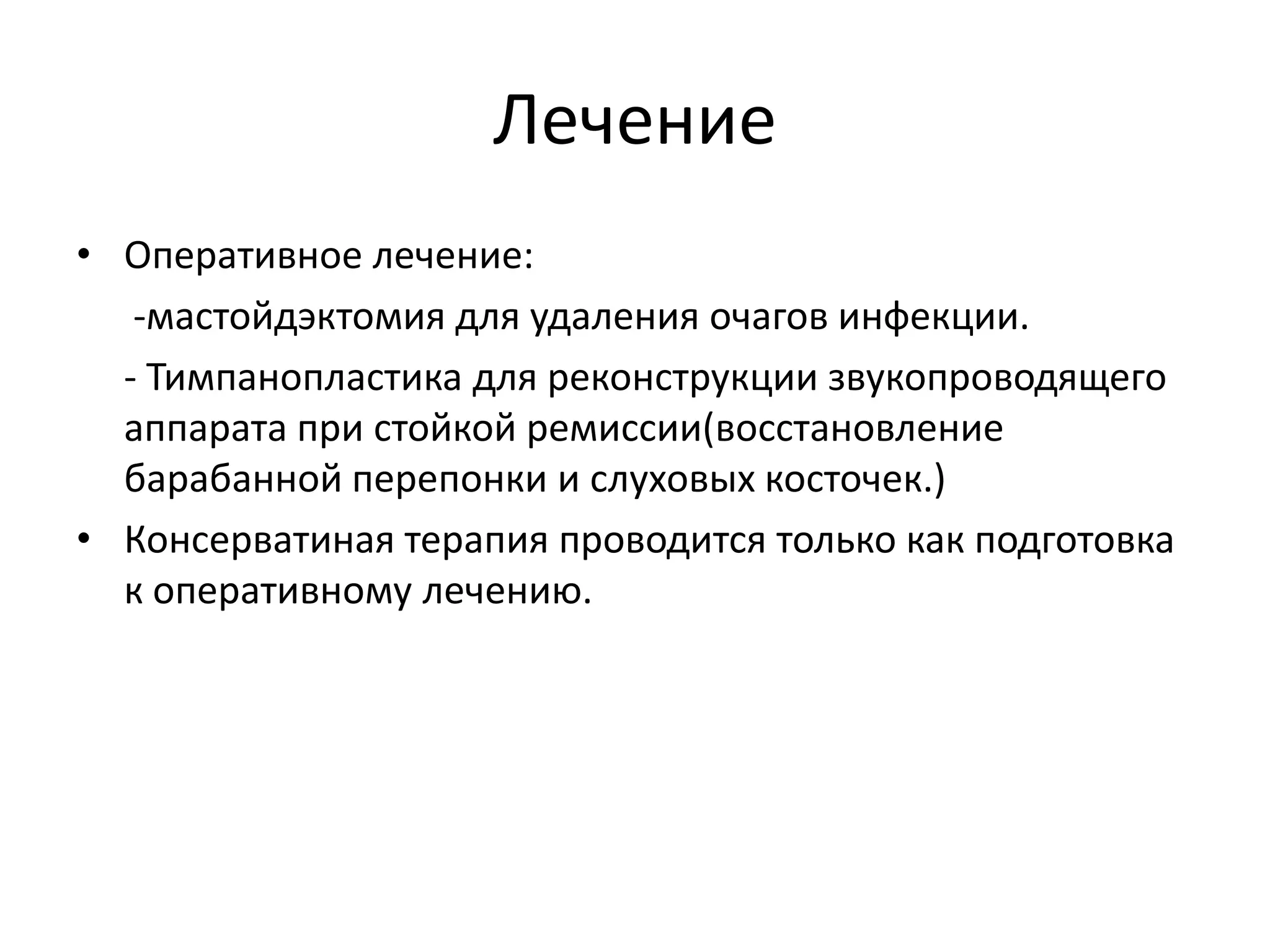
Pemphigoid is a relatively uncommon autoimmune disease that affects the skin and oral mucosa. There are two main forms - bullous pemphigoid and mucous membrane pemphigoid. Mucous membrane pemphigoid predominantly involves oral mucosal ulceration and desquamative gingivitis. Diagnosis involves a biopsy showing subepithelial blistering and direct immunofluorescence detecting linear deposition of IgG along the basement membrane. Treatment typically involves an initial course of systemic steroids like prednisolone followed by long-term topical steroid maintenance therapy.
24 04-2020 Bds third year lecture on smoking and periodontiumDr. Mamta Singh
Periodontitis is a group of inflammatory diseases affecting the supporting tissues of the tooth (periodontium). The periodontium consists of four tissues : gingiva, alveolar bone and periodontal ligaments. Tobbaco use is one of the modifiable risk factors and has enormous influance on the development, progres and tretmen results of periodontal disease. The relationship between smoking and periodontal health was investigated as early as the miiddle of last century. Smoking is an independent risk factor for the initiation, extent and severity of periodontal disease. Additionally, smoking can lower the chances for successful tretment. Tretmans in patients with periodontal disease must be focused on understanding the relationship between genetic and environmental factors. Only with individual approach we can identify our pacients risks and achieve better results.
Dry socket is a common complication after tooth extraction where the blood clot in the extraction socket fails to form properly. It causes severe throbbing pain 2-5 days after extraction. Risk factors include smoking, single extractions, and surgical extractions. Treatment involves removing any infection in the socket, prescribing antibiotics and painkillers.
This document discusses desquamative gingivitis, which is characterized by intense redness, peeling, and ulceration of the gums. It is not a specific disease but rather a gum condition associated with various underlying causes. The document goes on to classify, describe clinically, and discuss the diagnosis and management of desquamative gingivitis. Several diseases that can clinically present as desquamative gingivitis are described in detail, including lichen planus, mucous membrane pemphigoid, bullous pemphigoid, pemphigus vulgaris, dermatitis herpetiformis, and linear IgA disease. Histopathological features of these conditions are also summarized
This document provides an overview of halitosis (bad breath) including its classification, etiology, diagnosis, and treatment. Halitosis can be genuine (caused by intraoral or extraoral factors) or pseudo (no actual malodor detected). Diagnosis involves organoleptic measurement, gas chromatography, microbial tests, and medical exams to determine the source. Treatment aims to reduce oral microbial load and malodorous compounds through mechanical cleaning, antimicrobial mouthwashes, and addressing any underlying medical conditions causing halitosis.
Операция атипичного удаления зуба: показания и противопоказания, инструментарий, методика проведения, подготовка больного. Особенности операции удаления зуба у лиц с сопутствующими заболеваниями. Осложнения во время и после операции удаления зубов.
This document discusses periodontal response to external forces on teeth. It defines trauma from occlusion as injury to tissues from forces exceeding their adaptive capacity. Trauma can be acute or chronic, and primary (normal support) or secondary (reduced support). It discusses classifications and concepts around how gingival inflammation spreads to bone. Glickman's concept describes zones of irritation and co-destruction that are differentially affected. Waerhaug's concept is that bone loss is due exclusively to plaque inflammation. The stages of tissue response to increased forces are injury, repair, and adaptive remodeling.
Acute necrotizing ulcerative gingivitis (ANUG), also known as Vincent's gingivitis or Vincent's infection, is a polymicrobial infection of the gums that results in ulceration, necrosis, and pain. It is caused by an overgrowth of oral bacteria like spirochetes and fusiform bacteria when the immune system is weakened by factors like stress, malnutrition, or HIV infection. ANUG begins as small ulcers on the gums that can spread and deepen over time if left untreated, potentially resulting in bone and tissue loss. Symptoms include bleeding, foul taste, and pain when eating. Treatment focuses on improving oral hygiene and nutrition to restore immune function and resolve the
Mandibular Radiolucencies; A Systematic Approach to DiagnosisAhmed Adawy
Dr. Ahmed M. Adawy, Professor Emeritus, Dep. Oral & Maxillofacial Surgery. Former Dean, Faculty
of Dental Medicine, Al-Azhar University. Conventional radiography may revel a variety of
radiolucent legions in the mandible. Interpretation of such radiolucencies can be challenging either
because the clinical presentation may be non specific or because the ;legion is detected
incidentally. Further, interpretation may vary from one examiner to another. thus, systemic
approach is necessary to diagnose the legion or at least provide a meaningful deferential
diagnosis. This approach should focus on specific radiographic parameters. Initially, the legion
should be placed in the category of either normal or abnormal. The presented parameters includes
describing the legion in terms of: 1- Location, 2- Margins, 3- Size and shape, 4- Effect on
surrounding structures. Obviously, however diagnosis of a legion should never be made
exclusively on the bases of radiographic interpretation. Radiographic interpretation should be used
along with clinical information and other tests to formulate a deferential diagnosis.
Periodontal probes have evolved over time and are now used to measure pocket depths, assess bone and furcation levels, and detect early inflammation. Standard probes are calibrated stainless steel rods but newer versions include computerized probes that can detect the cementoenamel junction and thermal probes that can measure subgingival temperature changes as a marker of inflammation. Proper probing technique involves walking the probe parallel to the tooth and exploring all surfaces and potential pockets to determine the deepest level of penetration.
The Indian Dental Academy is the Leader in continuing dental education , training dentists in all aspects of dentistry and
offering a wide range of dental certified courses in different formats.
This document discusses different types of gingival enlargement, including classifications based on etiology and location. Key types discussed include inflammatory enlargement (both chronic and acute), drug-induced enlargement caused by medications like anticonvulsants and calcium channel blockers, and enlargement associated with systemic conditions like pregnancy, puberty, and vitamin C deficiency. False enlargement and neoplastic enlargements are also mentioned. Clinical features, etiology, and pathology of several conditions are provided.
The document reports on a case study of a 65-year-old male patient who presented with diffuse erythematous patches on the hard palate and grooves that bled upon probing, and was diagnosed with oral erythroplasia based on a positive toluidine blue staining test and histological examination showing epithelial atrophy and mild dysplasia. Oral erythroplasia is a potentially malignant disorder strongly associated with tobacco and alcohol use that requires surgical excision due to its high risk of transforming into carcinoma.
Alveolar bone forms tooth sockets and provides attachment for the periodontal ligament. It consists of inner and outer cortical plates, cancellous bone, and bone lining the socket. These structures meet at the alveolar crest, usually 2 mm below the cementoenamel junction. The bone lining the socket, also known as bundle bone, cribriform plate, or lamina dura, provides direct support for teeth. Bone remodeling involves the interdependent action of osteoblasts and osteoclasts to replace old bone.
The document discusses various salivary gland disorders including infections, inflammatory conditions, cysts, tumors and other pathologies. It provides details on:
- Acute and chronic bacterial sialadenitis, most commonly caused by retrograde infection from the mouth. Acute infections more often affect the parotid gland.
- Viral infections like mumps can cause acute non-suppurative sialadenitis. Mumps is spread through droplets and involves the parotid glands.
- Sjögren's syndrome is an autoimmune condition characterized by lymphocytic destruction of exocrine glands causing dry mouth and eyes. Diagnosis involves labial biopsy.
- Common benign sal
DRUG INDUCED GINGIVAL ENLARGEMENT / dental crown & bridge coursesIndian dental academy
The Indian Dental Academy is the Leader in continuing dental education , training dentists in all aspects of dentistry and
offering a wide range of dental certified courses in different formats.for more details please visit
www.indiandentalacademy.com
The Indian Dental Academy is the Leader in continuing dental education , training dentists in all aspects of dentistry and
offering a wide range of dental certified courses in different formats.
this ppt depicts pattern of bone destruction. its a very good slide show showing the process of bone formation, bone destruction and their patterns in periodontal diseases.
Trotter's triad is a set of three symptoms seen in nasopharyngeal carcinoma patients: middle ear effusion, palatal asymmetry, and a nasopharyngeal tumor. An example case showed a patient with left-sided middle ear effusion, palatal asymmetry, and a nasopharyngeal tumor that had extended intracranially through the foramen ovale and invaded the pterygoid muscles.
When the primary EBV infection mainly occurs in childhood, often it is without symptoms. However, when teens and young adults get infected, because of their aggressive and active immune systems, they get the set of symptoms we call "mono (Masucci et al., 1994).
The EBV virus develops its symptoms quiet slowly. A patient has had the virus for as many as 30-60 days before the first mild symptoms develop: fatigue, mild headaches and loss of appetite are common (Schuster et al., 1992). While sometimes these are the only symptoms, commonly the patient follows in about a week with fever, sore throat (often mild to chronic, with exudates or "white spots" on the tonsils), and swollen glands in the neck, aggrandizing fatigue, muscle aches and occasionally a skin rash. In most cases, the spleen can enlarge and the liver can become inflamed (Tattevin et al.,2006). Diagnosing Infectious Mononucleosis
Healthcare cateres typically diagnose infectious mononucleosis based on symptoms.
Laboratory tests are not mainly needed to diagnose infectious mononucleosis. However, specific laboratory tests may be needed to identify the specific cause of illness in people who do not have a typical case of infectious mononucleosis.
The blood work of the patients who have infectious mononucleosis due to EBV infection may show (Ramagopalan et al., 2011)
more white blood cells (lymphocytes) than normal
unusual looking white blood cells (atypical lymphocytes)
fewer than the normal neutrophils or platelets
abnormal liver function
2. это хроническое гнойное воспаление среднего уха,
характеризующееся триадой признаков:
* наличием стойкой перфорации барабанной
перепонки,
*постоянным или периодически повторяющимся
гноетечением из уха, кот. приводить к деструкции
костных структур среднего уха
* выраженным снижением слуха, постепенно
прогрессирующим при длительном течении
заболевания
3. Эпидемиология:
• Во всем мире ХГСО страдают от 1 до 46%
человек, проживающих в развитых и
развивающихся странах. Это примерно 65–330
млн человек, 60% из них имеют значительное
снижение слуха.
• Распространенность ХГСО в РФ. составляет от 8,4
до 39,2 на 1000 населения.
4. Этиология:
• Это следствие нелеченного острого
среднего отита, особенно в первые 5 лет
жизни, когда сформировавшиеся
поствоспалительные изменения в
слизистой оболочке и структурах среднего
уха способствуют хронизации процесса.
• Результат разрыва барабанной перепонке
при травме.
5. Возбудители
• Часто встречающиеся микобактерии:
-грам + Staphylococcus aureus*
-грам - Pseudomonas aeruginosa*, Proteus
mirabilis, Klebsiella pneumoniae
-грибы: р.Candida, Aspergillus, Mucor
* Встречается в 70-90%.
6. Предрасполагающие факторы
• Тип возбудителей, их патогенность и резистентности к А/Б
терапии.
• Ославленный организм при общих заболеваниях:
аллергические,иммунные, сахарный диабет, кахексия.
• анатомические особенности строения среднего уха (узкие
пространства барабанной полости, адитуса, карманы
наружного аттика)
• хроническая патология носоглотки и полости носа приводят к
стойкому отрицательному давлению в барабанной
полости, ретротимпанальном пространстве, клетках
сосцевидного отростка и к затянувшемуся острому процессу в
среднем ухе с тенденцией перехода его в секреторную и
фиброзирующую формы
• Хронический процесс развивается постепенно вследствие
частых рецидивов острого воспаления
7. Патогенез ХГСО
Нарушение дренажной и
вентиляционной функции
слуховой трубы,
Нарушение аэрации
полостей среднего уха
Ведут к
затруднению
эвакуации
содержимого,
происходит его
застой
Является
Питательной средой
для микроорганизмов
8. Морфологические формы ХГСО
• Катаральная форма: Воспаление поверхностных слоев
эпителии(отек,метаплазия эпителия, гиперсекреция слизь)
• Гнойная форма: воспаление всей слизистой
оболочки(метаплазия эпителии, клеточная
инфильтрация, экссудат гнойного характера)
• Гранулационная форма: воспаление до подслизистого
слоя.
• Кариозная форма: воспаление до костных стенок среднего
уха.
• Холестеатомная форма: бесструктурная аморфная масса.
• Полипозная форма: образование полипов.
• Сочетанная форма: чаще кариозно-холестеатомная
12. Диагностические мероприятие при
ХГСО
Общий оториноларингологический осмотр с
применением отомикроскопии
Аудиологическое обследование, в том числе
аудиометрию, которая позволяет оценить функцию
слухобой трубы.
Маневр Вальсальвы для выталкивания отделяемого в
слуховой проход.
Обязательное исследование флоры и ее чувствительности
к антибиотикам
Фистульные пробы
КТ височных костей
13. Классификация ХГСО:В соотвествии с МКБ-10 по
характеру патологического процесса в среднем ухе, по особенностям
клинического течения и тяжести заболевания делят на две формы.
Хронический туботимпанальной гнойный средний отит-- Мезотимпанит
Хронический эпитимпано-антральный гнойный средний отит--Эпитимпанит
Мезотимпанит имеет относительно
благоприятное течение, перфорация барабанной
перепонки располагается в натянутой ее части.
Хронический воспалительный процесс ограничен
слизистой оболочкой, но он может привести со
временем к остеиту т.е. К деструкции слуховых
косточек. Ранее считалось, что для
мезотимпанита холестеатомный процесс не
характерен, однако в последнее время все чаще во
время операции выявляется холестеатома
мезотимпанума, распространяющаяся в другие
отделы барабанной полости. Согласно статистике
отдела микрохирургии уха МНПЦО за период с 2009
по 2012 г., в 12,4% случаев среди всех
оперированных пациентов с диагнозом
«мезотимпанит» была обнаружена холестеатома
мезотимпанума.
Для эпитимпанита характерно
недоброкачественное тяжелое
течение, а перфорация
локализуется в ненатянутой части
барабанной перепонки.При
эпитимпаните происходит более
глубокое поражение тканей и в
деструктивный(кариозный) процесс
вовлечены костные структуры
среднего уха.
Часто формируются глубокие
ретракционные карманы с
деструкцией латеральной
стенки аттика, эпидермизацией
аттика и ретротимпанальных
отделов, образованием
холестеатомы
14. Мезотимпанит
Локализация процесса в среднем и нижним этаже барабанной
полости
Выделение из уха носят слизистый или слизисто-гнойный характер и
обычно не имеют запаха, но при наличии грануляций или полипов
можно наблюдать кровянисто-гнойные выделения.
При отоскопии: перфорация в натянутой части и может быть:
-центральная перфорация не достигает костного кольца
- краевая перфорация достигает костного кольца.
-Рубцовые изменение в натянутой части.
Снижение слуха по кондуктивному тупу
Слух может быть снижен на 15-20 дБ или оставаться в пределах
нормы.
При обострение быстро купируется, редко приводит к осложнениям.
15. Эпитимпанит
Патологическое изменение локализуются преимущественно в
надбарабанном пространстве и сосцевидном отростке.
В зависимости от того, какая патология преобладает, кариозное поражение
костной ткани, или изменения , вызванные образованием холестеатомы
различают гнойно-кариозную и холестеатомную формы.
Выделение гнойные с неприятным запахом, иногда с примесью крови или
крошковидных масс.
При отоскопии в начальной стадии можно увидеть небольшую перфорацию в
ненатянутой части б/перепонки. Постепенно увеличиваясь, перфорацию
польностью занимает латеральную стенку аттика.
Снижение слуха более выраженное чем при мезотимпаните.
Боль в ухе, головная боль.
16. Холестеатома
• Это разрастание орогевающего эпителия
среднего уха. Обычно развивается из-за
рецидивирующей инфекции, которая
вызывает врастание кожи, выстилающей
барабанную перепонку в полость среднего
уха.
17. Причины возникновения
Нарушение аэрации полостей среднего уха
Наличие ретракций барабанной перепонки
Состояние мукоцилиарного транспорта в
среднем ухе (его слабая выраженность в
области адитуса и антрума)
18.
19.
20.
21. Стадии ХГСО
• Ст. Обострение: -боль в ухе не интенсивная, т.к. Имеется
отток
-Оторея гноевидного характера
- ухудшение слуха
• Ст. Внеобострения: периодическое гноетечения из
уха, может быть выделения скудно-слизистого характера а
также м.б. Сухое ухо.
22. Лечение
• Оперативное лечение:
-мастойдэктомия для удаления очагов инфекции.
- Тимпанопластика для реконструкции звукопроводящего
аппарата при стойкой ремиссии(восстановление
барабанной перепонки и слуховых косточек.)
• Консерватиная терапия проводится только как подготовка
к оперативному лечению.