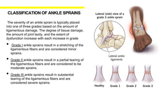
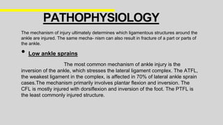
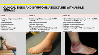
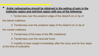
DIPTENDU BHATTACHARJEE
MPT (SPORTS)
ADHUNIK INSTITUTE OF EDUCATION
AND RESEARCH,DUHAI,GHAZIABAD
• Ankle sprains are common injuries in active individuals, with an
estimated incidence of 61 ankle sprains per 10,000 persons each
year (Maffulli and Ferran 2008)
• They are the most common injury sustained by high school and
collegiate athletes, accounting for up to 30% of sports injuries
(Hass et al. 2010). An age of 10 to 19 years old is associated with
higher rates of ankle sprains. Half of all ankle sprains occur during
athletic activity.
• Malliaropoulos et al. (2009)
In a study of 202 elite track and field athletes with
lateral ankle sprain,found that 18% sustained a second sprain within
24 months; low-grade acute ankle sprains (grade I or II) resulted in a
higher risk of reinjury than high-grade (grade III) sprains.
Because of the potential for reinjury and chronic dysfunction
and the importance of a normally functioning ankle in active people, it
is important that ankle sprains be managed correctly with a thorough
rehabilitation and reconditioning program.
INTRODUCTION
The ankle, or talocrural joint, is a junction of the
tibia, fibula, and talus.
• Lateral support to the ankle joint
A. Anterior Talofibular ligament (ATFL)
B. Calcaneofibular ligament (CFL)
C. Posterior Talofibular ligament (PTFL)
The ATFL is the most commonly injured ligament,
followed by the CFL. The CFL is usually injured in
combination with the ATFL. Sprains to both the
ATFL and CFL are a result of a combined
inversion and plantar-flexion mechanism
RELEVANT
ANATOMY
• whereas the deltoid ligament complex (DLC), made up of
the anterior and posterior tibiotalar ligaments, the
tibiocalcaneal ligament, and the tibionavicular ligament,
provides medial support.
• Additional support to the Talocrural joint provided by:
A. The inferior anterior and posterior tibiofibular
ligaments B.
The interosseous membrane
• The peroneal muscle group, composed of the peroneus
brevis, longus, and tertius muscles that attach to and
act upon the foot provide dynamic control of the ankle.
They are responsible for everting the ankle and,
therefore, resisting inversion.
The severity of an ankle sprain is typically placed
into one of three grades based on the amount of
ligamentous damage. The degree of tissue damage,
the amount of joint laxity, and the extent of
dysfunction increase with each increase in grade
• Grade I ankle sprains result in a stretching of the
ligamentous fibers and are considered minor
sprains.
• Grade II ankle sprains result in a partial tearing of
the ligamentous fibers and are considered to be
moderate sprains.
• Grade III ankle sprains result in substantial
tearing of the ligamentous fibers and are
considered severe sprains.
CLASSIFICATION OF ANKLE SPRAINS
• Grade I partial rupture of ATFL ,inconclusive Anterior Drawer Test and negative
Talar tilt test.
• Grade II complete rupture of ATFL,sprained CFL, positive Anterior Drawer Test and
normal talar tilt test .
• Grade III complete rupture of all three lateral ankle ligaments, positive Anterior
Drawer Test and talar tilt test. Present of ankle instability.
HAMILTON
CLASSIFICATION
The mechanism of injury ultimately determines which ligamentous structures around the
ankle are injured. The same mecha- nism can also result in fracture of a part or parts of
the ankle.
• Low ankle sprains
The most common mechanism of ankle injury is the
inversion of the ankle, which stresses the lateral ligament complex. The ATFL,
the weakest ligament in the complex, is affected in 70% of lateral ankle sprain
cases.The mechanism primarily involves plantar flexion and inversion. The
CFL is mostly injured with dorsiflexion and inversion of the foot. The PTFL is
the least commonly injured structure.
PATHOPHYSIOLOGY
• High ankle sprains
Injuries to the distal
tibiofibular syndesmosis, known as ‘high ankle
sprains’, are much less common than low-
grade ankle sprains. The most common
mechanism of high ankle injuries is external
rotation and/or ankle dorsiflexion. These
injuries are high-energy injuries compared to
low ankle sprains and tend to occur in athletes
There are several factors that increase the risk of ankle sprains in an adult. These
factors are classified as either intrinsic or extrinsic.
• Intrinsic risk factors
1. Body Mass Index 7. Previous history of ankle sprain
2. Range of Motion 8. Female sex
3. Muscle strength
4. Postural stability
5. Proprioception
6. Muscle Reaction Time
PREDISPOSING FACTORS
• Extrinsic risk factors
1. Type of sports practiced
Highest incidence with aeroball, basketball, indoor
volleyball, field sports and climbing.
2. Male athletes
CLINICAL SIGNS AND SYMPTOMS ASSOCIATED WITH ANKLE
SPRAINS
ASSESSMENT
History and physical examination are crucial steps in planning the treatment
for ankle sprains.
• History
A. The time and mechanism of injury
B. weight-bearing status.
C. whether the injury is improving.
D. any previous trauma to the ankle are important components of the
history.
HISTORY AND PHYSICAL EXAMINATION
• Physical Examination:
The physical examination includes assessing the site, severity of swelling, bruising, and
pain, as well as the range of movement of the ankle joint. It is important to thoroughly
examine the joint beyond the bruised area to rule out any other injury.
A. Palpation of the Lateral Collaterals (Anterior Talofibular Ligament and Calcaneofibular
Ligament)
B. Medial palpation of the deltoid ligament
C. Palpation of the proximal fibula close to the knee to rule out a Maisonneuve fracture
(tearing of the interosseous membrane and proximal fibula fracture)
D. Squeeze test to rule out ankle syndesmosis tearing with resultant ankle mortise instability
E. External rotation (Cotton) test to test for syndesmosis injury
F. Anterior Drawer and Inversion (Talar Tilt) Stress Testing
G. Palpation of the proximal (base) fifth metatarsal to rule out avulsion fracture from
peroneus brevis pull
Ankle fractures, specifically Maisonneuve fractures, should be excluded when
assessing patients with a high ankle sprain. Maisonneuve injuries are rare
proximal fibula fractures accompanied by syndesmotic injuries (rupture of the
AITFL and anterior deltoid ligaments). This can be determined by radio- graphic
images of the knee and weight-bearing ankle syn- desmotic views
• Ottawa Ankle rules
The Ottawa Ankle rules determine the need for radiographs in acute
ankle injuries in the emergency department. They are reported to have
sensitivity of 98.5% in patients above 6 years of age.
IMAGINING :
• Ankle radiographsa should be obtained in the setting of pain in the
malleolar region and mid-foot region with any of the following:
1. Tenderness over the posterior edge of the distal 6 cm or tip of
the lateral malleolus
2. Tenderness over the posterior edge of the distal 6 cm or tip of
the medial malleolus
3. Tenderness of the base of the fifth metatarsal
4. Tenderness over the navicular bone
5. Inability to bear weight immediately after the injury and for four steps
at the time of evaluation
ANKLE SPRAIN
REHABILITATION
SUB-ACUTE PHASE
• To Prevent further injury continue taping and bracing
• Gradually progress into rehabilitation and reconditioning
activities
• Introduce thermotherapy (hot packs, warm whirlpool
baths),Ultrasound (progressing to continuous cycle) and
Massage (flushing techniques in early stages, cross-friction
techniques in later stages) to promote tissue healing
• Electrical stimulation
• Grade I to II joint mobilizations
• Progress with pain-free PROM, AAROM, AROM
• Joint mobilizations (progressing to grade II–III as needed)
• Soft tissue techniques (massage, myofascial release, etc.)
• Towel curls,Marble pick ups
• Isometric strengthening exercises
• Progressing to isotonic strengthening exercises
ACUTE PHASE
• PRICE protocol
• Pulsed ultrasound after 3 days
• Electrical stimulation
• Ankle pump with ankle elevated
• Grade 1 joint mobilisations(after 3 days)
• Manual therapy technique to address positional fault of
talus or fibula
• Ankle pumps
• Heel cord stretches
• Towel curls,toe pick ups
• Partial weight bearing or full weight bearing as tolerated
• Stationary bike
• Open kinetic chain knee flexion and extension exercises
• OKC hip flexion,extension,abduction,adduction exercises
• Trunk exercises
MATURATION PHASE
• Low-load,long-durationstaticstretching,Dynamic stretching activities
• Joint mobilizations (grade III–IV as needed) To talus and fibula
• Continue exercises from subacute stage emphasizing isotonics, proprioceptive
neuromuscular facilitation (PNF), closed kinetic chain (CKC) exercises
• Functional exercises (jumping, running, changes of direction)
• Plyometrics
• Stances with perturbations(playing catch)
• Single-legged stance, Lunges/squats on an unstable surface, Exercises with eyes closed
• Jumping rope, Four-square hops/side to side hops
• SEMO drill, “Shadow boxing”
• Forward running, backward running, lateral shuffles, carioca, figure-eight running, cutting,
hopping, skipping
1. S . Brent Brotzman, MD . Robert C . Manske, PT - Clinical orthopaedic
Rehabilitation 3rd edition
2. Brukner khan’s Clinical sports medicine 5th edition
3. Intrinsic Risk Factors of Lateral Ankle Sprain: A Systematic Review and Meta-
analysis - Takumi Kobayashi 2015
4. Diagnosis, treatment and prevention of ankle sprains: update of an evidence-based
clinical guideline 2018
Gwendolyn Vuurberg,Alexander Hoorntje, Lauren M Wink,
5. Ankle sprains: a review of mechanism, pathoanatomy and management Wajha
Zahira ,Hannah Meacher , Catriona heaver 2023
REFERENCE:
THANK YOU

Lateral ankle sprain presentation, Details about LAS

  • 1.
    DIPTENDU BHATTACHARJEE MPT (SPORTS) ADHUNIKINSTITUTE OF EDUCATION AND RESEARCH,DUHAI,GHAZIABAD
  • 2.
    • Ankle sprainsare common injuries in active individuals, with an estimated incidence of 61 ankle sprains per 10,000 persons each year (Maffulli and Ferran 2008) • They are the most common injury sustained by high school and collegiate athletes, accounting for up to 30% of sports injuries (Hass et al. 2010). An age of 10 to 19 years old is associated with higher rates of ankle sprains. Half of all ankle sprains occur during athletic activity. • Malliaropoulos et al. (2009) In a study of 202 elite track and field athletes with lateral ankle sprain,found that 18% sustained a second sprain within 24 months; low-grade acute ankle sprains (grade I or II) resulted in a higher risk of reinjury than high-grade (grade III) sprains. Because of the potential for reinjury and chronic dysfunction and the importance of a normally functioning ankle in active people, it is important that ankle sprains be managed correctly with a thorough rehabilitation and reconditioning program. INTRODUCTION
  • 3.
    The ankle, ortalocrural joint, is a junction of the tibia, fibula, and talus. • Lateral support to the ankle joint A. Anterior Talofibular ligament (ATFL) B. Calcaneofibular ligament (CFL) C. Posterior Talofibular ligament (PTFL) The ATFL is the most commonly injured ligament, followed by the CFL. The CFL is usually injured in combination with the ATFL. Sprains to both the ATFL and CFL are a result of a combined inversion and plantar-flexion mechanism RELEVANT ANATOMY
  • 4.
    • whereas thedeltoid ligament complex (DLC), made up of the anterior and posterior tibiotalar ligaments, the tibiocalcaneal ligament, and the tibionavicular ligament, provides medial support. • Additional support to the Talocrural joint provided by: A. The inferior anterior and posterior tibiofibular ligaments B. The interosseous membrane • The peroneal muscle group, composed of the peroneus brevis, longus, and tertius muscles that attach to and act upon the foot provide dynamic control of the ankle. They are responsible for everting the ankle and, therefore, resisting inversion.
  • 5.
    The severity ofan ankle sprain is typically placed into one of three grades based on the amount of ligamentous damage. The degree of tissue damage, the amount of joint laxity, and the extent of dysfunction increase with each increase in grade • Grade I ankle sprains result in a stretching of the ligamentous fibers and are considered minor sprains. • Grade II ankle sprains result in a partial tearing of the ligamentous fibers and are considered to be moderate sprains. • Grade III ankle sprains result in substantial tearing of the ligamentous fibers and are considered severe sprains. CLASSIFICATION OF ANKLE SPRAINS
  • 6.
    • Grade Ipartial rupture of ATFL ,inconclusive Anterior Drawer Test and negative Talar tilt test. • Grade II complete rupture of ATFL,sprained CFL, positive Anterior Drawer Test and normal talar tilt test . • Grade III complete rupture of all three lateral ankle ligaments, positive Anterior Drawer Test and talar tilt test. Present of ankle instability. HAMILTON CLASSIFICATION
  • 7.
    The mechanism ofinjury ultimately determines which ligamentous structures around the ankle are injured. The same mecha- nism can also result in fracture of a part or parts of the ankle. • Low ankle sprains The most common mechanism of ankle injury is the inversion of the ankle, which stresses the lateral ligament complex. The ATFL, the weakest ligament in the complex, is affected in 70% of lateral ankle sprain cases.The mechanism primarily involves plantar flexion and inversion. The CFL is mostly injured with dorsiflexion and inversion of the foot. The PTFL is the least commonly injured structure. PATHOPHYSIOLOGY
  • 8.
    • High anklesprains Injuries to the distal tibiofibular syndesmosis, known as ‘high ankle sprains’, are much less common than low- grade ankle sprains. The most common mechanism of high ankle injuries is external rotation and/or ankle dorsiflexion. These injuries are high-energy injuries compared to low ankle sprains and tend to occur in athletes
  • 9.
    There are severalfactors that increase the risk of ankle sprains in an adult. These factors are classified as either intrinsic or extrinsic. • Intrinsic risk factors 1. Body Mass Index 7. Previous history of ankle sprain 2. Range of Motion 8. Female sex 3. Muscle strength 4. Postural stability 5. Proprioception 6. Muscle Reaction Time PREDISPOSING FACTORS
  • 10.
    • Extrinsic riskfactors 1. Type of sports practiced Highest incidence with aeroball, basketball, indoor volleyball, field sports and climbing. 2. Male athletes
  • 11.
    CLINICAL SIGNS ANDSYMPTOMS ASSOCIATED WITH ANKLE SPRAINS
  • 12.
  • 13.
    History and physicalexamination are crucial steps in planning the treatment for ankle sprains. • History A. The time and mechanism of injury B. weight-bearing status. C. whether the injury is improving. D. any previous trauma to the ankle are important components of the history. HISTORY AND PHYSICAL EXAMINATION
  • 14.
    • Physical Examination: Thephysical examination includes assessing the site, severity of swelling, bruising, and pain, as well as the range of movement of the ankle joint. It is important to thoroughly examine the joint beyond the bruised area to rule out any other injury. A. Palpation of the Lateral Collaterals (Anterior Talofibular Ligament and Calcaneofibular Ligament) B. Medial palpation of the deltoid ligament C. Palpation of the proximal fibula close to the knee to rule out a Maisonneuve fracture (tearing of the interosseous membrane and proximal fibula fracture) D. Squeeze test to rule out ankle syndesmosis tearing with resultant ankle mortise instability E. External rotation (Cotton) test to test for syndesmosis injury F. Anterior Drawer and Inversion (Talar Tilt) Stress Testing G. Palpation of the proximal (base) fifth metatarsal to rule out avulsion fracture from peroneus brevis pull
  • 15.
    Ankle fractures, specificallyMaisonneuve fractures, should be excluded when assessing patients with a high ankle sprain. Maisonneuve injuries are rare proximal fibula fractures accompanied by syndesmotic injuries (rupture of the AITFL and anterior deltoid ligaments). This can be determined by radio- graphic images of the knee and weight-bearing ankle syn- desmotic views • Ottawa Ankle rules The Ottawa Ankle rules determine the need for radiographs in acute ankle injuries in the emergency department. They are reported to have sensitivity of 98.5% in patients above 6 years of age. IMAGINING :
  • 16.
    • Ankle radiographsashould be obtained in the setting of pain in the malleolar region and mid-foot region with any of the following: 1. Tenderness over the posterior edge of the distal 6 cm or tip of the lateral malleolus 2. Tenderness over the posterior edge of the distal 6 cm or tip of the medial malleolus 3. Tenderness of the base of the fifth metatarsal 4. Tenderness over the navicular bone 5. Inability to bear weight immediately after the injury and for four steps at the time of evaluation
  • 17.
  • 18.
    SUB-ACUTE PHASE • ToPrevent further injury continue taping and bracing • Gradually progress into rehabilitation and reconditioning activities • Introduce thermotherapy (hot packs, warm whirlpool baths),Ultrasound (progressing to continuous cycle) and Massage (flushing techniques in early stages, cross-friction techniques in later stages) to promote tissue healing • Electrical stimulation • Grade I to II joint mobilizations • Progress with pain-free PROM, AAROM, AROM • Joint mobilizations (progressing to grade II–III as needed) • Soft tissue techniques (massage, myofascial release, etc.) • Towel curls,Marble pick ups • Isometric strengthening exercises • Progressing to isotonic strengthening exercises • Progressing to isotonic strengthening exercises ACUTE PHASE • PRICE protocol • Pulsed ultrasound after 3 days • Electrical stimulation • Ankle pump with ankle elevated • Grade 1 joint mobilisations(after 3 days) • Manual therapy technique to address positional fault of talus or fibula • Ankle pumps • Heel cord stretches • Towel curls,toe pick ups • Partial weight bearing or full weight bearing as tolerated • Stationary bike • Open kinetic chain knee flexion and extension exercises • OKC hip flexion,extension,abduction,adduction exercises • Trunk exercises
  • 19.
    MATURATION PHASE • Low-load,long-durationstaticstretching,Dynamicstretching activities • Joint mobilizations (grade III–IV as needed) To talus and fibula • Continue exercises from subacute stage emphasizing isotonics, proprioceptive neuromuscular facilitation (PNF), closed kinetic chain (CKC) exercises • Functional exercises (jumping, running, changes of direction) • Plyometrics • Stances with perturbations(playing catch) • Single-legged stance, Lunges/squats on an unstable surface, Exercises with eyes closed • Jumping rope, Four-square hops/side to side hops • SEMO drill, “Shadow boxing” • Forward running, backward running, lateral shuffles, carioca, figure-eight running, cutting, hopping, skipping
  • 20.
    1. S .Brent Brotzman, MD . Robert C . Manske, PT - Clinical orthopaedic Rehabilitation 3rd edition 2. Brukner khan’s Clinical sports medicine 5th edition 3. Intrinsic Risk Factors of Lateral Ankle Sprain: A Systematic Review and Meta- analysis - Takumi Kobayashi 2015 4. Diagnosis, treatment and prevention of ankle sprains: update of an evidence-based clinical guideline 2018 Gwendolyn Vuurberg,Alexander Hoorntje, Lauren M Wink, 5. Ankle sprains: a review of mechanism, pathoanatomy and management Wajha Zahira ,Hannah Meacher , Catriona heaver 2023 REFERENCE:
  • 21.