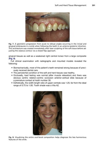
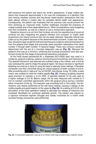
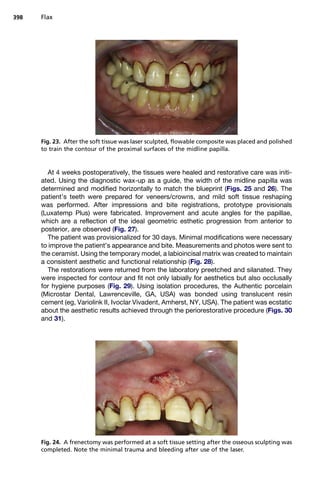
This document discusses the use of lasers in soft and hard tissue management for esthetic dental procedures. It provides background on the history and mechanisms of dental lasers. The erbium laser wavelength is highlighted as it allows conservative, less invasive treatment of both hard tissues like enamel and bone as well as soft tissues. Specific techniques described include laser-assisted cavity preparation and restoration, as well as cosmetic procedures like gingival contouring and osseous crown lengthening to enhance the smile. Case studies demonstrate how these laser techniques can be used to adjust gingival levels and proportions for esthetic outcomes.


![SUMMARY
With expanding emphasis on minimally invasive care (eg, Botox, mesotherapy),
dentistry has as amazing an ally in laser technology. The literature demonstrating
the health benefits is being augmented with the cosmetic advantages as well. Care-
fully using the hard and soft tissue lasers gives cosmetic dentistry a patient friendly
tool to predictably and comfortably compliment the many advances in smile design.
These are truly exciting times for patients and professionals alike!
REFERENCES
1. Coluzzi D. Fundamentals of dental lasers: science and instruments. Dent Clin
North Am 2004;48:750.
2. Van As G. Erbium lasers in dentistry. Dent Clin North Am 2004;48:1018–9, 1028.
3. Wigdor H, Walsh J, Featherstone J, et al. Lasers in dentistry. Lasers Surg Med
1995;16:103–33.
4. Schwartz F, Rothamel D, Becker J, et al. Influence of an Er: YAG laser on the
surface structure of titanium implants. Schweiz Monatsschr Zahnmed 2003;
113(6):660–71 [in French, German].
5. Freiberg RJ, Cozean C. Pulsed erbium laser ablation of hard dental tissue: the
effects of atomized water spray versus water surface film. Lasers in dentistry
VIII. Proc SPIE 2002;4610:74–84.
6. Bornstein E, Lomke MA. The safety and effectiveness of dental Er: YAG lasers:
a literature review with specific reference to bone. Dent Today 2003;22(10):
129–33.
7. Neev J. Modern optics and dentistry. In: Miserdino L, Pick R, editors. Lasers in
dentistry. Carol Stream (IL): Quintessence Publishing Co; 1995. p. 287.
8. Hadley J, Young DA, Eversole LR, et al. Laser powered hydrokinetic system for
caries removal and cavity preparation. J Am Dent Assoc 2000;131:777–85.
9. Glockner K, Rumpler J, Ebeleseder K, et al. Intrapulpal pressure during preparation
with a Er:YAG laser compared to the conventional bur: an in vitro study. Lasers Surg
Med 1999;17:153–7.
10. Hibst R. Lasers for caries removal and cavity preparation: state of the art and
future directions. J Oral Laser Appl 2002;2:203–12.
11. Apel C, Schafer C, Gutknercht N. Demineralization of Er:YAG and ErCr:YSGG
laser-prepared enamel cavities in vitro. Caries Res 2003;37(1):34–7.
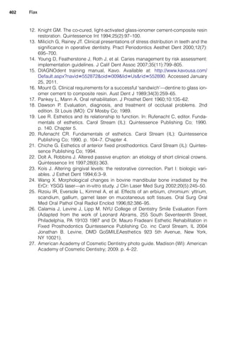
Fig. 31. A beautiful and believe smile created in a nontraditional but predictable manner.
Soft and Hard Tissue Management 401](https://image.slidesharecdn.com/flax2011dental-clinics-of-north-america-161031025737/85/Soft-And-Hard-Tissue-Management-Using-Lasers-19-320.jpg)


![Laser basics[26239].pptx](https://cdn.slidesharecdn.com/ss_thumbnails/laserbasics26239-231026124741-3b010082-thumbnail.jpg?width=640&height=640&fit=bounds)


