This document provides information about the 4th Annual 3D Cell Culture Conference on February 18-20, 2020 in London. The focus day on February 18th will focus on 3D bioprinting and feature speakers from companies like GSK, Merck, Novo Nordisk, and Newcastle University discussing various applications of 3D bioprinting. The main conference on February 19-20th will discuss topics like organ-on-a-chip models, 3D cancer models, 3D infectious disease models, and the adoption of new 3D cell culture technologies. It provides an agenda with over 30 speakers from industry and academia as well as information on registration discounts if signing up before certain dates.
(October 12, 2021) Webinar: Clinical Field MRI As A Measurement Instrument fo...Scintica Instrumentation
Watch our webinar where Professor Marc-Andre Fortin presented about the 3D printing of hydrogels and hydrated substances that have been introduced in various fields of biomedical research including regenerative medicine, cosmetic surgery, orthopedics, and medical physics.
However, one of the main challenges faced by 3D printing and bioprinting is geometrical conformity. In this presentation, studies requiring hydrogel 3D printing in the fields of ophthalmology, regenerative medicine, and medical physics, were described. MRI scanning procedures were developed and optimized for these specific applications.
The presentation highlighted the potential role of MRI in the development of more accurate, more precise 3D-printed hydrogel objects.
(June 28th, 2021) Webinar: Applications Overview Using Low Field MRI To Study...Scintica Instrumentation
About Rutgers Molecular Imaging Center and the Aspect M2
The Rutgers Molecular Imaging Center received the Aspect M2 in the summer of 2012. With the versatility of the M2, a wide range of studies and over 85000 scans have been completed at the center. We have had conventional scans such as cancers, traumatic brain injuries, contrast agents, and other biologicals. The center has also reviewed items such as flow measurements of toothpaste, water filters in action, and pills breaking down during digestion. Please join us as we review examples of the various scans completed here at Rutgers University.
The Rutgers Molecular Imaging Center received the Aspect M2 in 2012. Since the M2’s arrival, it has been one of the most utilized instruments at our center. As of May 1st, 2021, the instrument has seen over 6900 hours of use generating 450 projects/patient files comprising a total of over 85,000 scans. Since the M2 was installed, a combination of 60 departments (over 100 researchers) from multiple universities and pharmaceutical companies have had scans completed at Rutgers. At least ten users from five different departments have been trained over the years to independently run the MRI. This webinar will highlight some of the scans completed here at the center.
One major focus of the center has been the brain and central nervous system. Some examples are the following: Longevity scans of Traumatic Brain Injury have been completed using the Mouse Brain Coil. For another TBI investigator project, leakage into the ventricles post-injury has been tracked with the use of positive contrast agents. Negative contrast agents have been utilized to track stem cells traveling from the lower vertebrae up the spine to points of injury or accumulating around damage in the brain. Changes to the brain following low levels of radiation exposure have been observed using the rat coil. Spontaneous paralysis was reviewed when phenotyping a new mouse strain. A wound healing model tracked the progress of recovery within a spinal injury model.
A second important focus involves cancer models actively scanned at Rutgers. Multiple investigators track the progress of lung cancer and its metastasis. Another investigator tracked cancers in the liver through an interesting negative contrast technique. One Cancer Institute of NJ investigator tracked prostate growth in the same mice for up to two years. Yet another research group tracked an aggressive lymphoma on a weekly basis. Other interesting cancer scans that have been recorded are tumors in the bone, under the tongue, breast duct, uterus, just to name a few of the ones scanned at the center.
(December 2, 2021) The Bench to Bedside Series: Preclinical Cancer Research w...Scintica Instrumentation
Overview:
The goal of this webinar will be to provide a high-level overview of the various stages of preclinical cancer research and discuss the role that innovative instrumentation can play in moving science forward.
To better understand how to treat and control cancer, researchers start by investigating the basics – the cells, molecules, and genes that make up the human body. This type of study, which is often referred to as basic or discovery research, aims to understand the underlying mechanisms contributing to cancer growth and spread. This knowledge is an essential starting point for developing future diagnostic tests and treatment strategies.
After finding an innovative idea that works in cells, researchers need to take their studies to the next level by employing animal models that have similar biology to humans. Animal models have helped scientists make some of the most important cancer discoveries over the years. Furthermore, preclinical imaging technologies allow researchers to perform longitudinal animal studies that are noninvasive leaving the underlying biology intact so that one can track changes throughout the entire disease process.
It was previously thought that the journey from bench to bedside was unidirectional, starting with discovery research and moving towards clinical trials. However, in the last decade, it has become crucial for basic scientists and clinicians to work together towards finding innovative solutions that will positively impact patient care.
After attending this webinar, we hope you will have a better understanding of the preclinical workflow needed to push an idea from bench to bedside as well as some of the key equipment that is needed along the way.
This webinar series will be hosted by Drs. Katie Parkins and Tyler Lalonde, both of which have extensive experience in translational research areas including oncology, neuroscience, molecular imaging, and drug development.
In this webinar we will discuss the following topics:
• Introduction To Cancer Research
• What does “Bench to Bedside” mean?
• In vitro characterization
• Rapid throughput screening
• Quantitative tools
• Moving towards translation
Regular cancer treatment focuses on killing the cancer cells through large doses of medicine, but it also kills other cells in the body and causes significant side effects and potential long-term effects. UCalgary researcher David Cramb – who looks at using nanoparticles to deliver the drugs solely to the tumor in much smaller, more effective quantities shares the potential breakthroughs that can be made possible through nanomedicine both in the treatment and diagnosis of cancer. Watch the full webinar recording: http://www.ucalgary.ca/explore/nanomedicine-new-way-detect-and-treat-cancer
Towards second generation expert systems in telepathology for aid in diagnosisTouradj Ebrahimi
Slides of my invited plenary talk at 10th European Congress on Telepathology and 4th International Congress on Virtual Microscopy, in Vilnius, Lithuania, 1-3 July 2010.
Nano medicine is a field of new research area and this presentation for my research and describe about nano medicines, pros and cons, suggestions to development.
This is the first of a 4-part series introducing Scintica’s newly formed relationship with IVIM Technology and their IntraVital Microscopy platform (IVM).
In this session, we introduced the fundamentals of fluorescence microscopy, review some example images, and focus on this technique's intravital imaging applications. This webinar focused on formulating a basic understanding of the imaging modality to further understand the IVM system's capabilities throughout the rest of the webinar series.
First, the fundamental principles of fluorescence imaging were explained, along with their advantages and challenges with applied in an in vivo setting. Next, we highlighted intravital microscopy's advantages and its role in oncology research and other scientific areas. We also provided an overview of the most commonly used animal models for intravital imaging. Finally, we focused on the importance of acquiring quantitative imaging data and navigate around some pitfalls. Key examples from the research field were collected in this webinar.
After attending this webinar, attendees will have:
a basic understanding of the fundamentals of fluorescence microscopy,
an overview of intravital imaging advantages and applications,
an overview of the most commonly used intravital imaging animal models,
an understanding of what to pay attention to in order to acquire quantitative imaging data.
Data-integration platform for cancer research:cBioPortal demoCORBEL
Participants will be introduced to the data-integration platform cBioPortal. Here, different sources of research data (clinical, imaging, biosample and experimental) of a study are integrated, enabling viewing, querying and analysis.
This webinar is aimed at data managers, researchers, PhD students and postdocs involved in clinical, translational and biomedical research.
Improvements in sequencing technologies have led to a deluge of genomics data in many fields of research. Specifically, the increasing size of cancer-related genomics datasets require comprehensive software solutions that remain accessible to clinical researchers. Clearly, there is an obvious need for tools that integrate genomics and other molecular biology results with the phenotypic and clinical outcome data. During this webinar, the cBio Cancer Genomics Portal (cBioPortal) will be introduced through a practical use case.
The cBioPortal is an open source data integration platform that enables researchers to view, query, analyse and share complex genomic cancer datasets in a user-friendly manner. The platform was originally developed by Memorial Sloan Kettering Cancer Center (New York, USA)1 and is actively maintained and further developed by an international community. The original instance of cBioPortal (http://cbioportal.org) currently provides access to data from almost 83000 tumor samples from 273 public studies.
The demo will include:
· short introduction on the FAIR principles (Findable, Accessible, Interoperable, Reusable)
· navigation through a public study on the data-integration platform cBioPortal
· recreation of select plots from publications of interest using cBioPortal functionalities
The CORBEL webinar series aims to address challenges and share best practice between biological and medical research infrastructures. The series is aimed at technical operators of RIs and is aligned with the CORBEL competency framework.
Building on the sell-out success of the launch event, SMi Group is delighted to announce the return of 3D Cell Culture, taking place on 21st and 22nd of February 2018, in London UK.
3D Cell Culture is rapidly growing with incredible potential for industrial application and a widespread reach that can be seen across many different fields, such as 3D bioprinting and microfluidics.
The 2nd annual conference will explore these overlapping areas and will combine pioneering breakthroughs with scientific research to strengthen your commercial success. Join us for exclusive insight into key topics such as disease models, organoids, organ-on-a-chip technologies, Ipsc advances and CRISPR technology. Notable speakers on the agenda for 2018 will include experts from Aurelia Bioscience, ReInnervate Ltd, Cell and Gene Therapy Catapult, University College London, Novartis Institutes for Biomedical Research, Kugelmeiers, GSK, AstraZeneca, Roche and more!
Tumour models London 1-3 December 2015 AgendaDiane McKenna
Tumour Models London 2015, now in its 4th year,is the leading meeting dedicated to improve preclinical predictability and translational success of oncology discoveries. Tackling clinical failures rates, preclinical strategies and translational challenges, this Summit will enable you to translate your discoveries from model to human studies with superior predictability to future proof clinical success. [Read More…]
5 Best Companies in Digital Pathology Market, March 2023insightscare
latest edition 5 Best Companies in Digital Pathology Market 2023 walks you through the new dawn in diagnostics, promising improved patient outcomes, accelerated research, and heightened accessibility to quality healthcare.
The conference will provide an interactive networking forum to both further develop and answer your queries through a vibrant exhibition room full of technology providers showcasing their technologies and other solutions, poster presentation sessions, expert led case study presentations, a high-level panel discussion, a round table discussion session, and interactive Q&A sessions from a 40-strong speaker faculty examining topics on 4 separate tracks outlined below.
We have 13 research and development projects within:
• Research
• Oncology
• Respiratory, Inflammation and Autoimmunity
• Cardiovascular and Metabolic Disease
• Antibody Discovery and Protein Engineering
• Pathology
• Biopharmaceutical Development
• Cell Culture and Fermentation Sciences
• Formulation Sciences
• Analytical Biotechnology Science
- Discover new methods for managing clinical next-gen data with insights from Pfizer, Boston Children’s Hospital and AstraZeneca
- Uncover and critique the latest technologies out there for you to use in clinical trials. Mayo Clinic, Merck and Harvard Medical School let you into their trade secrets
- Hear the genomics strategies that Roche, Millennium and Regeneron are using for discovery and validation of clinically actionable biomarkers
-Bristol-Myers Squibb, Takeda and Partners Healthcare the role that NGS can play when implementing an effective strategy in the lab to speed up CDx development
- Learn how to integrate molecular details into medical decision making, with fresh data from Washington University School of Medicine and Genzyme
Honest Reviews of Tim Han LMA Course Program.pptxtimhan337
Personal development courses are widely available today, with each one promising life-changing outcomes. Tim Han’s Life Mastery Achievers (LMA) Course has drawn a lot of interest. In addition to offering my frank assessment of Success Insider’s LMA Course, this piece examines the course’s effects via a variety of Tim Han LMA course reviews and Success Insider comments.
Normal Labour/ Stages of Labour/ Mechanism of LabourWasim Ak
Normal labor is also termed spontaneous labor, defined as the natural physiological process through which the fetus, placenta, and membranes are expelled from the uterus through the birth canal at term (37 to 42 weeks
How to Make a Field invisible in Odoo 17Celine George
It is possible to hide or invisible some fields in odoo. Commonly using “invisible” attribute in the field definition to invisible the fields. This slide will show how to make a field invisible in odoo 17.
Read| The latest issue of The Challenger is here! We are thrilled to announce that our school paper has qualified for the NATIONAL SCHOOLS PRESS CONFERENCE (NSPC) 2024. Thank you for your unwavering support and trust. Dive into the stories that made us stand out!
The French Revolution, which began in 1789, was a period of radical social and political upheaval in France. It marked the decline of absolute monarchies, the rise of secular and democratic republics, and the eventual rise of Napoleon Bonaparte. This revolutionary period is crucial in understanding the transition from feudalism to modernity in Europe.
For more information, visit-www.vavaclasses.com
Introduction to AI for Nonprofits with Tapp NetworkTechSoup
Dive into the world of AI! Experts Jon Hill and Tareq Monaur will guide you through AI's role in enhancing nonprofit websites and basic marketing strategies, making it easy to understand and apply.
Unit 8 - Information and Communication Technology (Paper I).pdfThiyagu K
This slides describes the basic concepts of ICT, basics of Email, Emerging Technology and Digital Initiatives in Education. This presentations aligns with the UGC Paper I syllabus.
Operation “Blue Star” is the only event in the history of Independent India where the state went into war with its own people. Even after about 40 years it is not clear if it was culmination of states anger over people of the region, a political game of power or start of dictatorial chapter in the democratic setup.
The people of Punjab felt alienated from main stream due to denial of their just demands during a long democratic struggle since independence. As it happen all over the word, it led to militant struggle with great loss of lives of military, police and civilian personnel. Killing of Indira Gandhi and massacre of innocent Sikhs in Delhi and other India cities was also associated with this movement.
Embracing GenAI - A Strategic ImperativePeter Windle
Artificial Intelligence (AI) technologies such as Generative AI, Image Generators and Large Language Models have had a dramatic impact on teaching, learning and assessment over the past 18 months. The most immediate threat AI posed was to Academic Integrity with Higher Education Institutes (HEIs) focusing their efforts on combating the use of GenAI in assessment. Guidelines were developed for staff and students, policies put in place too. Innovative educators have forged paths in the use of Generative AI for teaching, learning and assessments leading to pockets of transformation springing up across HEIs, often with little or no top-down guidance, support or direction.
This Gasta posits a strategic approach to integrating AI into HEIs to prepare staff, students and the curriculum for an evolving world and workplace. We will highlight the advantages of working with these technologies beyond the realm of teaching, learning and assessment by considering prompt engineering skills, industry impact, curriculum changes, and the need for staff upskilling. In contrast, not engaging strategically with Generative AI poses risks, including falling behind peers, missed opportunities and failing to ensure our graduates remain employable. The rapid evolution of AI technologies necessitates a proactive and strategic approach if we are to remain relevant.
Safalta Digital marketing institute in Noida, provide complete applications that encompass a huge range of virtual advertising and marketing additives, which includes search engine optimization, virtual communication advertising, pay-per-click on marketing, content material advertising, internet analytics, and greater. These university courses are designed for students who possess a comprehensive understanding of virtual marketing strategies and attributes.Safalta Digital Marketing Institute in Noida is a first choice for young individuals or students who are looking to start their careers in the field of digital advertising. The institute gives specialized courses designed and certification.
for beginners, providing thorough training in areas such as SEO, digital communication marketing, and PPC training in Noida. After finishing the program, students receive the certifications recognised by top different universitie, setting a strong foundation for a successful career in digital marketing.
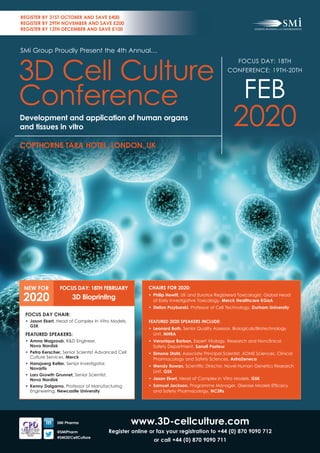
1. www.3D-cellculture.com
register online or fax your registration to +44 (0) 870 9090 712
or call +44 (0) 870 9090 711
SMi Pharma
@SMiPharm
#SMi3DCellCulture
CHAIrS for 2020:
• Philip Hewitt, UK and Eurotox Registered Toxicologist, Global Head
of Early Investigative Toxicology, Merck Healthcare KGaA
• Stefan Przyborski, Professor of Cell Technology, Durham university
feAtureD 2020 SPeAKerS InCluDe:
• Leonard Both, Senior Quality Assessor, Biologicals/Biotechnology
Unit, MHrA
• Veronique Barban, Expert Virology, Research and Nonclinical
Safety Department, Sanofi Pasteur
• Simone Stahl, Associate Principal Scientist, ADME Sciences, Clinical
Pharmacology and Safety Sciences, AstraZeneca
• Wendy Rowan, Scientifi c Director, Novel Human Genetics Research
Unit, GSK
• Jason Ekert, Head of Complex In Vitro Models, GSK
• Samuel Jackson, Programme Manager, Disease Models Effi cacy
and Safety Pharmacology, nC3rs
FOCUS DAY: 18TH
CONFERENCE: 19TH-20TH
FEB
2020
COPTHORNE TARA HOTEL, LONDON, UK
SMi Group Proudly Present the 4th Annual…
3D Cell Culture
Conference
Development and application of human organs
and tissues in vitro
REGISTER BY 31ST OCTOBER AND SAVE £400
REGISTER BY 29TH NOVEMBER AND SAVE £200
REGISTER BY 13TH DECEMBER AND SAVE £100
FOCUS DAY CHAIR:
• Jason Ekert, Head of Complex In Vitro Models,
GSK
feAtureD SPeAKerS:
• Amna Magzoub, R&D Engineer,
Novo Nordisk
• Petra Kerscher, Senior Scientist Advanced Cell
Culture Services, Merck
• Hansjoerg Keller, Senior Investigator,
novartis
• Lars Growth Grunnet, Senior Scientist,
Novo Nordisk
• Kenny Dalgarno, Professor of Manufacturing
Engineering, newcastle university
NEW FOR
2020
FOCUS DAY: 18TH FEBRUARY
3D Bioprinting
2. 3D Cell Culture ConferenCe
focus Day | Tuesday 18th February 2020 www.3D-cellculture.com
3D Bioprinting
Copthorne Tara Hotel, London, UK
Chaired by Jason Ekert, Head of Complex In Vitro Models, GSK
08.30 registration & Coffee
09.00 Chair’s Opening Remarks
Jason Ekert, Head Complex In Vitro Models, GSK
oPenInG ADDreSS
09.10 First Applications of 3D Bioprinted Tissue Models for
Drug Screening
• Use of 3D bioprinting in Pharma industry
• Validation of printed tissue models
• The importance of biomaterials for 3D bioprinting
• Outlook of 3D bioprinting for drug screening applications
Petra Kerscher, Senior Scientist, Merck KGaA
09.50 Designing 3D Scaffolds for Stem Cell Therapy
• Proof of principle using INS-1E and NN stem-cell derived Beta
Cells
• Modeling the heart with stem-cell derived CMs.
• Other aspirations for target diseases: Stroke, AMD, etc
Amna Magzoub, R&D Engineer, Novo Nordisk A/S
Lars Growth Grunnet, Senior Scientist, Novo Nordisk
10.30 Morning Coffee
11.00 3D Printing of Medicines: A Digital Pharmacy Era
• An introduction on the need for personalised medicines
• Discussion on the clinical and wider healthcare impact of 3D
printing
• Explanation of the various 3D printing technologies, and their
benefi ts / drawbacks for medicine production
• Discussion on the current challenges to the clinical integration of
3D printing
Sarah Trenfield, Director of Innovation, FabRx Ltd
11.40 3D Bioprinted Human Skeletal Muscle Models for In Vitro
Physiological Drug Screening
• In vitro human microphysiological assays boost drug
development
• 3D bioprinting enables the fabrication of complex human tissue
in vitro models for drug discovery
• 3D bioprinted human skeletal muscle models mimic
pharmacological regulation of muscle contractile force
• 3D bioprinted contractile human skeletal muscle models allow
functional screening of test compounds
Hansjoerg Keller, Senior Investigator I, Musculoskeletal Disease
Area, novartis
13.20 networking lunch
PAnel DISCuSSIon
13.20 The use of bioprinting for screening
• Opportunities for bioprinted organoids for screening purposes
• Challenges in compatibility of HTS for 3D models
• How we can improve 3D modelling for effi cacy testing and
further applications
Panel Moderator: Jason Ekert, Head Complex In Vitro Models, GSK
Panelists: Petra Kerscher, Senior Scientist, Merck
Hansjoerg Keller, Senior Investigator I, novartis
14.00 Reactive Jet Impingement: A New 3D Printing Process for High Cell
Density Gels
• Introduction to the reactive jet impingement process
• Outline value of high cell density gels
• Case studies on printing musculoskeletal micro-tissues
Kenny Dalgarno, Professor of Manufacturing Engineering,
newcastle university
14.40 Afternoon tea
15.10 3D Extracellular matrix scaffolds and hydrogels for target discovery
and drug profiling
• The ECM exhibits an important functional role in the control of
key cellular events
• The contribution of ECM is often overlooked in target and drug
discovery efforts
• Engitix has developed unique technologies allowing the
development of human tissue-specifi c and disease-specifi c ECM
biomaterials for target discovery and drug profi ling purposes
Gino Van Heeke, CSO, engitix
15.50 3D Bioprinting Engineering Artificial Respiratory Tract Tissue
• Ethos and background of why the need to develop and improve
upon the current gold-standard in vitro models of the respiratory
epithelium exists
• The primary research aim of this project is to create the fi rst
bioprinted, multi-cellular, 3D model of the upper respiratory
mucosa; with various collaborations involved in the project
• Data outlining key stages towards a new ALI culture
methodology involving in vitro primary human bronchial epithelial
cell culture on collagen layers
• Hydrogels used for extended culture and differentiation of
primary HBECs at an air-liquid interface
• Preliminary bioprinting of primary human lung fi broblasts
Naheem Yaqub, PhD Candidate, University College London in
Collaboration with GSK
16.30 Chair’s Closing Remarks and Close of Focus Day
register online at www.3D-cellculture.com
focus Day overview
Bioprinting has now become an effi cient and accurate method to build in vitro
tissue models with the potential to provide pathologically relevant responses
and thus model human disease mechanisms. The clinical applications of
3D printing are rapidly moving from the research to production phases and
will certainly continue to grow, with ever increasing numbers of therapies
becoming commercialized.
TOPICS COVERED WILL INCLUDE:
• Learn how leaders in big Pharma are incorporating bioprinting into cell
culture research
• Explore novel applications of 3D bioprinting for in vitro models and
regenerative medicine
• Network with the leading pharmaceutical and biotech companies
developing the use of bioprinting with the pharma industry
3. 3D Cell Culture Conference
Day One | Wednesday 19th February 2020 www.3D-cellculture.com
08.30 Registration & Coffee
09.00 Chairs’ Opening Remarks
Stefan Przyborski, Professor of Cell Technology, University of Durham
Philip Hewitt, UK and Eutrotox Registered Toxicologist, Head of Early
Investigative Technology, Merck
CO-CHAIR OPENING ADDRESS
09.10 The Development and Application of 3D Culture
Techniques to Construct Models of Human Tissues
• Overview of 3D cell culture technologies
• Impact and importance of 3D cell culture
• Beyond 3D cell culture as a technique, applications to building
human tissues in vitro
• New innovations and developments to further enhance 3D
technology
Stefan Przyborski, Professor of Cell Technology, University of Durham
09.50 Organ-on-a-chip: Can we finally replace animals in
pharmaceutical research
• Development of human, translatable in vitro MPS models
• Status of OOAC field; current models and industry “validation”
• Context of use: focus on safety testing
• Future: can we replace animals or even clinical trials?
Philip Hewitt, UK and Eutrotox Registered Toxicologist, Head of Early
Investigative Technology, Merck
10.30 Morning Coffee
INNOVATIONS IN 3D CELL CULTURE MODELS
11.00 Complex In Vitro Models for preclinical Oncology drug
development
• Characterization and validation criteria for Oncology models
• Case studies -
- Avascular and vascular microfluidic tumor models
- Tumor Organoids for combination drug screening
• Future direction
Jason Ekert, Head of Complex In Vitro Models, GSK
11.40 3D-models for infectious diseases and vaccines
• Potential and Limits of 3D-models for industrial development of
human vaccines
• Case study: 2D and 3D liver models for yellow fever virus
viscerotropism assessment
Veronique Barban, Expert Virology, Research and Nonclinical
Safety Department, Sanofi Pasteur
12.20 Networking Lunch
APPLICATIONS OF TECHNOLOGY AND CASE STUDIES
PANEL DISCUSSION
13.20 Adopting new technologies for 3D models
• An outlook of new and developing technologies for 3D cell
culture models
• Challenges and barriers in the adoption of new technology
• What is required to make a decision on adoption and what is
needed to promote adoption in industry?
Panel Moderator: Stefan Przyborski, Professor of Cell Technology,
University of Durham
Panelists: Philip Hewitt, UK and Eutrotox Registered Toxicologist,
Head of Early Investigative Technology, Merck
Floriane Groell, Senior Scientist, Novartis Pharma AG
Samuel Jackson, Programme Manager, Disease Models Efficacy
and Safety Pharmacology, NC3Rs
14.00 Printing 3D Cell Cultures in Osteoarthritis Research
• Outline the use of bioprinting in two major projects which focus
on osteoarthritis:
- Tissue Engineering Regenerative Therapies Centre Versus
Arthritis
- CRACK-IT Challenge Project: Printing Osteoarthritis on a Chip
• Outline the printing approaches taken, and approaches to
(i) integration with downstream processes, and (ii) scale-up
Kenny Dalgarno, Professor of Manufacturing Engineering,
Newcastle University
14.40 Afternoon Tea
15.10 C-Stem® technology: 3D microfluidics enabled scale up of culture
and differentiation of pluripotent stem cells organoids
• In vivo like environment allows recapitulation of correct PSC
topology
• Strongly diminished cell death contributes to increased yields
• Protection from bioreactor mechanical stress allows scale up of
PSC culture in standard liquid bioreactors
• Differentiation of PSC can be achieved within C-Stem™ by
leveraging 2D protocols
Maxime Feyeux, Co-Founder, President, CSO, TreeFrog Therapeutics
15.50 Microfluidic technologies for precision medicine in oncology
• Maximising screening of patient tissue
• Physiological tumour models
• Microfluidic technologies
• Precision medicine
Michele Zagnoni, Reader, University of Strathclyde
16.30 Chairs’ Closing Remarks and Close of Day One
Alternatively fax your registration to +44 (0)870 9090 712 or call +44 (0)870 9090 711
4. 3D Cell Culture Conference
www.3D-cellculture.com Day Two | Thursday 20th February 2020
08.30 Registration Coffee
09.00 Chairs’ Opening Remarks
Stefan Przyborski, Professor of Cell Technology, University of Durham
Philip Hewitt, UK and Eutrotox Registered Toxicologist, Head of Early
Investigative Technology, Merck
A Future Outlook of 3D Cell Culture
OPENING ADDRESS
09.10 Regulatory outlook on 3D Cell Culture Models
• An overview of the applicable regulatory guidelines
• Validating new technology, relevant quality considerations and
regulatory processes
• Challenges for the traditional regulatory framework
Leonard Both, Senior Quality Assessor, Biologicals/Biotechnology
Unit, MHRA
09.50 Addressing unmet needs in 3D cell systems to drive replacement of
animal modelling
• Much progress has been made in 3D cell model development,
to the point that many sub-organ level systems can now be
effectively recapitulated in vitro
• However, there are several key areas which have yet to be
effectively tackled, such as the immune system and tissue
vascularization and innervation
• Given that these systems or structures are implicated in most
physiological processes and many disease states, making
progress on these difficult to tackle topics is key to the future
success of 3D modelling approaches
• By addressing these areas of unmet need, animal modelling
paradigms will increasingly be replaced or augmented with
human cell-based systems
Samuel Jackson, Programme Manager, Disease Models Efficacy
and Safety Pharmacology, NC3Rs
10.30 Morning Coffee
11.00 Reducing attrition in drug discovery through the use of human
translational cellular models
• The challenge in drug discovery to reduce clinical attrition
• The new generation of human translational models for use in drug
discovery
• The application and challenges in application of human
translational models for drug discovery
• Looking to the future
Wendy Rowan, Scientific Director, Novel Human Genetics Research
Unit, GSK
INNOVATIONS IN 3D CELL CULTURE MODELS
11.40 Application of 3D models to ADME research
• Importance of physiological features in 3D models for ADME
research with a focus on drug transporters
• Examples of current landscape of ADME 3D models and their
applications
• Transporter characterisation of renal 3D models such as
organoids or microfluidic systems
• Outlook of how ADME 3D models can be applied to drug
projects
Simone Stahl, Associate Principal Scientist, ADME Sciences, Clinical
Pharmacology and Safety Sciences, AstraZeneca
12.20 Networking Lunch
13.20 3D cell culture model to mimic the human subcutaneous tissue
• Development of a 3D cell culture model of the human
subcutaneous tissue, allowing the immunogenicity prediction of
subcutaneously injected therapeutic proteins
• Use of hydrogels as a scaffold to mimic the visco-elastic
properties of the human subcutaneous tissue in vitro.
Measurement of hydrogels elastic Young’s moduli and
comparison with ex vivo human samples
• Investigation of the hydrogels cytocompatibility with a human
myelomonocytic cell line (MUTZ-3) and their influence on cellular
phenotype changes
Floriane Groell, (Research Associate of the) Biopharmaceutical
Sciences department, University of Geneva
3D IMAGING AND HIGH THROUGHPUT SCREENING
14.00 Visualising the unexpected
• Advanced 3D imaging can lead to unexpected scientific
discoveries
• Imaging developments and application in advanced 3D culture
systems
• Understanding the efficacy and tumor targeting behavior of
immune cell therapy through 3D imaging
• Future perspective
Ellen J. Wehrens, Scientific Writer, Imaging Department, Princess
Máxima Center for Pediatric Oncology
14.40 Afternoon Tea
15.10 Development of a high throughput and drug responsive 3D model
of white adipose tissue
• Conception and optimization of a novel biofabrication workflow
for the development of 3D tissue models i.e. the bioprinting and
dispensing of matrix-based cellular constructs under oil prior to
media transfer
• A joint collaboration between OxSyBio and MRC Harwell led to
the development of a 3D white adipose tissue (WAT) model for
mechanistic studies of metabolic diseases
• Extensive characterization of the adipogenic spheroid (i.e. the 3D
WAT) revealed the model was reproducible, highly differentiated,
had active lipid metabolism and was drug-responsive
• Establishment of high throughput monitoring of drug-effects
on 3D WAT models on a 96-well scale. Specifically, using high
content-imaging of lipid phenotype and time-course metabolite
measurements
Rajesh Pandey, Postdoctoral Training Fellow, MRC Harwell Institute
Alexander D. Graham, Investigator Scientist, MRC Harwell Institute
15.50 Co-Chairs’ Closing Remarks and Close of Day Two
Register online at www.3D-cellculture.com
5. Geo Breakdown - Past Attendees:
3D Cell Culture ConferenCe
Focus Day: 18th February 2020 | Conference: 19th-20th February 2020 www.3D-cellculture.com
MArKetInG PArtnerSHIP oPPortunItIeS
SMi Group is offering companies the opportunity to partner on our dedicated events in order to help raise your company profile,
add value, create awareness of your products/services to our key audience within the pharmaceutical industry. Interested in partnering?
Contact Simi Sapal, SMi Marketing on +44 (0) 207 827 6162 or email: ssapal@smi-online.co.uk
SPONSORSHIP AND EXHIBITION OPPORTUNITIES
SMi offer sponsorship, exhibition, advertising and branding packages, uniquely tailored to complement your company’s marketing strategy. Prime
networking opportunities exist to entertain, enhance and expand your client base within the context of an independent discussion specific to your industry.
Should you wish to jointhe increasing number of companies benefiting from sponsoring our conferences please call:
Alia Malick on +44 (0) 20 7827 6168 or email: amalick@smi-online.co.uk
register online at www.3D-cellculture.com
NOVEMBER 2019
Ophthalmic Drugs
18th - 19th November 2019, London, UK
DECEMBER 2019
Respiratory Drug Delivery
5th - 6th December 2019, London, UK
JANUARY 2020
Pre-Filled Syringes UK
15th - 16th January 2020, London, UK
Pharmaceutical Microbiology UK
20th - 21st January 2020, London, UK
Disruptive Technologies in Pharma
20th - 21st January 2020, London, UK
FEBRUARY 2020
Parallel Trade
4th - 5th February 2020, London, UK
3D Cell Culture
19th - 20th February 2020, London, UK
RNAi Therapeutics
19th - 20th February 2020, London, UK
MArCH 2020
Superbugs Superdrugs 2020
16th - 17th March 2020, London, UK
Artificial Intelligence in Drug Discovery
16th - 17th March 2020, London, UK
APrIl 2020
Pre-Filled Syringes East Coast
27th - 28th April 2020, Boston, USA
Pharmaceutical Microbiology East Coast
29th - 30th April 2020, Boston, USA
MAY 2020
Pain Therapeutics
11th - 12th May 2020, London, UK
Highly Potent Active Pharmaceutical
Ingredients
11th - 12th May 2020, London, UK
Injectable Drug Delivery
13th - 14th May 2020, London, UK
JUNE 2020
Aseptic Processing
3rd - 4th June 2020, London, UK
Pre-Filled Syringes West Coast
15th - 16th June 2020, San Diego, USA
Pharmaceutical Microbiology West Coast
18th - 19th June 2020, San Diego, USA
SMi PHARMACEUTICAL EVENT PLANNER 2019-2020
Official Media Partners: Supported by:
Who Should Attend:
• Biotech / Pharma Companies - Team Leader, Junior Principle investigator, Cell and
Molecular Scientist, Chief Scientifi c Offi cer, Deputy Director, Manager, Founder, CEO,
Head, Director, Programme Manager, Lab Head, Toxicologist
• Academia Professor - Cell Technology, Stem Cell Sciences, Biochemistry, Protein
Technology and Tissue Engineering
• Solution Providers - Manager, Head of, CEO, Director, Founder, Account Manager,
President, CSO, Executive, Managing Director, Vice President, COO
• Organisations / Regulatory - Director, Senior Science Advisor, Scientifi c Offi cer
Why to Attend:
• leArn from leading pharma companies to benchmark
against their applications of 3D technology
• eXPlore case studies of 3D cell culture, high throughput
screening, imaging and microphysiological systems in
industry
• HeAr about the latest technology advances allowing
3D bioprinting to revolutionize in vitro models
• NETWORK with the key industry players developing the
use of bioprinting as part of their RD
• unDerStAnD novel applications of 3D bioprinting for in
vitro models, regenerative medicine and screeningSnap Shot of Past Attendees:
• Adaptimmune Ltd
• Aptuit (Verona)
• Asterand Bioscience
• AstraZeneca
• Aurelia Bioscience
• Barts Cancer Institute
• Benevolent AI
• Biogelx Ltd
• Bio-Techne
• BioTek Instruments
• Boehringer-Ingelheim
• Cell and Gene Therapy
Catapult
• Cellesce Ltd
• Cellon
• Cellular Dynamics
International
• Corning
• Enplas Europe
• F. Hoffmann-La Roche
• GE Healthcare
• GSK
• Hubrecht Institute
• Idorsia Pharmaceuticals
• Immunocore Ltd
• Integra Biosciences
• Jellagen
• JSR Life Sciences
• Kugelmeiers
• Lonza
• Medical Research
Council
• MedImmune
• MHRA
• Microduits
• Neem Biotech
• Nexcelom Bioscience
• Novartis Institutes For
Biomedical Research
• Novartis Pharmaceuticals
• Pfi zer
• Plasticell
• Poietis
• Progen Biotechnik
• Public Health England
• Roche Diagnostics
• Sanofi
• Sigma Aldrich
• Stemtek Therapeutics
• Tebu-Bio
• The Francis Crick
Institute
• Thermo Fisher Scientifi c
• TissUse
• UK Regenerative
Medicine Platform
• Unilever Research
Colworth
• Ventana Medical
System
InfoGrAPHICS
Asia
2%
USA
North
America
4%
UK
53%
Europe
41%
6. 3D Cell Culture Conference 2020
Copthorne Tara Hotel, London, UK
Conference: 19th - 20th February 2020
Focus Day: 18th February 2020
4 WAYS TO REGISTER
If you have any further queries please call the Events Team on tel +44 (0) 870 9090 711 or you can email them at events@smi-online.co.uk
Unique Reference Number
Our Reference P-310
Documentation
I cannot attend but would like to Purchase access to the following Document Portal/
Paper Copy documentation. Price Total
□ Access to the conference documentation
on the Document Portal £499.00 + VAT £598.80
□ The Conference Presentations – paper copy £499.00 - £499.00
(or only £300 if ordered with the Document Portal)
Payment must be made to SMi Group Ltd, and received before the event, by one of
the following methods quoting reference P-310 and the delegate’s name. Bookings
made within 7 days of the event require payment on booking, methods of payment:
□ UK BACS Sort Code 300009, Account 00936418
□ Wire Transfer Lloyds TSB Bank plc, 39 Threadneedle Street, London, EC2R 8AU
Swift (BIC): LOYDGB21013, Account 00936418
IBAN GB48 LOYD 3000 0900 9364 18
□ Cheque We can only accept Sterling cheques drawn on a UK bank.
□ Credit Card □ Visa □ MasterCard □ American Express
SMi Group will apply surcharges to commercial cards
Please tick here □ if the card provided is not a commercial card
Card No: □□□□ □□□□ □□□□ □□□□
Valid From □□/□□ Expiry Date □□/□□
CVV Number □□□□ 3 digit security on reverse of card, 4 digits for AMEX card
Cardholder’s Name:
Signature: Date:
I agree to be bound by SMi’s Terms and Conditions of Booking.
Card Billing Address (If different from above):
PAYMENT
vat
VAT at 20% is charged on the attendance fees for all delegates. VAT is also charged
on Document portal and literature distribution for all UK customers and for those EU
Customers not supplying a registration number for their own country here
CONFERENCE Prices GROUP DISCOUNTS AVAILABLE
I would like to attend: (Please tick as appropriate) Fee TOTAL
□ Conference and Focus Day £2398.00 + VAT £2877.60
□ Conference only £1499.00 + VAT £1798.80
□ Focus Day only £899.00 + VAT £1078.80
PROMOTIONAL LITERATURE DISTRIBUTION
□ Distribution of your company’s promotional
literature to all conference attendees £999.00 + VAT £1198.80
The conference fee includes refreshments, lunch, conference papers, and access
to the Document Portal. Presentations that are available for download will be
subject to distribution rights by speakers. Please note that some presentations may
not be available for download. Access information for the document portal will be
sent to the e-mail address provided during registration. Details are sent within 24
hours post conference.
□ Book by 31st October to receive £400 off the conference price
□ Book by 29th November to receive £200 off the conference price
□ Book by 13th December to receive £100 off the conference price
EARLY BIRD
DISCOUNT
Please complete fully and clearly in capital letters. Please photocopy for additional delegates.
Title: Forename:
Surname:
Job Title:
Department/Division:
Company/Organisation:
Email:
If you would like to continue to receive email updates about our
events, please tick □
Company VAT Number:
Address:
Town/City:
Post/Zip Code: Country:
Direct Tel: Direct Fax:
Mobile:
Switchboard:
Signature: Date:
I agree to be bound by SMi’s Terms and Conditions of Booking.
ACCOUNTS DEPT
Title: Forename:
Surname:
Email:
Address (if different from above):
Town/City:
Post/Zip Code: Country:
Direct Tel: Direct Fax:
Payment: If payment is not made at the time of booking, then an invoice will be issued and must
be paid immediately and prior to the start of the event. If payment has not been received then
credit card details will be requested and payment taken before entry to the event. Bookings within
7 days of event require payment on booking. Access to the Document Portal will not be given until
payment has been received.
Substitutions/Name Changes: If you are unable to attend you may nominate, in writing, another
delegate to take your place at any time prior to the start of the event. Two or more delegates may
not ‘share’ a place at an event. Please make separate bookings for each delegate.
Cancellation: If you wish to cancel your attendance at an event and you are unable to send
a substitute, then we will refund/credit 50% of the due fee less a £50 administration charge,
providing that cancellation is made in writing and received at least 28 days prior to the start of the
event. Regretfully cancellation after this time cannot be accepted. We will however provide the
conferences documentation via the Document Portal to any delegate who has paid but is unable
to attend for any reason. Due to the interactive nature of the Briefings we are not normally able to
provide documentation in these circumstances. We cannot accept cancellations of orders placed
for Documentation or the Document Portal as these are reproduced specifically to order. If we
have to cancel the event for any reason, then we will make a full refund immediately, but disclaim
any further liability.
Alterations: It may become necessary for us to make alterations to the content, speakers, timing,
venue or date of the event compared to the advertised programme.
Privacy policy / Opt Out: For full details on our privacy policy please go to
http://smi-online.co.uk/privacy-legals/privacy-policy. If you no longer wish to receive email
updates you can opt out by going to the following webpage http://www.smi-online.co.uk/opt-out
Terms and Conditions of Booking
Delegate Details
venue Copthorne Tara Hotel, London, UK
□ Please contact me to book my hotel
Alternatively call us on +44 (0) 870 9090 711,
email: events@smi-online.co.uk or fax +44 (0) 870 9090 712
1 Online at www.3D-cellculture.com
2 FAX your booking form to +44 (0) 870 9090 712
3 PHONE on +44 (0) 870 9090 711
4 POST your booking form to: Events Team, SMi Group Ltd,
India House, 45 Curlew Street, London SE1 2ND