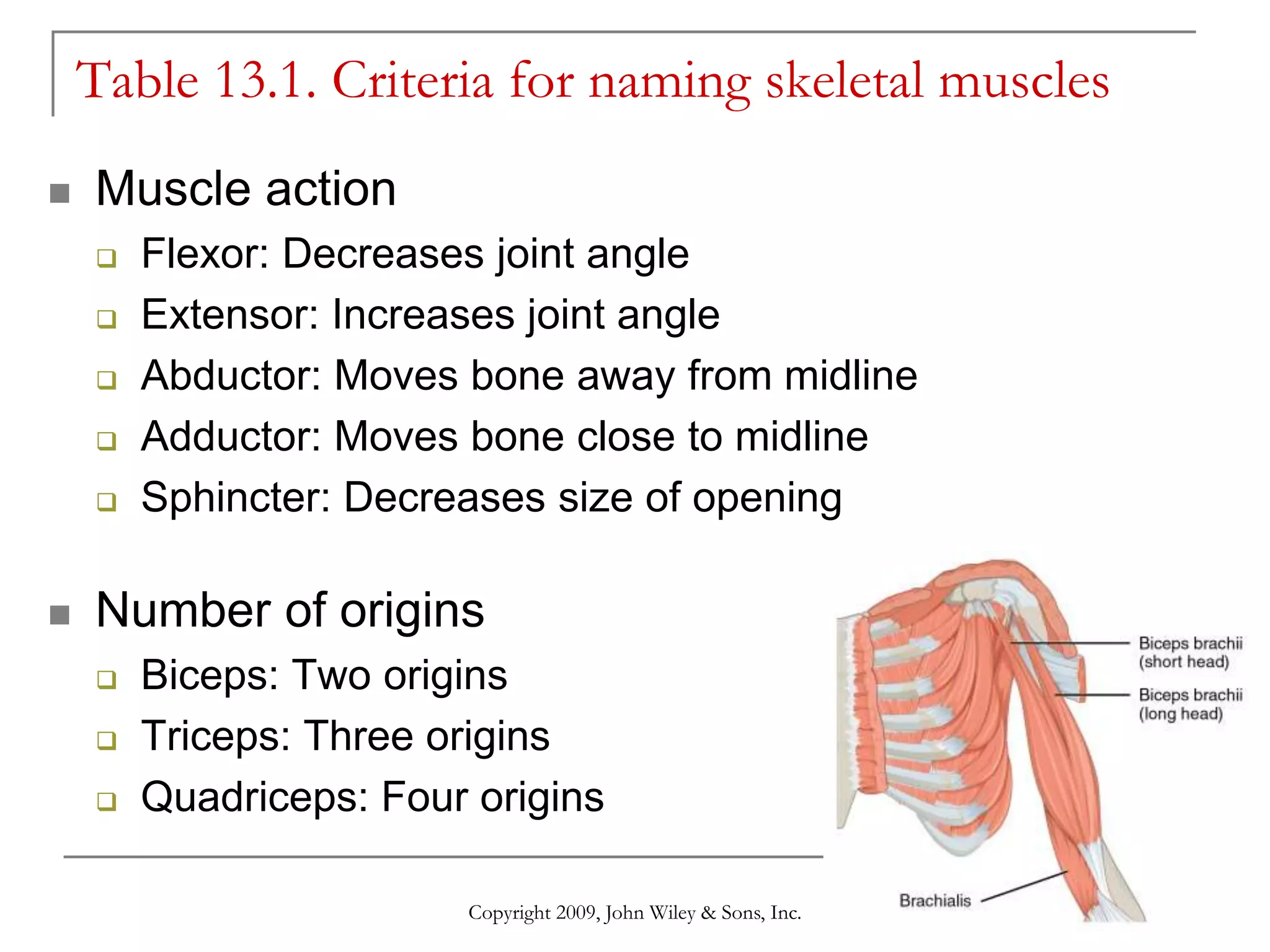
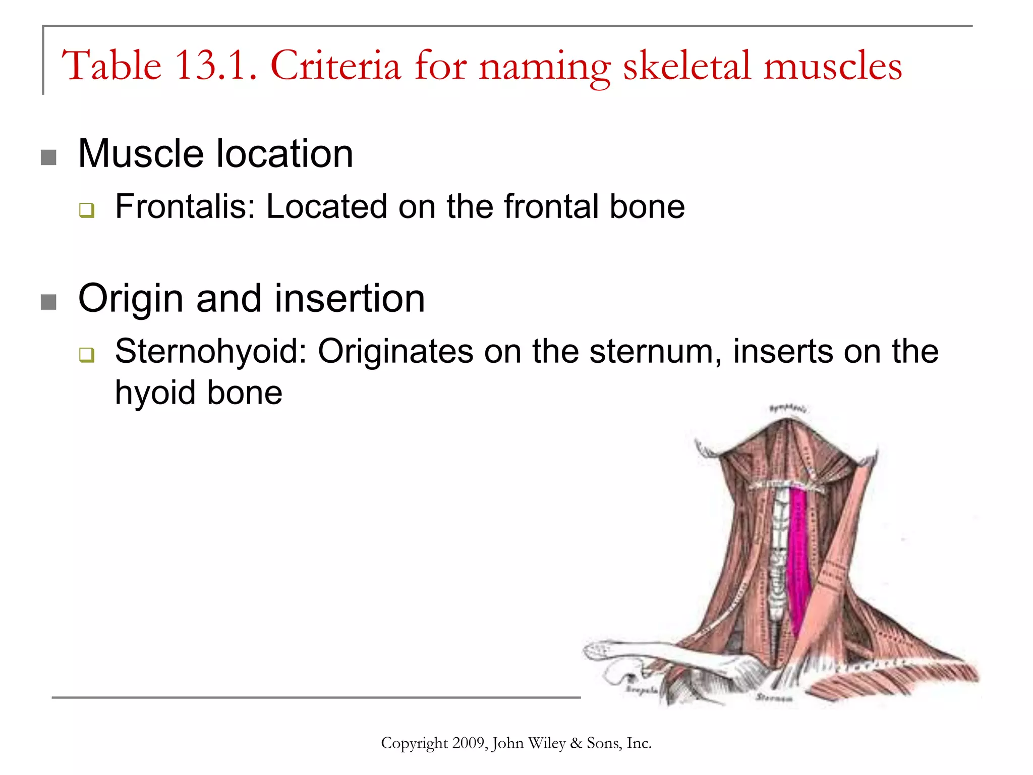
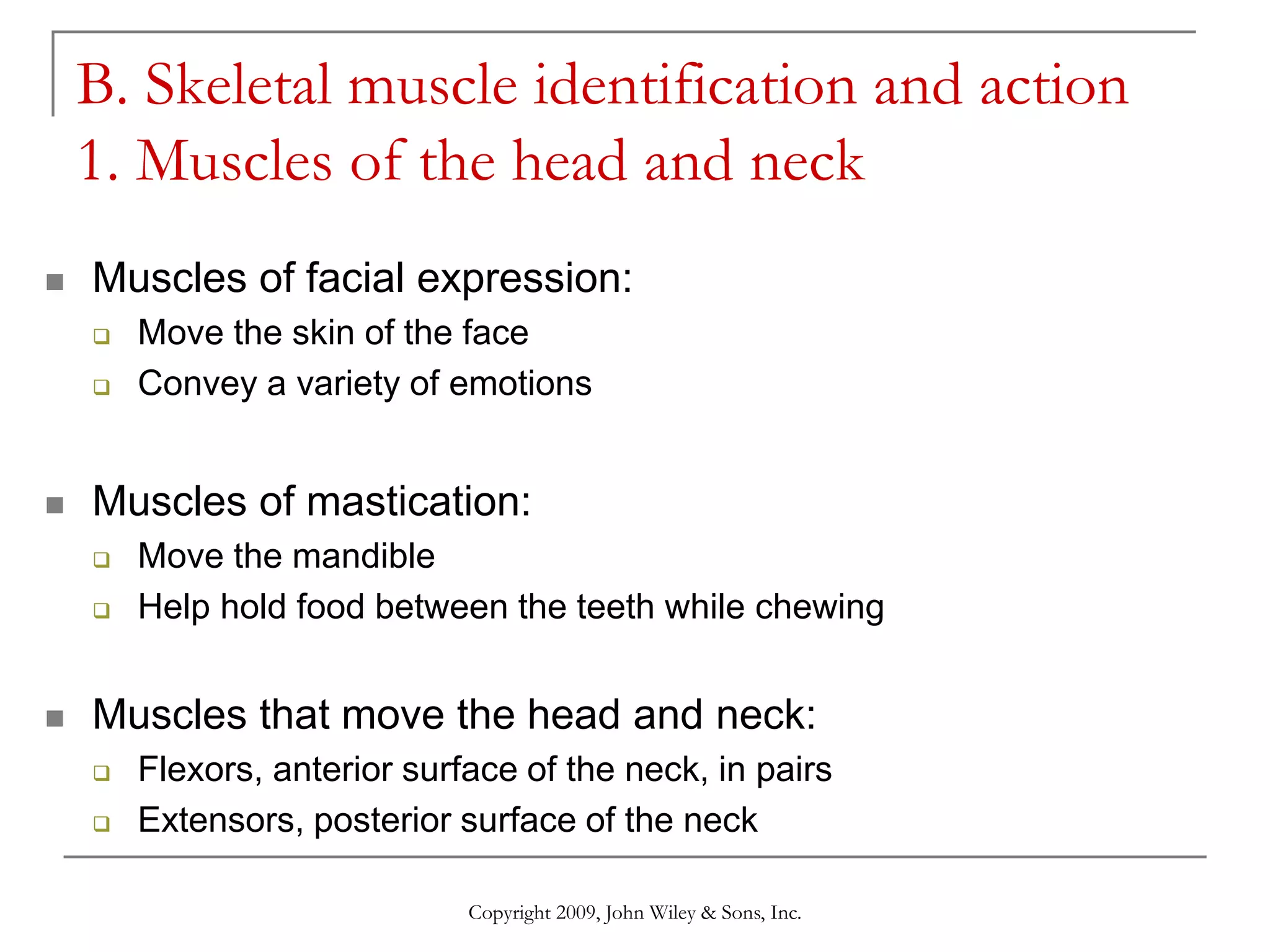
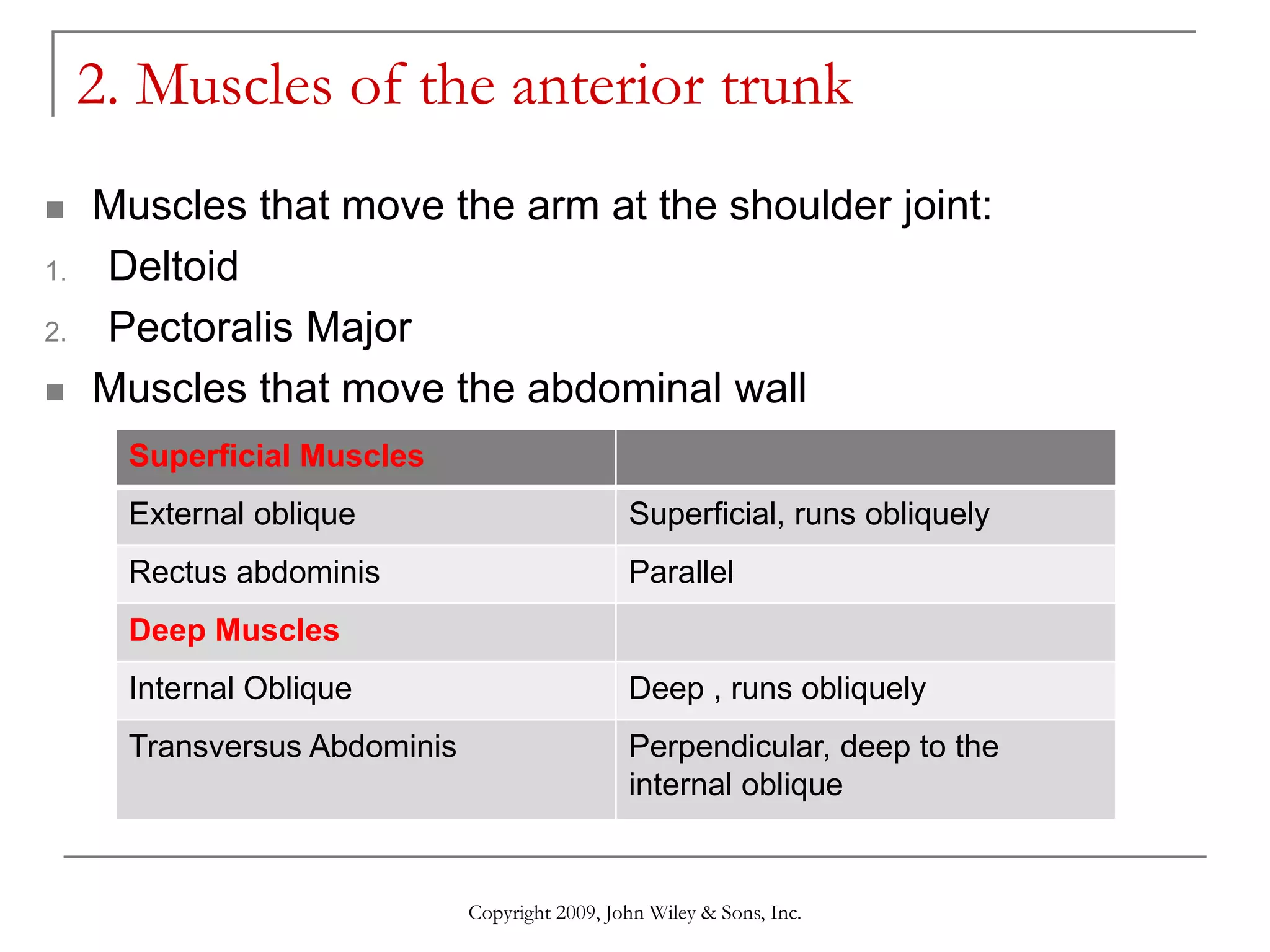
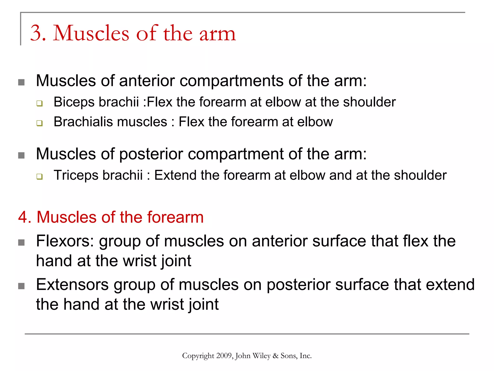
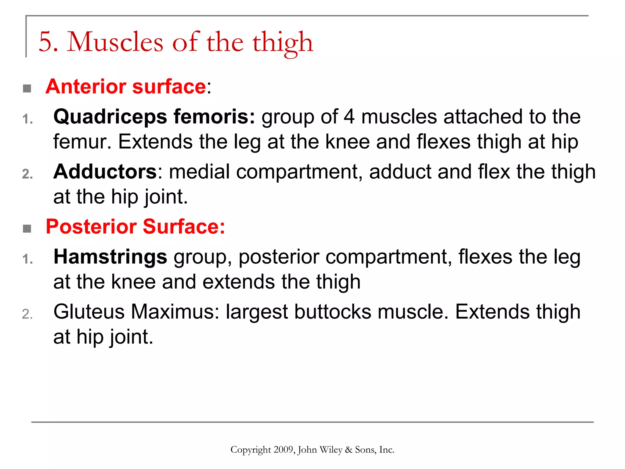
Copyright 2009, John Wiley & Sons, Inc.
Skeletal muscles
and their actions
Objectives
 Naming of skeletal muscles
 Identify major skeletal muscles
 Actions of major skeletal muscles
Copyright 2009, John Wiley & Sons, Inc.
A. Criteria for naming skeletal muscles
 Orientation, relative to body midline
 Size
 Shape
 Action
 Number of origins
 Location
 Attachment points:
 Origin: Nonmoving point of attachment
 Insertion: Moves toward the origin
Copyright 2009, John Wiley & Sons, Inc.
Table 13.1. Criteria for naming skeletal muscles
 Orientation, relative to body midline
 Rectus: Muscle parallel to midline
 Transverse: Muscle perpendicular to midline
 Oblique: Muscle diagonal to midline
Copyright 2009, John Wiley & Sons, Inc.
Table 13.1. Criteria for naming skeletal muscles
 Muscle size
 Maximus: Largest
 Medius: Middle
 Minimus: Smallest
 Latissimus: Wide
 Major: Larger
 Muscle shape
 Deltoid: Triangular
 Trapezius: Trapezoid
 Orbicularis: Circular
Copyright 2009, John Wiley & Sons, Inc.
Table 13.1. Criteria for naming skeletal muscles
 Muscle action
 Flexor: Decreases joint angle
 Extensor: Increases joint angle
 Abductor: Moves bone away from midline
 Adductor: Moves bone close to midline
 Sphincter: Decreases size of opening
 Number of origins
 Biceps: Two origins
 Triceps: Three origins
 Quadriceps: Four origins
Copyright 2009, John Wiley & Sons, Inc.
Table 13.1. Criteria for naming skeletal muscles
 Muscle location
 Frontalis: Located on the frontal bone
 Origin and insertion
 Sternohyoid: Originates on the sternum, inserts on the
hyoid bone
Copyright 2009, John Wiley & Sons, Inc.
B. Skeletal muscle identification and action
1. Muscles of the head and neck
 Muscles of facial expression:
 Move the skin of the face
 Convey a variety of emotions
 Muscles of mastication:
 Move the mandible
 Help hold food between the teeth while chewing
 Muscles that move the head and neck:
 Flexors, anterior surface of the neck, in pairs
 Extensors, posterior surface of the neck
Copyright 2009, John Wiley & Sons, Inc.
Muscle Location Function
Muscles of facial
expressions
Frontalis Over the frontal bone Raises eyebrows and
wrinkles forehead
Orbicularis oculi Encircles the eyes Closes the eye
Orbicularis oris Encircles the mouth Closes and purses lips
Occipitalis Over occipital bone Pulls the scalp posteriorly
Zygomaticus major Between zygomatic bone
and corner of the mouth
Raises the corners of the
mouth(smiling muscle)
Muscles of mastication
Temporalis Over temporal bone Moves the mandible
Masseter Cheek area Moves the mandible
Muscles that move the
head
Sternocleidomastoid
(SCM)
Between sternum,clavicle,
and mastoid process
(temporal bone)
Both:flex the head(prayer
muscle)
One:rotates the head to
the sides
Trapezius Upper back and posterior
neck
Extends the head
(a) Right lateral superficial view
Epicranial aponeurosis
Occipitalis
Temporalis
Zygomatic arch
Mandible
Masseter
Sternocleidomastoid (SCM)
Trapezius
Frontalis
Orbicularis oculi
Nasalis
Zygomasticus
major
Buccinator
Orbicularis oris
Depressor labii inferioris
Platysma
Muscles of Facial Expression
Muscles of mastication
Muscles that move the head Table 13.2. Muscles of the head and neck
2. Muscles of the anterior trunk
 Muscles that move the arm at the shoulder joint:
1. Deltoid
2. Pectoralis Major
 Muscles that move the abdominal wall
Copyright 2009, John Wiley & Sons, Inc.
Superficial Muscles
External oblique Superficial, runs obliquely
Rectus abdominis Parallel
Deep Muscles
Internal Oblique Deep , runs obliquely
Transversus Abdominis Perpendicular, deep to the
internal oblique
(a) Anterior superficial view
(b) Anterior deep view
Rectus abdominis
External oblique
External oblique (cut)
Rectus abdominis
Transversus abdominis
Internal oblique
Deltoid
Pectoralis
major
Muscles that move the arm at the shoulder joint
Muscles that move the abdominal wall
Table 13.3. Muscles of the anterior trunk
Muscles of the Posterior Trunk
Muscles that move the arm at the shoulder
joint:
1. Deltoid
2. Latissimus Dorsi
Muscles that move the scapula:
1. Trapezius
Copyright © 2015 John Wiley & Sons, Inc. All rights reserved.
Muscles that move the arm at the shoulder joint
Muscles that move the scapula
Table 13.4. Muscles of the posterior trunk
3. Muscles of the arm
 Muscles of anterior compartments of the arm:
 Biceps brachii :Flex the forearm at elbow at the shoulder
 Brachialis muscles : Flex the forearm at elbow
 Muscles of posterior compartment of the arm:
 Triceps brachii : Extend the forearm at elbow and at the shoulder
4. Muscles of the forearm
 Flexors: group of muscles on anterior surface that flex the
hand at the wrist joint
 Extensors group of muscles on posterior surface that extend
the hand at the wrist joint
Copyright 2009, John Wiley & Sons, Inc.
Table 13.5. Muscles of the arm
5. Muscles of the thigh
 Anterior surface:
1. Quadriceps femoris: group of 4 muscles attached to the
femur. Extends the leg at the knee and flexes thigh at hip
2. Adductors: medial compartment, adduct and flex the thigh
at the hip joint.
 Posterior Surface:
1. Hamstrings group, posterior compartment, flexes the leg
at the knee and extends the thigh
2. Gluteus Maximus: largest buttocks muscle. Extends thigh
at hip joint.
Copyright 2009, John Wiley & Sons, Inc.
(a) Anterior superficial view
Adductors
Quadriceps femoris:
Rectus femoris (cut)
Vastus intermedius
Vastus lateralis
Vastus medialis
Patella
(a) Posterior superficial view
Gluteus medius
Gluteus maximus
Hamstrings:
Semitendinosus
Biceps femoris
Semimembranosus
Gastrocnemius
Copyright © 2015 John Wiley & Sons, Inc. All rights reserved.
Transverse plane
View
Copyright 2009, John Wiley & Sons, Inc.
5. Muscles of the leg
 Tibialis anterior, anterior compartment of the leg, lateral to
tibia. Moves the foot
 Gastrocnemius(superficial) and soleus(deep), posterior
compartment of the leg, moves the foot.
 Achilles tendon formed by tendons of gastrocnemius and
soleus, attach the muscle to the calcaneal bone(heel of the
foot)
(d) Anterior superficial view
(e) Right lateral superficial view
Patella
Fibula
Tibia
Tibialis anterior
Gastrocnemius
Soleus
Calcaneal (Achilles)
tendon
Fibula
(a) Posterior superficial view (b) Posterior deep view
Tibia
Gastrocnemius (cut)
Tibia
Gastrocnemius
Soleus (cut)
Fibula
Soleus
Fibularis longus
Flexor digitorum longus
Calcaneal (Achilles) tendon (cut)
Femur
Fibula

Skeletal muscles ppt.pptx

  • 1.
    Copyright 2009, JohnWiley & Sons, Inc. Skeletal muscles and their actions
  • 2.
    Objectives  Naming ofskeletal muscles  Identify major skeletal muscles  Actions of major skeletal muscles Copyright 2009, John Wiley & Sons, Inc.
  • 3.
    A. Criteria fornaming skeletal muscles  Orientation, relative to body midline  Size  Shape  Action  Number of origins  Location  Attachment points:  Origin: Nonmoving point of attachment  Insertion: Moves toward the origin Copyright 2009, John Wiley & Sons, Inc.
  • 4.
    Table 13.1. Criteriafor naming skeletal muscles  Orientation, relative to body midline  Rectus: Muscle parallel to midline  Transverse: Muscle perpendicular to midline  Oblique: Muscle diagonal to midline Copyright 2009, John Wiley & Sons, Inc.
  • 5.
    Table 13.1. Criteriafor naming skeletal muscles  Muscle size  Maximus: Largest  Medius: Middle  Minimus: Smallest  Latissimus: Wide  Major: Larger  Muscle shape  Deltoid: Triangular  Trapezius: Trapezoid  Orbicularis: Circular Copyright 2009, John Wiley & Sons, Inc.
  • 6.
    Table 13.1. Criteriafor naming skeletal muscles  Muscle action  Flexor: Decreases joint angle  Extensor: Increases joint angle  Abductor: Moves bone away from midline  Adductor: Moves bone close to midline  Sphincter: Decreases size of opening  Number of origins  Biceps: Two origins  Triceps: Three origins  Quadriceps: Four origins Copyright 2009, John Wiley & Sons, Inc.
  • 7.
    Table 13.1. Criteriafor naming skeletal muscles  Muscle location  Frontalis: Located on the frontal bone  Origin and insertion  Sternohyoid: Originates on the sternum, inserts on the hyoid bone Copyright 2009, John Wiley & Sons, Inc.
  • 8.
    B. Skeletal muscleidentification and action 1. Muscles of the head and neck  Muscles of facial expression:  Move the skin of the face  Convey a variety of emotions  Muscles of mastication:  Move the mandible  Help hold food between the teeth while chewing  Muscles that move the head and neck:  Flexors, anterior surface of the neck, in pairs  Extensors, posterior surface of the neck Copyright 2009, John Wiley & Sons, Inc.
  • 9.
    Copyright 2009, JohnWiley & Sons, Inc. Muscle Location Function Muscles of facial expressions Frontalis Over the frontal bone Raises eyebrows and wrinkles forehead Orbicularis oculi Encircles the eyes Closes the eye Orbicularis oris Encircles the mouth Closes and purses lips Occipitalis Over occipital bone Pulls the scalp posteriorly Zygomaticus major Between zygomatic bone and corner of the mouth Raises the corners of the mouth(smiling muscle) Muscles of mastication Temporalis Over temporal bone Moves the mandible Masseter Cheek area Moves the mandible Muscles that move the head Sternocleidomastoid (SCM) Between sternum,clavicle, and mastoid process (temporal bone) Both:flex the head(prayer muscle) One:rotates the head to the sides Trapezius Upper back and posterior neck Extends the head
  • 10.
    (a) Right lateralsuperficial view Epicranial aponeurosis Occipitalis Temporalis Zygomatic arch Mandible Masseter Sternocleidomastoid (SCM) Trapezius Frontalis Orbicularis oculi Nasalis Zygomasticus major Buccinator Orbicularis oris Depressor labii inferioris Platysma Muscles of Facial Expression Muscles of mastication Muscles that move the head Table 13.2. Muscles of the head and neck
  • 11.
    2. Muscles ofthe anterior trunk  Muscles that move the arm at the shoulder joint: 1. Deltoid 2. Pectoralis Major  Muscles that move the abdominal wall Copyright 2009, John Wiley & Sons, Inc. Superficial Muscles External oblique Superficial, runs obliquely Rectus abdominis Parallel Deep Muscles Internal Oblique Deep , runs obliquely Transversus Abdominis Perpendicular, deep to the internal oblique
  • 12.
    (a) Anterior superficialview (b) Anterior deep view Rectus abdominis External oblique External oblique (cut) Rectus abdominis Transversus abdominis Internal oblique Deltoid Pectoralis major Muscles that move the arm at the shoulder joint Muscles that move the abdominal wall Table 13.3. Muscles of the anterior trunk
  • 13.
    Muscles of thePosterior Trunk Muscles that move the arm at the shoulder joint: 1. Deltoid 2. Latissimus Dorsi Muscles that move the scapula: 1. Trapezius Copyright © 2015 John Wiley & Sons, Inc. All rights reserved.
  • 14.
    Muscles that movethe arm at the shoulder joint Muscles that move the scapula Table 13.4. Muscles of the posterior trunk
  • 15.
    3. Muscles ofthe arm  Muscles of anterior compartments of the arm:  Biceps brachii :Flex the forearm at elbow at the shoulder  Brachialis muscles : Flex the forearm at elbow  Muscles of posterior compartment of the arm:  Triceps brachii : Extend the forearm at elbow and at the shoulder 4. Muscles of the forearm  Flexors: group of muscles on anterior surface that flex the hand at the wrist joint  Extensors group of muscles on posterior surface that extend the hand at the wrist joint Copyright 2009, John Wiley & Sons, Inc.
  • 16.
    Copyright 2009, JohnWiley & Sons, Inc. Table 13.5. Muscles of the arm
  • 17.
    5. Muscles ofthe thigh  Anterior surface: 1. Quadriceps femoris: group of 4 muscles attached to the femur. Extends the leg at the knee and flexes thigh at hip 2. Adductors: medial compartment, adduct and flex the thigh at the hip joint.  Posterior Surface: 1. Hamstrings group, posterior compartment, flexes the leg at the knee and extends the thigh 2. Gluteus Maximus: largest buttocks muscle. Extends thigh at hip joint. Copyright 2009, John Wiley & Sons, Inc.
  • 18.
    (a) Anterior superficialview Adductors Quadriceps femoris: Rectus femoris (cut) Vastus intermedius Vastus lateralis Vastus medialis Patella (a) Posterior superficial view Gluteus medius Gluteus maximus Hamstrings: Semitendinosus Biceps femoris Semimembranosus Gastrocnemius Copyright © 2015 John Wiley & Sons, Inc. All rights reserved. Transverse plane View
  • 19.
    Copyright 2009, JohnWiley & Sons, Inc. 5. Muscles of the leg  Tibialis anterior, anterior compartment of the leg, lateral to tibia. Moves the foot  Gastrocnemius(superficial) and soleus(deep), posterior compartment of the leg, moves the foot.  Achilles tendon formed by tendons of gastrocnemius and soleus, attach the muscle to the calcaneal bone(heel of the foot)
  • 20.
    (d) Anterior superficialview (e) Right lateral superficial view Patella Fibula Tibia Tibialis anterior Gastrocnemius Soleus Calcaneal (Achilles) tendon Fibula
  • 21.
    (a) Posterior superficialview (b) Posterior deep view Tibia Gastrocnemius (cut) Tibia Gastrocnemius Soleus (cut) Fibula Soleus Fibularis longus Flexor digitorum longus Calcaneal (Achilles) tendon (cut) Femur Fibula

Editor's Notes

  • #4 When skeletal muscles contract, they pull their point of insertion toward their point of origin.
  • #9 Contraction of both flexors causes flexion. Contraction of one turns the head to the side. Unilateral contraction: cervical spine: neck ipsilateral flexion, neck contralateral rotation
  • #16 Compartment: muscles of the appendages that perform similar functions, surrounded by fascia
  • #18 Gluteus maximus extends the thigh