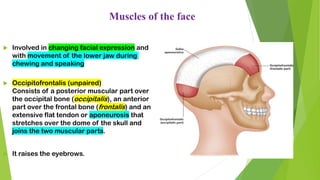
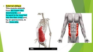
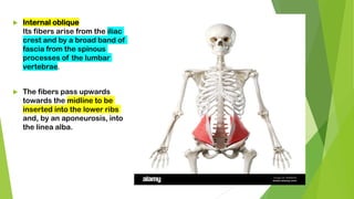
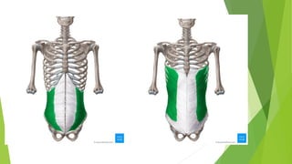
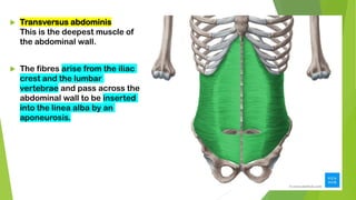
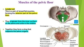
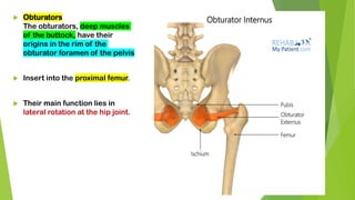
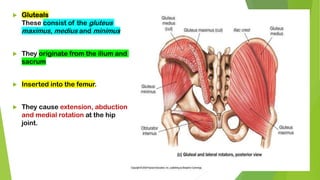
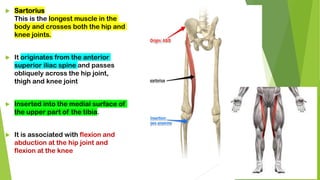
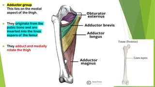
MUSCULAR SYSTEM
SUDESHNA BANERJI DUTTA
ASSISTANT PROFESSOR
Muscles of the face
 Involved in changing facial expression and
with movement of the lower jaw during
chewing and speaking
 Occipitofrontalis (unpaired)
Consists of a posterior muscular part over
the occipital bone (occipitalis), an anterior
part over the frontal bone (frontalis) and an
extensive flat tendon or aponeurosis that
stretches over the dome of the skull and
joins the two muscular parts.
 It raises the eyebrows.
 Levator palpebrae superioris
This muscle extends from the posterior part of the orbital cavity to the upper eyelid.
 It raises the eyelid
 Orbicularis oculi
This muscle surrounds the eye,
eyelid and orbital cavity.
 It closes the eye and when strongly
contracted ‘screws up’ the eyes.
➢ Buccinator
This flat muscle of the cheek draws the cheeks in towards the teeth in
chewing and in forcible expulsion of air from the mouth (‘the trumpeter’s
muscle’)
 Orbicularis oris (unpaired)
This muscle surrounds the mouth and
blends with the muscles of the cheeks.
 It closes the lips and, when strongly
contracted, shapes the mouth for whistling
 Masseter
This is a broad muscle,
extending from the zygomatic
arch to the angle of the jaw.
 In chewing it draws the
mandible up to the maxilla,
closing the jaw, and exerts
considerable pressure on the
food
 Temporalis
This muscle covers the squamous part
of the temporal bone.
 It closes the mouth and assists with
chewing.
 Pterygoid
This muscle extends from the
sphenoid bone to the mandible.
 It closes the mouth and pulls the
lower jaw forward.
Muscles of the neck
 Sternocleidomastoid
This muscle arises from the manubrium of
the sternum and the clavicle and extends
upwards to the mastoid process of the
temporal bone.
 It assists in turning the head from side to
side.
 Trapezius
This muscle covers the shoulder and the back
of the neck.
 The upper attachment is to the occipital
protuberance, the medial attachment is to the
transverse processes of the cervical and
thoracic vertebrae and the lateral attachment
is to the clavicle and to the spinous and
acromion processes of the scapula.
 It pulls the head backwards, and controls the
movements of the scapula when the shoulder
joint is in use
Muscles of the trunk
 These muscles stabilise the
association between the
appendicular and axial skeletons at
the pectoral girdle
 Stabilise and allow movement of
the shoulders and upper arms.

 Latissimus dorsi
This arises from the posterior part
of the iliac crest and the spinous
processes of the lumbar and
lower thoracic vertebrae. It
adducts, medially rotates and
extends the arm.
Muscles of the back
Muscles of the back
 Teres major
This originates from the
inferior angle of the
scapula and is inserted
into the humerus just
below the shoulder joint
 It extends, adducts and
medially rotates the
arm.
Muscles of the back
 Quadratus lumborum
This muscle originates from the
iliac crest, then it passes
upwards, parallel and close to the
vertebral column and it is inserted
into the 12th rib.
 Together the two muscles fix the
lower rib during respiration and
cause extension of the vertebral
column (bending backwards).
Muscles of the back
➢ Sacrospinalis (erector spinae)
This is a group of muscles lying
between the spinous and
transverse processes of the
vertebrae.
➢ They originate from the sacrum
and are finally inserted into the
occipital bone.
➢ Their contraction causes
extension of the vertebral
column.
Muscles of the abdominal wall
 Rectus abdominis
It is broad and flat,
originating from the
transverse part of the pubic
bone then passing upwards
to be inserted into the lower
ribs and the xiphoid
process of the sternum.
 External oblique
This muscle extends
from the lower ribs
downwards and
forward to be inserted
into the iliac crest and,
by an aponeurosis, to
the linea alba.
 Internal oblique
Its fibers arise from the iliac
crest and by a broad band of
fascia from the spinous
processes of the lumbar
vertebrae.
 The fibers pass upwards
towards the midline to be
inserted into the lower ribs
and, by an aponeurosis, into
the linea alba.
 Transversus abdominis
This is the deepest muscle of
the abdominal wall.
 The fibres arise from the iliac
crest and the lumbar
vertebrae and pass across the
abdominal wall to be inserted
into the linea alba by an
aponeurosis.
Muscles of the pelvic floor
 Levator ani
This is a pair of broad flat muscles,
forming the anterior part of the pelvic
floor.
 They originate from the inner surface
of the pelvis and unite in the midline.
 Together they form a sling that
supports the pelvic organs.
 Coccygeus
This is a paired triangular sheet
of muscle and tendinous fibres
situated behind the levator ani.
 They originate from the medial
surface of the ischium and are
inserted into the sacrum and
coccyx.
 They complete the formation of
the pelvic floor
Muscles of the shoulder and upper limb
 Deltoid
These muscle fibres originate from the clavicle, acromion process and spine of scapula
and radiate over the shoulder joint to be inserted into the deltoid tuberosity of the
humerus. It forms the fleshy and rounded contour of the shoulder and the main function
is movement of the arm.
 Pectoralis major
This lies on the anterior thoracic wall. The fibres originate from the middle
third of the clavicle and from the sternum and are inserted into the lip of the
intertubercular groove of the humerus.
 Coracobrachialis
This lies on the upper medial aspect of the arm. It arises from the coracoid process of the
scapula, stretches across in front of the shoulder joint and is inserted into the middle third
of the humerus. It flexes the shoulder joint
 Biceps
This lies on the anterior aspect of the
upper arm.
 It is divided into two parts (heads), each
of which has its own tendon. The short
head rises from the coracoid process of
the scapula and passes in front of the
shoulder joint to the arm.
 The long head originates from the rim of
the glenoid cavity and its tendon passes
through the joint cavity and the bicipital
groove of the humerus to the arm.
 Brachialis
This lies on the anterior aspect of the
upper arm deep to the biceps.
 It originates from the shaft of the
humerus, extends across the elbow
joint and is inserted into the ulna just
distal to the joint capsule.
 It is the main flexor of the elbow joint.
 Triceps
This lies on the posterior aspect of
the humerus.
 It arises from three heads, one from
the scapula and two from the
posterior surface of the humerus.
 The insertion is by a common tendon
to the olecranon process of the ulna.
 It helps to stabilise the shoulder
joint, assists in adduction of the arm
and extends the elbow joint.
 Brachioradialis
The brachioradialis spans the
elbow joint, originating on the
distal end of the humerus and
inserts on the lateral epicondyle
of the radius.
 When it contracts, it flexes the
elbow joint
 Pronator quadratus
This square-shaped muscle is
the main muscle causing
pronation of the hand
 Has attachments on the lower
sections of both the radius and
the ulna.
 Pronator teres
This lies obliquely across the upper
third of the front of the forearm.
 It arises from the medial epicondyle of
the humerus and the coronoid process
of the ulna and passes obliquely across
the forearm to be inserted into the
lateral surface of the shaft of the
radius.
 It rotates the radioulnar joints,
changing the hand from the anatomical
to the writing position, i.e. pronation.
 Supinator
This lies obliquely across the
posterior and lateral aspects of the
forearm.
 Its fibers arise from the lateral
epicondyle of the humerus and the
upper part of the ulna
 Inserted into the lateral surface of
the upper third of the radius.
 It rotates the radioulnar joints, often
with help from the biceps, changing
the hand from the writing to the
anatomical position, i.e. supination.
 Flexor carpi radialis
This lies on the anterior surface of
the forearm.
 It originates from the medial
epicondyle of the humerus and is
inserted into the second and third
metacarpal bones.
 It flexes the wrist joint, and when
acting with the extensor carpi
radialis, abducts the joint.
 Flexor carpi ulnaris
This lies on the medial aspect of the
forearm.
 It originates from the medial epicondyle
of the humerus and the upper parts of
the ulna and is inserted into the
pisiform, the hamate and the fifth
metacarpal bones.
 It flexes the wrist, and when acting with
the extensor carpi ulnaris, adducts the
joint
 Extensor carpi radialis longus
and brevis
These lie on the posterior aspect
of the forearm.
 The fibers originate from the
lateral epicondyle of the humerus
and are inserted by a long tendon
into the second and third
metacarpal bones.
 They extend and abduct the wrist
 Extensor carpi ulnaris
This lies on the posterior surface of
the forearm.
 It originates from the lateral epicondyle
of the humerus and is inserted into the
fifth metacarpal bone.
 It extends and adducts the wrist
 Palmaris longus
Its origin is on the medial
epicondyle of the humerus, and it
inserts on tendons on the palm of
the hand
 Extensor digitorum
This muscle originates on the lateral
epicondyle of the humerus and spans
both the elbow and wrist joints; in the
wrist, it divides into four tendons, one
for each finger.
 Action of this muscle can extend any of
the joints across which it passes, i.e.
the elbow, wrist or finger joints.
Muscles of the hip and lower limb
 Psoas
This arises from the transverse
processes and bodies of the lumbar
vertebrae.
 It passes across the flat part of the
ilium and behind the inguinal ligament
to be inserted into the femur.
 Together with the iliacus it flexes the hip
joint
Iliacus
 It originates from the iliac crest,
passes over the iliac fossa and joins
the tendon of the psoas muscle
 Inserted into the lesser trochanter
of the femur.
 The combined action of the iliacus
and psoas flexes the hip joint.
 Quadriceps femoris
This is a group of four muscles lying on
the front and sides of the thigh. They
are the rectus femoris and three vasti
lateralis, medialis and intermedius.
 The rectus femoris originates from the
ilium and the three vasti from the upper
end of the femur.
 Inserted into the tibia by the patellar
tendon.
 Only the rectus femoris flexes the hip
joint. Together, the group acts as a very
strong extensor of the knee joint.
 Obturators
The obturators, deep muscles
of the buttock, have their
origins in the rim of the
obturator foramen of the pelvis
 Insert into the proximal femur.
 Their main function lies in
lateral rotation at the hip joint.
 Gluteals
These consist of the gluteus
maximus, medius and minimus
 They originate from the ilium and
sacrum
 Inserted into the femur.
 They cause extension, abduction
and medial rotation at the hip
joint.
 Sartorius
This is the longest muscle in the
body and crosses both the hip and
knee joints.
 It originates from the anterior
superior iliac spine and passes
obliquely across the hip joint,
thigh and knee joint
 Inserted into the medial surface of
the upper part of the tibia.
 It is associated with flexion and
abduction at the hip joint and
flexion at the knee
 Adductor group
This lies on the medial
aspect of the thigh.
 They originate from the
pubic bone and are
inserted into the linea
aspera of the femur.
 They adduct and medially
rotate the thigh
 Hamstrings
These lie on the posterior aspect of
the thigh.
 They originate from the ischium and
are inserted into the upper end of the
tibia.
 They are the biceps femoris,
semimembranosus and
semitendinosus muscles.
 They flex the knee joint
 Anterior tibialis
This originates from the upper end of
the tibia, lies on the anterior surface
of the leg
 Inserted into the middle cuneiform
bone by a long tendon.
 It is associated with dorsiflexion of the
foot.
 Soleus
This is one of the main muscles of the calf
of the leg
 It originates from the heads and upper parts
of the fibula and the tibia.
 Insertion into the calcaneus by the
calcanean (Achilles)
tendon.
 It causes plantarflexion at the ankle and
helps to stabilise the joint when standing.

MUSCLE anatomy bsc 1st sem classroom presentation

  • 1.
    MUSCULAR SYSTEM SUDESHNA BANERJIDUTTA ASSISTANT PROFESSOR
  • 2.
    Muscles of theface  Involved in changing facial expression and with movement of the lower jaw during chewing and speaking  Occipitofrontalis (unpaired) Consists of a posterior muscular part over the occipital bone (occipitalis), an anterior part over the frontal bone (frontalis) and an extensive flat tendon or aponeurosis that stretches over the dome of the skull and joins the two muscular parts.  It raises the eyebrows.
  • 3.
     Levator palpebraesuperioris This muscle extends from the posterior part of the orbital cavity to the upper eyelid.  It raises the eyelid
  • 4.
     Orbicularis oculi Thismuscle surrounds the eye, eyelid and orbital cavity.  It closes the eye and when strongly contracted ‘screws up’ the eyes.
  • 5.
    ➢ Buccinator This flatmuscle of the cheek draws the cheeks in towards the teeth in chewing and in forcible expulsion of air from the mouth (‘the trumpeter’s muscle’)
  • 6.
     Orbicularis oris(unpaired) This muscle surrounds the mouth and blends with the muscles of the cheeks.  It closes the lips and, when strongly contracted, shapes the mouth for whistling
  • 7.
     Masseter This isa broad muscle, extending from the zygomatic arch to the angle of the jaw.  In chewing it draws the mandible up to the maxilla, closing the jaw, and exerts considerable pressure on the food
  • 8.
     Temporalis This musclecovers the squamous part of the temporal bone.  It closes the mouth and assists with chewing.
  • 9.
     Pterygoid This muscleextends from the sphenoid bone to the mandible.  It closes the mouth and pulls the lower jaw forward.
  • 10.
    Muscles of theneck  Sternocleidomastoid This muscle arises from the manubrium of the sternum and the clavicle and extends upwards to the mastoid process of the temporal bone.  It assists in turning the head from side to side.
  • 11.
     Trapezius This musclecovers the shoulder and the back of the neck.  The upper attachment is to the occipital protuberance, the medial attachment is to the transverse processes of the cervical and thoracic vertebrae and the lateral attachment is to the clavicle and to the spinous and acromion processes of the scapula.  It pulls the head backwards, and controls the movements of the scapula when the shoulder joint is in use
  • 12.
    Muscles of thetrunk  These muscles stabilise the association between the appendicular and axial skeletons at the pectoral girdle  Stabilise and allow movement of the shoulders and upper arms. 
  • 13.
     Latissimus dorsi Thisarises from the posterior part of the iliac crest and the spinous processes of the lumbar and lower thoracic vertebrae. It adducts, medially rotates and extends the arm. Muscles of the back
  • 14.
    Muscles of theback  Teres major This originates from the inferior angle of the scapula and is inserted into the humerus just below the shoulder joint  It extends, adducts and medially rotates the arm.
  • 15.
    Muscles of theback  Quadratus lumborum This muscle originates from the iliac crest, then it passes upwards, parallel and close to the vertebral column and it is inserted into the 12th rib.  Together the two muscles fix the lower rib during respiration and cause extension of the vertebral column (bending backwards).
  • 16.
    Muscles of theback ➢ Sacrospinalis (erector spinae) This is a group of muscles lying between the spinous and transverse processes of the vertebrae. ➢ They originate from the sacrum and are finally inserted into the occipital bone. ➢ Their contraction causes extension of the vertebral column.
  • 17.
    Muscles of theabdominal wall
  • 18.
     Rectus abdominis Itis broad and flat, originating from the transverse part of the pubic bone then passing upwards to be inserted into the lower ribs and the xiphoid process of the sternum.
  • 19.
     External oblique Thismuscle extends from the lower ribs downwards and forward to be inserted into the iliac crest and, by an aponeurosis, to the linea alba.
  • 20.
     Internal oblique Itsfibers arise from the iliac crest and by a broad band of fascia from the spinous processes of the lumbar vertebrae.  The fibers pass upwards towards the midline to be inserted into the lower ribs and, by an aponeurosis, into the linea alba.
  • 22.
     Transversus abdominis Thisis the deepest muscle of the abdominal wall.  The fibres arise from the iliac crest and the lumbar vertebrae and pass across the abdominal wall to be inserted into the linea alba by an aponeurosis.
  • 23.
    Muscles of thepelvic floor  Levator ani This is a pair of broad flat muscles, forming the anterior part of the pelvic floor.  They originate from the inner surface of the pelvis and unite in the midline.  Together they form a sling that supports the pelvic organs.
  • 24.
     Coccygeus This isa paired triangular sheet of muscle and tendinous fibres situated behind the levator ani.  They originate from the medial surface of the ischium and are inserted into the sacrum and coccyx.  They complete the formation of the pelvic floor
  • 25.
    Muscles of theshoulder and upper limb  Deltoid These muscle fibres originate from the clavicle, acromion process and spine of scapula and radiate over the shoulder joint to be inserted into the deltoid tuberosity of the humerus. It forms the fleshy and rounded contour of the shoulder and the main function is movement of the arm.
  • 26.
     Pectoralis major Thislies on the anterior thoracic wall. The fibres originate from the middle third of the clavicle and from the sternum and are inserted into the lip of the intertubercular groove of the humerus.
  • 27.
     Coracobrachialis This lieson the upper medial aspect of the arm. It arises from the coracoid process of the scapula, stretches across in front of the shoulder joint and is inserted into the middle third of the humerus. It flexes the shoulder joint
  • 28.
     Biceps This lieson the anterior aspect of the upper arm.  It is divided into two parts (heads), each of which has its own tendon. The short head rises from the coracoid process of the scapula and passes in front of the shoulder joint to the arm.  The long head originates from the rim of the glenoid cavity and its tendon passes through the joint cavity and the bicipital groove of the humerus to the arm.
  • 30.
     Brachialis This lieson the anterior aspect of the upper arm deep to the biceps.  It originates from the shaft of the humerus, extends across the elbow joint and is inserted into the ulna just distal to the joint capsule.  It is the main flexor of the elbow joint.
  • 31.
     Triceps This lieson the posterior aspect of the humerus.  It arises from three heads, one from the scapula and two from the posterior surface of the humerus.  The insertion is by a common tendon to the olecranon process of the ulna.  It helps to stabilise the shoulder joint, assists in adduction of the arm and extends the elbow joint.
  • 33.
     Brachioradialis The brachioradialisspans the elbow joint, originating on the distal end of the humerus and inserts on the lateral epicondyle of the radius.  When it contracts, it flexes the elbow joint
  • 34.
     Pronator quadratus Thissquare-shaped muscle is the main muscle causing pronation of the hand  Has attachments on the lower sections of both the radius and the ulna.
  • 35.
     Pronator teres Thislies obliquely across the upper third of the front of the forearm.  It arises from the medial epicondyle of the humerus and the coronoid process of the ulna and passes obliquely across the forearm to be inserted into the lateral surface of the shaft of the radius.  It rotates the radioulnar joints, changing the hand from the anatomical to the writing position, i.e. pronation.
  • 36.
     Supinator This liesobliquely across the posterior and lateral aspects of the forearm.  Its fibers arise from the lateral epicondyle of the humerus and the upper part of the ulna  Inserted into the lateral surface of the upper third of the radius.  It rotates the radioulnar joints, often with help from the biceps, changing the hand from the writing to the anatomical position, i.e. supination.
  • 37.
     Flexor carpiradialis This lies on the anterior surface of the forearm.  It originates from the medial epicondyle of the humerus and is inserted into the second and third metacarpal bones.  It flexes the wrist joint, and when acting with the extensor carpi radialis, abducts the joint.
  • 38.
     Flexor carpiulnaris This lies on the medial aspect of the forearm.  It originates from the medial epicondyle of the humerus and the upper parts of the ulna and is inserted into the pisiform, the hamate and the fifth metacarpal bones.  It flexes the wrist, and when acting with the extensor carpi ulnaris, adducts the joint
  • 40.
     Extensor carpiradialis longus and brevis These lie on the posterior aspect of the forearm.  The fibers originate from the lateral epicondyle of the humerus and are inserted by a long tendon into the second and third metacarpal bones.  They extend and abduct the wrist
  • 41.
     Extensor carpiulnaris This lies on the posterior surface of the forearm.  It originates from the lateral epicondyle of the humerus and is inserted into the fifth metacarpal bone.  It extends and adducts the wrist
  • 42.
     Palmaris longus Itsorigin is on the medial epicondyle of the humerus, and it inserts on tendons on the palm of the hand
  • 43.
     Extensor digitorum Thismuscle originates on the lateral epicondyle of the humerus and spans both the elbow and wrist joints; in the wrist, it divides into four tendons, one for each finger.  Action of this muscle can extend any of the joints across which it passes, i.e. the elbow, wrist or finger joints.
  • 44.
    Muscles of thehip and lower limb  Psoas This arises from the transverse processes and bodies of the lumbar vertebrae.  It passes across the flat part of the ilium and behind the inguinal ligament to be inserted into the femur.  Together with the iliacus it flexes the hip joint
  • 45.
    Iliacus  It originatesfrom the iliac crest, passes over the iliac fossa and joins the tendon of the psoas muscle  Inserted into the lesser trochanter of the femur.  The combined action of the iliacus and psoas flexes the hip joint.
  • 46.
     Quadriceps femoris Thisis a group of four muscles lying on the front and sides of the thigh. They are the rectus femoris and three vasti lateralis, medialis and intermedius.  The rectus femoris originates from the ilium and the three vasti from the upper end of the femur.  Inserted into the tibia by the patellar tendon.  Only the rectus femoris flexes the hip joint. Together, the group acts as a very strong extensor of the knee joint.
  • 47.
     Obturators The obturators,deep muscles of the buttock, have their origins in the rim of the obturator foramen of the pelvis  Insert into the proximal femur.  Their main function lies in lateral rotation at the hip joint.
  • 48.
     Gluteals These consistof the gluteus maximus, medius and minimus  They originate from the ilium and sacrum  Inserted into the femur.  They cause extension, abduction and medial rotation at the hip joint.
  • 49.
     Sartorius This isthe longest muscle in the body and crosses both the hip and knee joints.  It originates from the anterior superior iliac spine and passes obliquely across the hip joint, thigh and knee joint  Inserted into the medial surface of the upper part of the tibia.  It is associated with flexion and abduction at the hip joint and flexion at the knee
  • 50.
     Adductor group Thislies on the medial aspect of the thigh.  They originate from the pubic bone and are inserted into the linea aspera of the femur.  They adduct and medially rotate the thigh
  • 51.
     Hamstrings These lieon the posterior aspect of the thigh.  They originate from the ischium and are inserted into the upper end of the tibia.  They are the biceps femoris, semimembranosus and semitendinosus muscles.  They flex the knee joint
  • 52.
     Anterior tibialis Thisoriginates from the upper end of the tibia, lies on the anterior surface of the leg  Inserted into the middle cuneiform bone by a long tendon.  It is associated with dorsiflexion of the foot.
  • 53.
     Soleus This isone of the main muscles of the calf of the leg  It originates from the heads and upper parts of the fibula and the tibia.  Insertion into the calcaneus by the calcanean (Achilles) tendon.  It causes plantarflexion at the ankle and helps to stabilise the joint when standing.