Downloaded 14 times
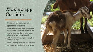
![Eimeria spp. Coccidia
• Can use McMaster technique to
determine fecal oocyte count
(FOC). [small egg]
• Small ruminants can be infected
by many different species of
Eimeria.
• Small ruminants harbor their
own species of Eimeria and there
is no cross-infection.
• Not all strains of coccidia are
disease causing or equally
pathogenic (speciation
warranted).
• A FOC of 5000 with appropriate
signs may be noteworthy, if](https://image.slidesharecdn.com/showmethesigns-240119201608-356fcf4e/85/Show-me-the-signs-27-320.jpg)

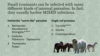
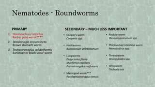
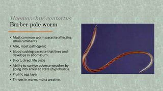
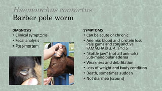
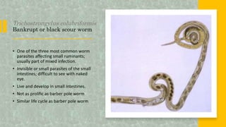
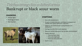
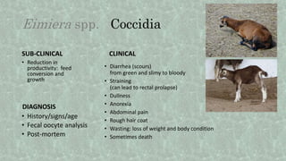
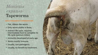
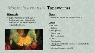
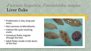
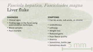
This document outlines the various internal parasites that can infest small ruminants, including helminths and protozoa such as nematodes, cestodes, trematodes, and coccidia, detailing their life cycles, symptoms, and diagnosis methods. Key parasites like Haemonchus contortus (barber pole worm), Trichostrongylus colubriformis (black scour worm), and Teladorsagia circumcincta (brown stomach worm) pose significant threats to animal health, leading to conditions such as anemia and reduced productivity. Recommendations for diagnosis include clinical signs, fecal analysis, and post-mortem evaluations to identify specific infections and their impact on the animals' health.


![Parasitology [Autosaved].pptx](https://cdn.slidesharecdn.com/ss_thumbnails/parasitologyautosaved-230930080756-f478f745-thumbnail.jpg?width=640&height=640&fit=bounds)