
Recommended
More Related Content
What's hot
What's hot (20)
Similar to Larval arrest and immunity in nematode parasites
Similar to Larval arrest and immunity in nematode parasites (20)
More from farhab dvm
More from farhab dvm (20)
Recently uploaded
Recently uploaded (20)
Larval arrest and immunity in nematode parasites
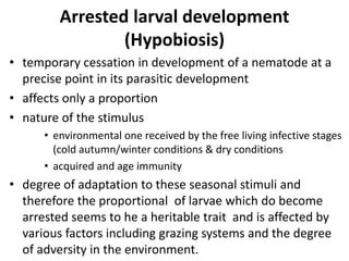
- 1. Arrested larval development (Hypobiosis) • temporary cessation in development of a nematode at a precise point in its parasitic development • affects only a proportion • nature of the stimulus • environmental one received by the free living infective stages (cold autumn/winter conditions & dry conditions • acquired and age immunity • degree of adaptation to these seasonal stimuli and therefore the proportional of larvae which do become arrested seems to he a heritable trait and is affected by various factors including grazing systems and the degree of adversity in the environment.
- 2. epidemiological importance • ensures the survival of the nematode during periods of adversity • subsequent maturation of arrested larvae increases the contamination of the environment and can sometimes result in clinical disease.
- 3. PERIPARTURIENT RISE (PPR) IN FAECAL EGG COUNTS • An increase in the numbers of nematode eggs in the faeces of animals around parturition. • Etiology: temporary relaxation in immunity (prolactin) • Source of the PPR: • Maturation of larvae arrested • An increased establishment of infections • An increased fecundity • Importance of the PPR: – new susceptible hosts – Increased contamination of the environment lead to clinical disease
- 4. Trichostrongyloidea • Small, often hair-like • Bursate group • Parasitize the alimentary tract except Dictyocaulus • Few cuticular appendages • Buccal capsule is vestigial • Well developed bursa and two spicules • Direct life cycle • Ensheathed L3 is infective stage • Ostertagia, Haemonchus, Trichostrongylus, Cooperia, Nematodirus
- 5. Ostertagia • Parasitic gastritis • Hosts: Ruminants • Site: Abomasum – Adult on surface of abomasum – larval stages occur in the gastric glands • Species: – Ostertagia ostertagi cattle – 0. circumcincta sheep and goats – 0. trifurcata sheep and goats
- 6. • Identification – slender reddish-brown – l.0 cm long • Species differentiation is based on the structure of the spicules • Direct life cycle • Eggs in feces----L3 ----2 weeks • L3 exsheathes in rumen---enter lumen of abomasal gland---- L4– L5– -- 18 days ----emerge from gland--- mature • Entire life cycle take 3 weeks • Early fourth larval stage
- 7. PATHOGENESIS • Reduction in the functional gastric gland – Failure to activate pepsinogen to pepsin – Loss of bacteriostatic effect • Enhanced permeability of the abomasal epithelium – Hypoalbuminaemia •
- 8. CLINICAL SIGNS • Profuse watery diarrhoea • Hind quarters heavily soiled with faeces • Dull coat • Inappetence • Weight loss (leakage of endogenous protein) Treatment • Benzimidazoles (albendazole, fenbendazole or Oxfendazole • Ivermectin • Febantel, Netobimin
- 9. HAEMONCHUS (Barber pole worm) • Blood sucking abomasal parasite • Host: cattle, sheep, goat • Species: – Haemonchus contortus – H. placei – H. similis • Size: 2.0-3.0 cm • Barber’s pole appearance: white ovaries winding spirally around the blood-filled intestine
- 10. • Microscopic: there are cervical papillae and a tiny lancet inside the buccal capsule – Female has vulval flap – Male has asymmetrical dorsal lobe • larvae moult twice in close apposition to the gastric glands • Prepatent period is 2-3 weeks in sheep and four weeks in cattle
- 11. Female - vulva flap Cervical papillae Buccal lancet
- 12. PATHOGENESIS • Acute haemorrhagic anaemia • Acute haemonchosis: –Acute haemorrhagic anaemia –Progressive and dramatic fall in the packed red cell volume –At necropsy 2000 and 20000 worms –Abomasal contents are fluid and dark brown • Chronic haemonchosis • Loss of weight, weakness and inappetence rather than marked anaemia
- 13. TRICHOSTRONGYLUS • Host: Ruminants, horse, fowl, pigs • Infection site: Small intestine, except T. axei and T. tenuis • Species – T. axei ruminants (abomasum), horse (stomach) – T. colubriformis ruminants – T. vitrinus sheep, goat – T. capricola sheep, goat – T. tenius small intestine and caeca of game birds
- 14. • Identification: small, hair like • Length…. 7.00mm • No buccal capsule • Distinct excretory notch in oesophageal region • Spicules are thick and unbranched • Life cycle: Direct • Exsheathment of L3 of intestinal species occurs in the abomasum • Parasitic phase is non-migratory • Prepatent period – Ruminants 2-3 weeks – Horse 25 days – Birds 10 days
- 15. • L3 of the intestinal species penetrate between the epithelial glands of the mucosa with formation of tunnels beneath the epithelium, but above the lamina propria • Haemorrhage, oedema • Loss of plasma proteins • Enteritis • Villi destroy
- 16. Clinical signs • rapid weight loss and diarrhoea • inappetence and poor growth rates
- 17. Cooperia • Host: Ruminants • Site: small intestine • Species: – C. oncophora – C. punctata – C. pectinata – C. surnobada – C. curticei • Identification: size similar to Ostertagia • Very large bursa
- 18. • Watch-spring like posture • Main generic features---small cephalic and the transverse cuticular striations in the oesophageal region • The spicules usually have a wing like expansion in the middle region and bear ridges • No gubernaculum • Females usually have a small vulval flap • Life cycle: direct
- 19. Pathogenesis • Penetrate the epithelial surface of the small intestine and cause a disruption • Villous atrophy • Reduction in the area available for absorption • Diarrhoea • Clinical Signs • Loss of appetite, • Poor weight gains • Diarrhoea • Suhmandibular oedema
- 20. Nematodirus • Host: Ruminants • Site: small intestine • Nematodirus battus sheep, occasionally calves • N. filicollis sheep and goats • N. spathigur sheep and goats
- 21. • Identification: Adults are slender • Size: 2.0 cm long • Intertwining of the thin, twisted worms produces an appearance similar to that of cotton wool • A small hut distinct cephalic vesicle • The spicules are long and slender with fused tips • Development to the L3 takes place within the egg shell
- 22. Pathogenesis • Disruption of the intestinal mucosa, particularly in the ileum • Severe damage to villi and erosion of the mucosa leading lo villous atrophy • Ability of the intestine to exchange fluids and nutrients is grossly reduced • Diarrhoea • Enteritis • Carcass has dehydrated appearance
- 23. Clinical signs • Diarrhoea • Thirsty • Infected ewes continue to graze • Inappetent and diarrhoeic lambs congregate round drinking places • Diagnosis • Clinical signs (Fecal egg count is of low value) • Treatment • levamisole, milbemycin
- 24. Dictyocaulus Parasitic bronchitis • husk, hoose, verminous pneumonia or dictyocaulosis • Host: Ruminants, horse & donkeys • Site: Trachea and bronchi, particularly of the diaphragmatic lobes • D. vivipurus cattle and deer • D. filaria sheep and goats • D. arnfieldi donkeys and horses
- 25. • Slender thread-like worms up to 8.0cm in length • Life cycle • Ovo-viviparous females • L1 migrate up the trachea, are swallowed and pass out in the faeces • Larvae: Sluggish, and their intestinal cells are filled with dark brown food granules • L3 stage is reached within five days • L3 penetrate the intestinal mucusa and pass to the mesenteric lymph nodes where they moult. • Then the L4, travel via the lymph and blood to the lungs. and break out of the capillaries into the alveoli about one week after infection. • The final moult occurs in the bronchioles a few days later and the young adults then move up the bronchi and mature. • The prepatent period is around 3-4 weeks.