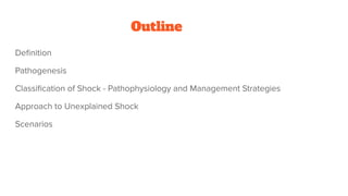
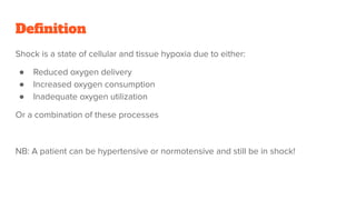
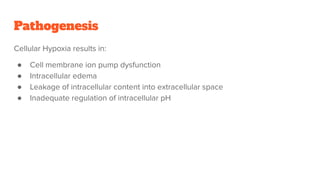
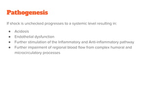
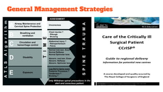
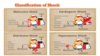
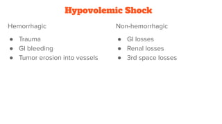
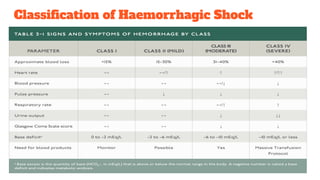
1. Shock is defined as cellular hypoxia due to reduced oxygen delivery, increased oxygen consumption, or inadequate utilization and can progress from cellular to systemic effects if unchecked.
2. Shock is classified based on pathophysiology into hypovolemic, cardiogenic, obstructive, distributive, and neurogenic types.
3. The document provides an overview of the pathogenesis, classification, treatment goals and strategies for each shock type to guide clinical management.
The document discusses various types of shock, including hypovolemic, cardiogenic, tension pneumothorax, cardiac tamponade, neurogenic, and septic shock. It describes the pathophysiology, clinical features, and basic management principles for each type of shock. The role of a pulmonary artery catheter in guiding resuscitation efforts by providing measured and calculated hemodynamic parameters is also summarized.
1) The document provides an overview of shock, including common clinical features, key hemodynamic parameters, and types of shock. It also reviews vasopressors commonly used to treat shock.
2) Emergency disorders in critical care are reviewed, including acute inhalational injuries, anaphylaxis, hypertensive emergencies, hyperthermic emergencies, hypothermic emergencies, and toxicology. Management strategies for these conditions are discussed.
3) Case examples are provided to demonstrate assessment and treatment of patients presenting with septic shock, acute liver failure, and altered mental status, and the appropriate next steps in management are outlined.
1. Shock is defined as inadequate tissue perfusion and cellular dysfunction due to an imbalance between oxygen delivery and demand.
2. There are several types of shock including hypovolemic, cardiogenic, obstructive, anaphylactic, and neurogenic shock.
3. Hypovolemic shock occurs due to loss of intravascular volume from hemorrhage, burns, or fluid losses. Cardiogenic shock results from cardiac failure leading to low cardiac output. Obstructive shock involves obstruction of venous return such as from tension pneumothorax.
This case presentation summarizes the management of a patient presenting with septic shock secondary to gastrointestinal loss and hypoglycemia. The 30-year-old male was referred from a primary hospital with diarrhea, vomiting, and altered mental status. On admission, he was found to be in septic shock with a low blood pressure, fast heart rate, and low blood sugar. He was treated aggressively with IV fluids, antibiotics, vasopressors, and glucose supplementation. Despite initial stabilization, his condition deteriorated with the development of aspiration pneumonia and bacterial meningitis. He was discharged against medical advice before further interventions could be pursued.
1) Acute coronary syndrome includes unstable angina and myocardial infarction, characterized by chest pain and elevated cardiac biomarkers.
2) Clinical features include severe, prolonged chest pain that may radiate to other areas, as well as syncope, vomiting, and arrhythmias.
3) Complications include heart failure, arrhythmias like ventricular fibrillation, cardiac rupture, and remodelling. Diagnosis involves ECG, cardiac enzymes, and angiography. Management focuses on reperfusion therapy, anticoagulation, and risk factor reduction.
Guidelines for management of acute strokesankalpgmc8
This document provides an overview of stroke types, pathophysiology, investigations, and management guidelines. It discusses the three main types of stroke: ischemic, intracerebral hemorrhage, and subarachnoid hemorrhage. For ischemic stroke, it describes the ischemic core and penumbra. It outlines the emergency evaluation of acute ischemic stroke including vital signs, blood tests, imaging, and scales like the NIH Stroke Scale. Management strategies discussed include thrombolysis, antiplatelet/anticoagulation drugs, neuroprotective agents, and surgical interventions. Complications like cerebral edema and their management are also summarized.
Anesthesia for spinal cord injury and scoliosis030Atef Salama
The potential risk factors for POVL that are listed are:
- Obesity
- Long Prone Cases
- Anemia
- Pressure on the globe
- Hypotension
- Glaucoma
Cataracts is not a risk factor for POVL.
This seminar covers different types of shock including definitions, pathophysiology, clinical features, investigations, and treatment. The main types discussed are hypovolaemic shock, traumatic shock, cardiogenic shock, neurogenic shock, septic shock, and crush syndrome. Hypovolaemic shock is the most common and results from sudden loss of blood or fluid volume. Treatment focuses on fluid resuscitation and controlling bleeding. Septic shock has a high mortality and is usually caused by gram-negative bacteria. Crush syndrome occurs after body portions are compressed by heavy weights.
A 75-year-old diabetic male presented with chest pain and other symptoms of acute coronary syndrome. The most probable diagnosis is myocardial infarction. Relevant investigations include ECG, biochemical markers like CK-MB and troponin, and echocardiogram. Management involves medical therapy in emergency, possible fibrinolysis or PCI, and long term preventative treatment. Complications can include heart failure, cardiogenic shock, arrhythmias if not properly managed.
shock is the state of insufficient blood flow to the tissues of the body .it contains introduction, definition, stages of shock, types of shock, diagnostic evaluation, prognosis ,prevention, care for each stage.
A 67-year-old man presents to the emergency department with sudden onset of chest pain and shortness of breath. On examination, he has signs of pulmonary edema and a loud heart murmur, and has a history of a recent heart attack. The most appropriate initial management is to provide standard treatment for pulmonary edema and consider the need for urgent valve replacement surgery given the acute presentation in the setting of known valvular heart disease.
This document provides an overview of the management of acute stroke. It defines stroke and transient ischemic attack, and discusses the epidemiology, classification, risk factors, pathophysiology, clinical presentation, diagnosis, management, complications and prognosis of stroke. The management involves resuscitation, reperfusion therapies like thrombolysis and thrombectomy, treating complications, secondary prevention including blood pressure and diabetes control, and rehabilitation. The document emphasizes the importance of specialized stroke units and timely management to improve outcomes for patients with acute stroke.
Shock is caused by a sudden drop in blood flow and can result from trauma that causes blood or fluid loss. There are several types but hypovolemic shock from blood loss is most common after trauma. Symptoms of hemorrhagic shock range from mild tachycardia with up to 15% blood loss to depressed mental status and organ dysfunction with over 40% blood loss. Treatment involves rapid fluid resuscitation, controlling bleeding, maximizing oxygen delivery, and determining the need for blood transfusion or surgery. Early recognition and treatment improve the prognosis for traumatic shock patients.
This document discusses the diagnosis and management of shock. It defines shock and outlines the 4 main categories: hypovolemic, cardiogenic, distributive, and obstructive. For each type of shock, it describes the pathophysiology, hemodynamic profile, and goals of treatment. The general principles of shock management are to improve oxygen delivery and utilization, treat the underlying cause, restore adequate perfusion through fluids and vasoactive agents if needed, and reduce oxygen demand. Timely antibiotics and reperfusion are crucial for septic and cardiac shock respectively. The overall goals are to prevent cellular and organ injury through optimized perfusion and supportive care.
This document discusses various neurological emergencies including acute ischemic stroke, intracranial hemorrhage, status epilepticus, Guillain-Barre syndrome, acute myelopathy, and myasthenic crisis. It provides details on the presentation, diagnosis, and treatment of acute ischemic stroke and intracranial hemorrhage, the two most common neurological emergencies. Key factors in evaluation and management are discussed such as use of CT, MRI, and thrombolytic therapy for ischemic stroke and controlling hypertension, treating coagulopathy, and managing intracranial pressure for intracranial hemorrhage.
1. ST elevation myocardial infarction (STEMI) occurs when there is ST elevation or new left bundle branch block on ECG due to acute coronary artery occlusion.
2. Diagnosis is based on symptoms, elevated cardiac biomarkers, and ECG changes showing ST elevation. Treatment involves stabilization, pain control, and reperfusion therapy.
3. Prognosis depends on factors like age, previous MI history, infarct location and size, and presence of heart failure or hypotension. Early reperfusion, beta-blockers, ACE inhibitors and risk factor modification can limit damage.
Septic shock is a type of distributive shock caused by systemic vasodilation from infection and sepsis. It is defined as sepsis with persisting hypotension requiring vasopressor support despite adequate fluid resuscitation. Diagnosis involves screening for signs of infection and organ dysfunction using criteria like SIRS, qSOFA and sepsis definitions. Management focuses on treating the underlying infection with antibiotics while providing hemodynamic support with fluids, vasopressors and inotropes to maintain perfusion.
Cardiogenic shock is defined as inadequate tissue perfusion due to cardiac dysfunction or hypo-perfusion of end organs due to cardiac failure. It has a high mortality rate of 50-80% and is most commonly caused by extensive acute myocardial infarction. Symptoms include cyanosis, decreased consciousness, and low blood pressure. Diagnosis involves identifying hypotension, low cardiac index, and signs of hypoperfusion on physical exam along with supportive tests like EKG, echocardiogram, and Swan-Ganz catheter. Treatment focuses on optimizing prefusion with vasopressors or inotropes, diuretics, emergent revascularization through cardiac catheterization, and mechanical circulatory support like IABP,
Cardiogenic shock is caused by severe impairment of myocardial performance resulting in diminished cardiac output and end-organ hypoperfusion. It presents clinically as hypotension refractory to fluids with signs of poor tissue perfusion. Acute myocardial infarction accounts for most cases of cardiogenic shock. Rapid diagnosis and treatment is needed to prevent end-organ damage. Management involves hemodynamic support, revascularization when possible, and mechanical circulatory support for refractory cases.
The document discusses the definition, pathophysiology, classification, clinical features, diagnosis, and management of shock in children. Shock is defined as a physiologic state characterized by a reduction in systemic tissue perfusion resulting in decreased oxygen delivery to tissues. The main types of shock are hypovolemic, cardiogenic, obstructive, and distributive shock, and treatment involves identifying the cause, restoring circulating volume and tissue perfusion through fluid resuscitation and vasoactive medications, and treating any underlying conditions.
This document discusses complications of spinal cord injury, including acute complications like pulmonary issues, cardiovascular problems, and pressure ulcers. Chronic complications include heterotopic ossification, gastrointestinal problems, and spasticity. Neurogenic shock, orthostatic hypotension, and autonomic dysreflexia are also covered. Respiratory complications are discussed along with prevention strategies. Bladder dysfunction, urinary tract infections, and thermal regulation challenges are also summarized.
This document provides an overview of radiological imaging in the management of stroke. It discusses:
1) Various imaging modalities used including unenhanced CT, CT angiography, MRI, and their benefits. Diffusion weighted MRI can detect acute ischemia within 30 minutes.
2) Examples of imaging findings for different stroke types like ischemic and hemorrhagic strokes. Ischemic strokes appear as bright lesions on DWI MRI.
3) Surgical interventions for acute stroke management include decompressive hemicraniectomy to reduce intracranial pressure for large hemispheric infarcts, and external ventricular drainage for intraventricular hemorrhage and hydrocephalus.
This document discusses the approach to evaluating and managing chest pain. It notes that chest pain is a common reason for emergency department visits and hospitalizations. A thorough history and physical exam is important to determine the likely cause, such as cardiac, pulmonary, gastrointestinal, or musculoskeletal origins. Initial testing may include an ECG, cardiac enzymes, chest x-ray, and echocardiogram. Life-threatening causes like myocardial infarction, pulmonary embolism, and aortic dissection require rapid diagnosis and treatment. Management depends on the identified condition but may include medications, procedures, or surgery.
This document discusses the management of coronary artery disease and acute coronary syndrome. It begins with the anatomy of the heart and coronary blood vessels. It then defines acute coronary syndrome as unstable angina or myocardial infarction caused by plaque rupture and thrombosis. Risk factors for coronary artery disease are outlined. The diagnostic approach involves assessing symptoms, signs, electrocardiogram changes and cardiac biomarker levels. Treatment focuses on reopening the blocked vessel with medications, fibrinolytics or percutaneous coronary intervention. Chronic stable angina from established coronary artery disease is also discussed.
The document provides information on myocardial infarction (MI or heart attack) including definitions, causes, pathophysiology, clinical manifestations, diagnostic tests, treatment, nursing management, and patient education. It defines MI as myocardial cell death due to prolonged ischemia. The main cause is sudden blockage of the coronary artery by a blood clot, causing irreversible damage to heart muscle. Clinical manifestations may include chest pain, shortness of breath, nausea, and changes in vital signs. Diagnostic tests include electrocardiogram, cardiac enzymes, and echocardiogram. Treatment focuses on reperfusion, reducing oxygen demand on the heart, and preventing complications. Nursing management involves monitoring for complications, relieving symptoms, promoting perfusion and respiratory function
The document discusses shock, including:
1. Circulatory shock occurs when the circulatory system is unable to provide adequate circulation and tissue perfusion, leading to cellular hypoxia and energy deficit.
2. Shock is classified as hypovolemic, cardiogenic, obstructive, or distributive. Hypovolemic shock results from blood or fluid loss while cardiogenic shock stems from heart problems reducing cardiac output.
3. During the reversible phase of hemorrhagic shock, compensatory mechanisms aim to maintain perfusion through vasoconstriction, tachycardia, fluid shifts, and hormonal responses. However, irreversible shock leads to organ dysfunction and failure without rapid intervention.
Neurogenic shock is a distributive type of shock caused by damage to the spinal cord, resulting in hypotension and bradycardia due to loss of sympathetic tone. It commonly occurs after high cervical or thoracic spinal cord injuries and is characterized by a hemodynamic triad of hypotension, bradycardia, and peripheral vasodilation. Immediate management involves C-spine immobilization, IV fluids, and vasopressors to maintain blood pressure, as well as monitoring of vital signs, oxygen levels, and neurological function.
This document discusses current treatment options for breast cancer, including both non-metastatic and metastatic disease. For non-metastatic cancer, local therapy involves surgical resection and lymph node removal, while systemic therapy includes pre-operative, postoperative, or both chemotherapy and endocrine therapy based on molecular subtype. Metastatic breast cancer focuses on prolonging life and palliating symptoms using the same systemic therapies. The document then covers specific topics like nipple discharge, cystic lesions, rare tumors, DCIS, mastectomy, breast-conserving therapy, and systemic therapies.
Emergency Management of Hip Dislocations & Pelvic Fractures.pdfBernard Fiifi Brakatu
Hip dislocations and pelvic fractures are common injuries seen in emergency departments. Hip dislocations, which are usually posterior, require prompt closed or open reduction to prevent osteonecrosis of the femoral head. Pelvic fractures are classified using the Young-Burgess system and range from stable patterns to more unstable anterior-posterior compression or lateral compression injuries which may lead to life-threatening hemorrhage. Clinical assessment focuses on signs of bleeding, neurovascular injury, or instability while radiological evaluation aids classification and guides management, which may involve hemorrhage control techniques, external or internal fixation, or angiography in unstable or bleeding patterns. Complications include bleeding, infection, instability, and nerve injuries requiring multidisciplinary care and early
More Related Content
Similar to Shock_ Pathophysiology & Current Management (1).pdf
A 75-year-old diabetic male presented with chest pain and other symptoms of acute coronary syndrome. The most probable diagnosis is myocardial infarction. Relevant investigations include ECG, biochemical markers like CK-MB and troponin, and echocardiogram. Management involves medical therapy in emergency, possible fibrinolysis or PCI, and long term preventative treatment. Complications can include heart failure, cardiogenic shock, arrhythmias if not properly managed.
shock is the state of insufficient blood flow to the tissues of the body .it contains introduction, definition, stages of shock, types of shock, diagnostic evaluation, prognosis ,prevention, care for each stage.
A 67-year-old man presents to the emergency department with sudden onset of chest pain and shortness of breath. On examination, he has signs of pulmonary edema and a loud heart murmur, and has a history of a recent heart attack. The most appropriate initial management is to provide standard treatment for pulmonary edema and consider the need for urgent valve replacement surgery given the acute presentation in the setting of known valvular heart disease.
This document provides an overview of the management of acute stroke. It defines stroke and transient ischemic attack, and discusses the epidemiology, classification, risk factors, pathophysiology, clinical presentation, diagnosis, management, complications and prognosis of stroke. The management involves resuscitation, reperfusion therapies like thrombolysis and thrombectomy, treating complications, secondary prevention including blood pressure and diabetes control, and rehabilitation. The document emphasizes the importance of specialized stroke units and timely management to improve outcomes for patients with acute stroke.
Shock is caused by a sudden drop in blood flow and can result from trauma that causes blood or fluid loss. There are several types but hypovolemic shock from blood loss is most common after trauma. Symptoms of hemorrhagic shock range from mild tachycardia with up to 15% blood loss to depressed mental status and organ dysfunction with over 40% blood loss. Treatment involves rapid fluid resuscitation, controlling bleeding, maximizing oxygen delivery, and determining the need for blood transfusion or surgery. Early recognition and treatment improve the prognosis for traumatic shock patients.
This document discusses the diagnosis and management of shock. It defines shock and outlines the 4 main categories: hypovolemic, cardiogenic, distributive, and obstructive. For each type of shock, it describes the pathophysiology, hemodynamic profile, and goals of treatment. The general principles of shock management are to improve oxygen delivery and utilization, treat the underlying cause, restore adequate perfusion through fluids and vasoactive agents if needed, and reduce oxygen demand. Timely antibiotics and reperfusion are crucial for septic and cardiac shock respectively. The overall goals are to prevent cellular and organ injury through optimized perfusion and supportive care.
This document discusses various neurological emergencies including acute ischemic stroke, intracranial hemorrhage, status epilepticus, Guillain-Barre syndrome, acute myelopathy, and myasthenic crisis. It provides details on the presentation, diagnosis, and treatment of acute ischemic stroke and intracranial hemorrhage, the two most common neurological emergencies. Key factors in evaluation and management are discussed such as use of CT, MRI, and thrombolytic therapy for ischemic stroke and controlling hypertension, treating coagulopathy, and managing intracranial pressure for intracranial hemorrhage.
1. ST elevation myocardial infarction (STEMI) occurs when there is ST elevation or new left bundle branch block on ECG due to acute coronary artery occlusion.
2. Diagnosis is based on symptoms, elevated cardiac biomarkers, and ECG changes showing ST elevation. Treatment involves stabilization, pain control, and reperfusion therapy.
3. Prognosis depends on factors like age, previous MI history, infarct location and size, and presence of heart failure or hypotension. Early reperfusion, beta-blockers, ACE inhibitors and risk factor modification can limit damage.
Septic shock is a type of distributive shock caused by systemic vasodilation from infection and sepsis. It is defined as sepsis with persisting hypotension requiring vasopressor support despite adequate fluid resuscitation. Diagnosis involves screening for signs of infection and organ dysfunction using criteria like SIRS, qSOFA and sepsis definitions. Management focuses on treating the underlying infection with antibiotics while providing hemodynamic support with fluids, vasopressors and inotropes to maintain perfusion.
Cardiogenic shock is defined as inadequate tissue perfusion due to cardiac dysfunction or hypo-perfusion of end organs due to cardiac failure. It has a high mortality rate of 50-80% and is most commonly caused by extensive acute myocardial infarction. Symptoms include cyanosis, decreased consciousness, and low blood pressure. Diagnosis involves identifying hypotension, low cardiac index, and signs of hypoperfusion on physical exam along with supportive tests like EKG, echocardiogram, and Swan-Ganz catheter. Treatment focuses on optimizing prefusion with vasopressors or inotropes, diuretics, emergent revascularization through cardiac catheterization, and mechanical circulatory support like IABP,
Cardiogenic shock is caused by severe impairment of myocardial performance resulting in diminished cardiac output and end-organ hypoperfusion. It presents clinically as hypotension refractory to fluids with signs of poor tissue perfusion. Acute myocardial infarction accounts for most cases of cardiogenic shock. Rapid diagnosis and treatment is needed to prevent end-organ damage. Management involves hemodynamic support, revascularization when possible, and mechanical circulatory support for refractory cases.
The document discusses the definition, pathophysiology, classification, clinical features, diagnosis, and management of shock in children. Shock is defined as a physiologic state characterized by a reduction in systemic tissue perfusion resulting in decreased oxygen delivery to tissues. The main types of shock are hypovolemic, cardiogenic, obstructive, and distributive shock, and treatment involves identifying the cause, restoring circulating volume and tissue perfusion through fluid resuscitation and vasoactive medications, and treating any underlying conditions.
This document discusses complications of spinal cord injury, including acute complications like pulmonary issues, cardiovascular problems, and pressure ulcers. Chronic complications include heterotopic ossification, gastrointestinal problems, and spasticity. Neurogenic shock, orthostatic hypotension, and autonomic dysreflexia are also covered. Respiratory complications are discussed along with prevention strategies. Bladder dysfunction, urinary tract infections, and thermal regulation challenges are also summarized.
This document provides an overview of radiological imaging in the management of stroke. It discusses:
1) Various imaging modalities used including unenhanced CT, CT angiography, MRI, and their benefits. Diffusion weighted MRI can detect acute ischemia within 30 minutes.
2) Examples of imaging findings for different stroke types like ischemic and hemorrhagic strokes. Ischemic strokes appear as bright lesions on DWI MRI.
3) Surgical interventions for acute stroke management include decompressive hemicraniectomy to reduce intracranial pressure for large hemispheric infarcts, and external ventricular drainage for intraventricular hemorrhage and hydrocephalus.
This document discusses the approach to evaluating and managing chest pain. It notes that chest pain is a common reason for emergency department visits and hospitalizations. A thorough history and physical exam is important to determine the likely cause, such as cardiac, pulmonary, gastrointestinal, or musculoskeletal origins. Initial testing may include an ECG, cardiac enzymes, chest x-ray, and echocardiogram. Life-threatening causes like myocardial infarction, pulmonary embolism, and aortic dissection require rapid diagnosis and treatment. Management depends on the identified condition but may include medications, procedures, or surgery.
This document discusses the management of coronary artery disease and acute coronary syndrome. It begins with the anatomy of the heart and coronary blood vessels. It then defines acute coronary syndrome as unstable angina or myocardial infarction caused by plaque rupture and thrombosis. Risk factors for coronary artery disease are outlined. The diagnostic approach involves assessing symptoms, signs, electrocardiogram changes and cardiac biomarker levels. Treatment focuses on reopening the blocked vessel with medications, fibrinolytics or percutaneous coronary intervention. Chronic stable angina from established coronary artery disease is also discussed.
The document provides information on myocardial infarction (MI or heart attack) including definitions, causes, pathophysiology, clinical manifestations, diagnostic tests, treatment, nursing management, and patient education. It defines MI as myocardial cell death due to prolonged ischemia. The main cause is sudden blockage of the coronary artery by a blood clot, causing irreversible damage to heart muscle. Clinical manifestations may include chest pain, shortness of breath, nausea, and changes in vital signs. Diagnostic tests include electrocardiogram, cardiac enzymes, and echocardiogram. Treatment focuses on reperfusion, reducing oxygen demand on the heart, and preventing complications. Nursing management involves monitoring for complications, relieving symptoms, promoting perfusion and respiratory function
The document discusses shock, including:
1. Circulatory shock occurs when the circulatory system is unable to provide adequate circulation and tissue perfusion, leading to cellular hypoxia and energy deficit.
2. Shock is classified as hypovolemic, cardiogenic, obstructive, or distributive. Hypovolemic shock results from blood or fluid loss while cardiogenic shock stems from heart problems reducing cardiac output.
3. During the reversible phase of hemorrhagic shock, compensatory mechanisms aim to maintain perfusion through vasoconstriction, tachycardia, fluid shifts, and hormonal responses. However, irreversible shock leads to organ dysfunction and failure without rapid intervention.
Neurogenic shock is a distributive type of shock caused by damage to the spinal cord, resulting in hypotension and bradycardia due to loss of sympathetic tone. It commonly occurs after high cervical or thoracic spinal cord injuries and is characterized by a hemodynamic triad of hypotension, bradycardia, and peripheral vasodilation. Immediate management involves C-spine immobilization, IV fluids, and vasopressors to maintain blood pressure, as well as monitoring of vital signs, oxygen levels, and neurological function.
Similar to Shock_ Pathophysiology & Current Management (1).pdf (20)
This document discusses current treatment options for breast cancer, including both non-metastatic and metastatic disease. For non-metastatic cancer, local therapy involves surgical resection and lymph node removal, while systemic therapy includes pre-operative, postoperative, or both chemotherapy and endocrine therapy based on molecular subtype. Metastatic breast cancer focuses on prolonging life and palliating symptoms using the same systemic therapies. The document then covers specific topics like nipple discharge, cystic lesions, rare tumors, DCIS, mastectomy, breast-conserving therapy, and systemic therapies.
Emergency Management of Hip Dislocations & Pelvic Fractures.pdfBernard Fiifi Brakatu
Hip dislocations and pelvic fractures are common injuries seen in emergency departments. Hip dislocations, which are usually posterior, require prompt closed or open reduction to prevent osteonecrosis of the femoral head. Pelvic fractures are classified using the Young-Burgess system and range from stable patterns to more unstable anterior-posterior compression or lateral compression injuries which may lead to life-threatening hemorrhage. Clinical assessment focuses on signs of bleeding, neurovascular injury, or instability while radiological evaluation aids classification and guides management, which may involve hemorrhage control techniques, external or internal fixation, or angiography in unstable or bleeding patterns. Complications include bleeding, infection, instability, and nerve injuries requiring multidisciplinary care and early
Steroid treatment for acute spinal cord injury (ASCI) has been studied extensively since the 1970s, but the evidence remains inconclusive. Early studies in cats found improved recovery with dexamethasone treatment. The 1985 NASCIS I trial found no difference with steroid treatment, but the 1992 NASCIS II trial found modest improved motor recovery when methylprednisolone was given within 8 hours. Subsequent trials found no benefit when treatment was given after 8 hours. While some meta-analyses found improved motor recovery, others and the most recent trials found no clear clinical benefits and increased risks of pneumonia and other complications. Current recommendations are that steroids are not a standard treatment but may be considered as an option if given
This document outlines a presentation on abdominal compartment syndrome (ACS) and intra-abdominal hypertension (IAH). It includes two case reports of patients who developed ACS after abdominal surgery or ingesting bleach. It defines ACS and IAH, describes how to grade and diagnose them, and outlines their epidemiology, etiology, pathophysiology, and management, including conservative measures and decompressive surgery if needed. Complications of decompressive laparotomy like enteroatmospheric fistulas are also discussed.
Peripheral arterial disease (PAD) is the obstruction or deterioration of arteries other than those supplying the heart and brain. PAD prevalence increases with age and is more common in Black individuals compared to Whites. A study in Ghana found higher PAD prevalence in Ghanaians living in Ghana compared to migrant Ghanaians in Europe. Risk factors for PAD include smoking, hypertension, diabetes, hyperlipidemia, and renal insufficiency. Treatment involves lifestyle modifications, medications, and revascularization procedures like angioplasty, stenting, or bypass surgery depending on the severity and location of arterial blockages. Amputations may be required for gangrenous or critically ischemic limbs.
This document discusses local anaesthesia techniques for common penile procedures. It describes relevant anatomy and different nerve block techniques like the dorsal penile nerve block and intracorpus spongiosal block. It also discusses a transillumination-guided urethral block technique for anterior urethral strictures that has been successfully used in 55 cases without needing additional sedation. The document emphasizes considering local anaesthesia as an option for penile procedures and calculating safe doses to minimize risks.
This document discusses pain management in oncology. It begins by outlining the objectives of understanding cancer pain management principles, categorizing pain, optimizing analgesia, using adjuvant analgesics, and recognizing when interventional therapies could help. It then provides background on cancer prevalence in Ghana and the high rates of pain in cancer patients. The rest of the document details approaches to assessing and treating cancer pain, including pharmacological and non-pharmacological options. It emphasizes a stepwise approach using the WHO analgesic ladder and treating the specific cause and type of cancer pain.
Keynote Presentation given to medical staff of the West Africa Rescue Association affiliated with International SOS, on management of organophosphate poisoning.
A middle-aged woman presented with generalized weakness, rashes, and sores. On examination, she had hyperpigmentation, edema, and rashes on her buttocks and groin. Tests found elevated muscle enzymes. She was diagnosed with dermatomyositis and admitted for IV fluids and steroids. Her symptoms worsened, and she developed difficulty raising her arms, confirming the diagnosis of dermatomyositis. She was started on immunosuppressants and referred for palliative care.
Systemic Lupus Erythematosus - Case Management and Literature ReviewBernard Fiifi Brakatu
A middle-aged woman presented with generalized weakness, dizziness and was found to have pancytopenia. Physical exam revealed signs of systemic lupus erythematosus including malar rash, oral ulcers, arthritis and hyperpigmented skin lesions. Laboratory tests confirmed pancytopenia and elevated inflammatory markers. A working diagnosis of SLE with pancytopenic anemia was made. She was started on steroids and other treatments. Further workup of her labs and signs are consistent with SLE. SLE is an autoimmune disease that commonly affects women and can involve multiple organ systems.
Personality types and choice of medical specialties among medical students in...Bernard Fiifi Brakatu
This document discusses personality types and their relationship to career choices in medicine. It describes a study conducted at the University of Ghana Medical School that assessed personality types of medical students using Jung's theory and the Myers-Briggs Type Indicator (MBTI). The study aimed to determine the distribution of personality types among students, their interests in different medical specialties, and whether certain personality types are correlated with choices in specialty. It provides background on Jung's theory of psychological types, the 16 MBTI personality types, and reviews literature on using personality typing to understand behaviors and classify individuals.
The document discusses the assessment and management of difficult airways. It begins with an introduction and overview of relevant anatomy. Assessment techniques are described, including patient history, physical exam findings like Mallampati score, and imaging. Management strategies for anticipated difficult intubation are outlined, such as specialized equipment, alternate airway devices, and surgical airway options if needed. Complications are noted and the importance of documentation and follow-up emphasized.
Osvaldo Bernardo Muchanga-GASTROINTESTINAL INFECTIONS AND GASTRITIS-2024.pdfOsvaldo Bernardo Muchanga
GASTROINTESTINAL INFECTIONS AND GASTRITIS
Osvaldo Bernardo Muchanga
Gastrointestinal Infections
GASTROINTESTINAL INFECTIONS result from the ingestion of pathogens that cause infections at the level of this tract, generally being transmitted by food, water and hands contaminated by microorganisms such as E. coli, Salmonella, Shigella, Vibrio cholerae, Campylobacter, Staphylococcus, Rotavirus among others that are generally contained in feces, thus configuring a FECAL-ORAL type of transmission.
Among the factors that lead to the occurrence of gastrointestinal infections are the hygienic and sanitary deficiencies that characterize our markets and other places where raw or cooked food is sold, poor environmental sanitation in communities, deficiencies in water treatment (or in the process of its plumbing), risky hygienic-sanitary habits (not washing hands after major and/or minor needs), among others.
These are generally consequences (signs and symptoms) resulting from gastrointestinal infections: diarrhea, vomiting, fever and malaise, among others.
The treatment consists of replacing lost liquids and electrolytes (drinking drinking water and other recommended liquids, including consumption of juicy fruits such as papayas, apples, pears, among others that contain water in their composition).
To prevent this, it is necessary to promote health education, improve the hygienic-sanitary conditions of markets and communities in general as a way of promoting, preserving and prolonging PUBLIC HEALTH.
Gastritis and Gastric Health
Gastric Health is one of the most relevant concerns in human health, with gastrointestinal infections being among the main illnesses that affect humans.
Among gastric problems, we have GASTRITIS AND GASTRIC ULCERS as the main public health problems. Gastritis and gastric ulcers normally result from inflammation and corrosion of the walls of the stomach (gastric mucosa) and are generally associated (caused) by the bacterium Helicobacter pylor, which, according to the literature, this bacterium settles on these walls (of the stomach) and starts to release urease that ends up altering the normal pH of the stomach (acid), which leads to inflammation and corrosion of the mucous membranes and consequent gastritis or ulcers, respectively.
In addition to bacterial infections, gastritis and gastric ulcers are associated with several factors, with emphasis on prolonged fasting, chemical substances including drugs, alcohol, foods with strong seasonings including chilli, which ends up causing inflammation of the stomach walls and/or corrosion. of the same, resulting in the appearance of wounds and consequent gastritis or ulcers, respectively.
Among patients with gastritis and/or ulcers, one of the dilemmas is associated with the foods to consume in order to minimize the sensation of pain and discomfort.
Computer in pharmaceutical research and development-Mpharm(Pharmaceutics)MuskanShingari
Statistics- Statistics is the science of collecting, organizing, presenting, analyzing and interpreting numerical data to assist in making more effective decisions.
A statistics is a measure which is used to estimate the population parameter
Parameters-It is used to describe the properties of an entire population.
Examples-Measures of central tendency Dispersion, Variance, Standard Deviation (SD), Absolute Error, Mean Absolute Error (MAE), Eigen Value
- Video recording of this lecture in English language: https://youtu.be/Pt1nA32sdHQ
- Video recording of this lecture in Arabic language: https://youtu.be/uFdc9F0rlP0
- Link to download the book free: https://nephrotube.blogspot.com/p/nephrotube-nephrology-books.html
- Link to NephroTube website: www.NephroTube.com
- Link to NephroTube social media accounts: https://nephrotube.blogspot.com/p/join-nephrotube-on-social-media.html
The skin is the largest organ and its health plays a vital role among the other sense organs. The skin concerns like acne breakout, psoriasis, or anything similar along the lines, finding a qualified and experienced dermatologist becomes paramount.
Spontaneous Bacterial Peritonitis - Pathogenesis , Clinical Features & Manage...Jim Jacob Roy
In this presentation , SBP ( spontaneous bacterial peritonitis ) , which is a common complication in patients with cirrhosis and ascites is described in detail.
The reference for this presentation is Sleisenger and Fordtran's Gastrointestinal and Liver Disease Textbook ( 11th edition ).
“Psychiatry and the Humanities”: An Innovative Course at the University of Mo...Université de Montréal
“Psychiatry and the Humanities”: An Innovative Course at the University of Montreal Expanding the medical model to embrace the humanities. Link: https://www.psychiatrictimes.com/view/-psychiatry-and-the-humanities-an-innovative-course-at-the-university-of-montreal
3. Definition
Shock is a state of cellular and tissue hypoxia due to either:
● Reduced oxygen delivery
● Increased oxygen consumption
● Inadequate oxygen utilization
Or a combination of these processes
NB: A patient can be hypertensive or normotensive and still be in shock!
4. Pathogenesis
Cellular Hypoxia results in:
● Cell membrane ion pump dysfunction
● Intracellular edema
● Leakage of intracellular content into extracellular space
● Inadequate regulation of intracellular pH
5. Pathogenesis
If shock is unchecked progresses to a systemic level resulting in:
● Acidosis
● Endothelial dysfunction
● Further stimulation of the Inflammatory and Anti-inflammatory pathway
● Further impairment of regional blood flow from complex humoral and
microcirculatory processes
10. Treatment Goals
Restore adequate organ perfusion and tissue oxygenation
● Early control of haemorrhage - employ damage control principles
● Adequate volume resuscitation - correct IVFs + blood products; massive
transfusion protocol (1:1:1)
● Avoid the lethal triad - acidosis, coagulopathy and hypothermia
Resus is complete when O2 debt is repaid, tissue acidosis corrected and aerobic
metabolism restored
11. Endpoints in resuscitation
Arterial BP (MAP), Heart rate, Urine output, CVP and Pulmonary artery occlusion
pressure are poor indicators of tissue perfusion
12. Obstructive Shock
Mechanical:
● Tension pneumothorax
● Massive Hemothorax (eg,
trauma, iatrogenic)
● Pericardial tamponade
● Severe dynamic hyperinflation
(eg, elevated intrinsic PEEP)
● Left or right ventricular outflow
tract obstruction
● Abdominal compartment
syndrome
● Aorto-caval compression (eg,
positioning, surgical retraction)
Pulmonary Vascular:
● Hemodynamically significant
pulmonary embolus
● Severe pulmonary hypertension
● Severe or acute obstruction of the
pulmonic or tricuspid valve
● Venous air embolus
13. Pathophysiology of Obstructive Shock
Mechanical obstruction of venous return
● Increased intrapleural pressure(tension pneumothorax)
● Increased intrapericadial pressure (cardiac tamponade) resulting in impeding
right atrial filling
Results in decreased CO with increased CVP
14. Rx
Tension Pneumo - Needle decompression (8cm needle depth) + ICD
Massive Haemothorax - ICD +/- urgent thoracotomy if initial drainage >1.5L or
200ml/hr for 2 to 4hrs or persistent need for blood transfusion
Pericardial tamponade - Pericardiocentesis or Pericardial window (subxiphoid or
transdiaphragmatic approach +/- exploration of heart (anterior thoracotomy, median
sternotomy or clamshell thoracotomy)
15. Cardiogenic Shock
Cardiomyopathic:
● Myocardial infarction
(involving >40% of the left
ventricle or with extensive
ischemia)
● Severe right ventricle
infarction
● Acute exacerbation of
severe heart failure from
dilated cardiomyopathy
● Stunned myocardium from
prolonged ischemia (eg,
cardiac arrest,
hypotension,
cardiopulmonary bypass)
● Advanced septic shock
● Myocarditis
● Myocardial contusion
● Drug-induced (eg, beta
blockers)
Arrhythmogenic:
● Tachyarrhythmia –
Atrial tachycardias
(fibrillation, flutter,
reentrant tachycardia),
ventricular tachycardia
and fibrillation
● Bradyarrhythmia –
Complete heart block,
Mobitz type II second
degree heart block
Mechanical:
● Severe valvular
insufficiency
● Acute valvular rupture
(papillary or chordae
tendineae rupture,
valvular abscess)
● Critical valvular stenosis
● Acute or severe
ventricular septal wall
defect
● Ruptured ventricular
wall aneurysm
● Atrial myxoma
16. Cardiogenic Shock
Failure of heart to deliver adequate cardiac output
Patient may have longstanding cardiac pathology
Rule out myocardial contusion in all chest trauma
● 12 lead ECG - if in keeping with MI changes, do cardiac enzymes and keep on monitor
Judicious use of IVFs followed by Inotropes - Dobutamine and Noradrenaline (or adrenaline
Infusion - titrate strength based on volume status)
Morphine
Anticoagulation
Broadspectrum anti-arrythymics (amiodarone)
Intra-aortic Balloon Pump, Impella Heart, ECMO
19. Pathophysiology of Distributive shock
Severe peripheral vasodilatation triggered by:
Septic shock - dysregulated host response to infection
SIRS - robust inflammatory response from a major body insult
Neurogenic shock - Interruption of autonomic pathways resulting in decreased
vascular resistance and altered vagal tone
Anaphylactic Shock -Severe IgE-mediated allergic response
20. Overview of the Autonomic Nervous System
Based on the reflex arc
● Afferent Limb - transmits info from periphery to spinal cord via dorsal root
ganglion to spinal cord or cranial nerves to brain
● Efferent Limb - pre- and post-ganglionic fibers + autonomic ganglion
● Central Integrating System - simple reflexes completed within organ system
and complex ones within hypothalamus and brain stem
21. Autonomic Ganglia
Parasympathetic - Craniosacral - CN3,7, 9, 10 and
S2-S4
Sympathetic
Pre-ganglionic -cell bodies in the grey matter of
lateral horns of T1 to L2/L3
Preganglionic fibers synapse with postganglionic
neurones in the ganglia of the sympathetic chain (2
paravertebral chains)
Postganglionic fibers leave ganglia to join spinal or
visceral nerves to innervate target organs
22. Paravertebral Sympathetic Chain
4 parts:
Cervical - 3 ganglia (superior, middle and inferior)
Inferior cervical ganglion fuses with T1 ganglion to form stellate ganglion
Thoracic - T1-T5 supply aortic, cardiac and pulmonary plexus
Lumbar - Prevertebral ganglia. Branches form the coeliac plexus
Pelvic - sacral ganglia
23. Neurogenic shock
Different from Spinal shock
SCI at or below T6 because of loss of thoracic sympathetic outflow
Decreased venous tone resulting in blood pooling in extremities and hypotension
Bradycardia - Decreased sympathetic tone to the heart
Core hypothermia with warm extremities - from peripheral pooling
Rx - Judicious IVFs + Inotropes (noradrenaline or adrenaline infusion) +/-atropine for
bradycardia
24.
25. Combined (Mixed) Shock
Patient with sepsis/pancreatitis(distributive shock) may have a
hypovolemic component(decreased intake, vomiting, diarrhea, etc)
Polytrauma patient with SCI (neurogenic) and abdominal or
extremity trauma (hypovolemic)
26. APPROACH TO UNEXPLAINED
SHOCK IN TRAUMA
ATLS Series - C-spine, CXR, AP Pelvis X-Rays +/- Lodox scan
CT Brain
Rule out the Deadly Dozen with a checklist!
FAST!
CT Abdo if FAST positive
Repeat neuro exam and document spinal level if any.
32. Q1
34yo M unrestrained driver in RTA with scalp laceration, bloody otorrhea, battle’s
sign and racoon eyes, limb lacerations and abdominal abrasions. Darkened skin
around upper thigh and waist, GCS - 13/15. Spine cleared by xrays. BP - 116/72 HR -
123 SpO2- 92% NRM at 15L/min. Cold, clammy extremities. What type of shock is
this?
A. Neurogenic
B. Distributive
C. Hypovolemic
D. Cardiogenic
E. Obstructive
33. Q2
27yo M motorcyclist presents with bilateral femur fractures, rib fractures and right
haemothorax. He is combative. SpO2 - 88% NRM at 15L/min, BP - 106/73 HR - 123
Temp - 38.2. You notice a petechial rash over his chest. What type of shock is this?
A. Hypovolemic
B. Obstructive
C. Distributive
D. Combined Hypovolemic & Distributive
E. Neurogenic
34. Q3
42yo F, helmeted pillion rider who fell from motorcycle after sudden deceleration.
Abrasions on chest and right breast. GCS-15/15. 2 rib fractures on the right with mild
chest contusion. Rest of exam unremarkable. FAST - Negative. BP - 78/51 HR - 54
SpO2 - 100% on 4L INO2. HO gives adequate IVFs but BPS don’t improve and
patient arrests at TSEU. What type of shock is this?
A. Hypovolemic
B. Distributive
C. Cardiogenic
D. Neurogenic
E. Obstructive
35. Q4
44yo M fell from a 3m height while painting. Abrasions on his upper back. ℅ pain
on upper body. No obvious external injuries. GCS 15/15. Warm extremities but
temperature 35.8deg. BP - 86/58, HR - 52, SpO2 99% on NRM 10L/min. Power is
5/5 in upper limbs but 1/5 in lower limbs. Altered sensation on lower limbs. Absent
anal wink and bulbocavernosus tests. What type of shock is this?
A.Spinal
B. Neurogenic
C. Combined Spinal & Neurogenic
D. Hypovolemic
E. Cardiogenic
36.
37. Resources
ATLS Manual - 10th Edition
Barber AE, Shires GT. Cell damage after shock. New Horiz. 1996 May;4(2):161-7. PMID: 8774792.
Cannon JW. Hemorrhagic Shock. N Engl J Med. 2018 Jan 25;378(4):370-379. doi: 10.1056/NEJMra1705649. PMID: 29365303.
Sabiston Textbook of Surgery - 21st Edition
Schwartz’s Principles of Surgery - 11th Edition