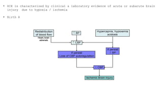
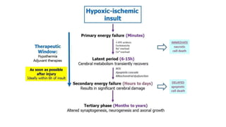
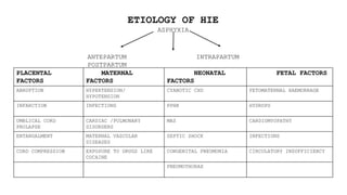
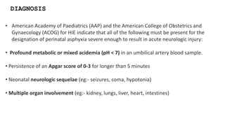
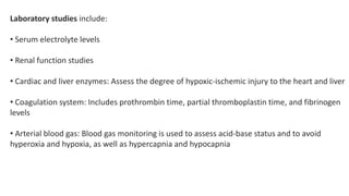
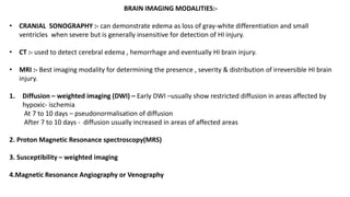
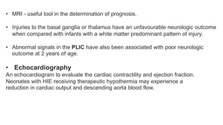
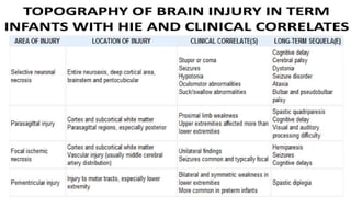
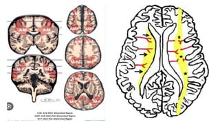
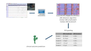
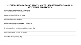
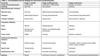
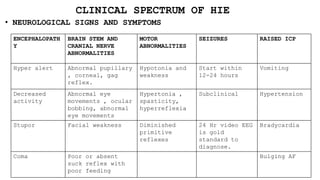
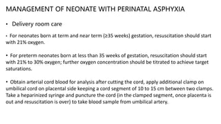
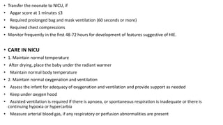
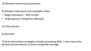
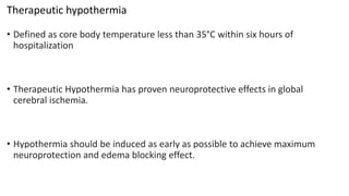
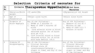
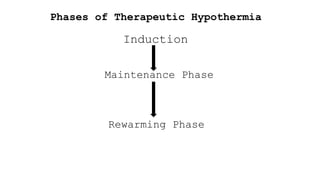
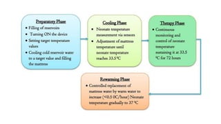
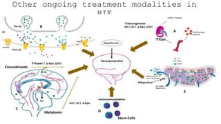
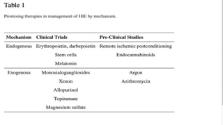
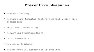
HYPOXIC ISCHEMIC
ENCEPHALOPATHY(HIE)
BY- Dr. SHIVANJAN GOYAL(JR3)
GUIDE- Dr. SUVIDHA SARDAR
• HIE is characterized by clinical & laboratory evidence of acute or subacute brain
injury due to hypoxia / ischemia
• Birth Asphyxia causes 8,40,000 or 23% neonatal deaths worldwide .
ETIOLOGY OF HIE
ASPHYXIA
ANTEPARTUM INTRAPARTUM
POSTPARTUM
PLACENTAL
FACTORS
MATERNAL
FACTORS
NEONATAL
FACTORS
FETAL FACTORS
ABRUPTION HYPERTENSION/
HYPOTENSION
CYANOTIC CHD FETOMATERNAL HAEMORRAGE
INFARCTION INFECTIONS PPHN HYDROPS
UMBLICAL CORD
PROLAPSE
CARDIAC /PULMONARY
DISORDERS
MAS CARDIOMYOPATHY
ENTANGALMENT MATERNAL VASCULAR
DISEASES
SEPTIC SHOCK INFECTIONS
CORD COMPRESSION EXPOSURE TO DRUGS LIKE
COCAINE
CONGENITAL PNEUMONIA CIRCULATORY INSUFFICIENCY
PNEUMOTHORAX
• American Academy of Paediatrics (AAP) and the American College of Obstetrics and
Gynaecology (ACOG) for HIE indicate that all of the following must be present for the
designation of perinatal asphyxia severe enough to result in acute neurologic injury:
• Profound metabolic or mixed acidemia (pH < 7) in an umbilical artery blood sample.
• Persistence of an Apgar score of 0-3 for longer than 5 minutes
• Neonatal neurologic sequelae (eg:- seizures, coma, hypotonia)
• Multiple organ involvement (eg:- kidney, lungs, liver, heart, intestines)
DIAGNOSIS
Laboratory studies include:
• Serum electrolyte levels
• Renal function studies
• Cardiac and liver enzymes: Assess the degree of hypoxic-ischemic injury to the heart and liver
• Coagulation system: Includes prothrombin time, partial thromboplastin time, and fibrinogen
levels
• Arterial blood gas: Blood gas monitoring is used to assess acid-base status and to avoid
hyperoxia and hypoxia, as well as hypercapnia and hypocapnia
BRAIN IMAGING MODALITIES:-
• CRANIAL SONOGRAPHY :- can demonstrate edema as loss of gray-white differentiation and small
ventricles when severe but is generally insensitive for detection of HI injury.
• CT :- used to detect cerebral edema , hemorrhage and eventually HI brain injury.
• MRI :- Best imaging modality for determining the presence , severity & distribution of irreversible HI brain
injury.
1. Diffusion – weighted imaging (DWI) – Early DWI –usually show restricted diffusion in areas affected by
hypoxic- ischemia
At 7 to 10 days – pseudonormalisation of diffusion
After 7 to 10 days - diffusion usually increased in areas of affected areas
2. Proton Magnetic Resonance spectroscopy(MRS)
3. Susceptibility – weighted imaging
4.Magnetic Resonance Angiography or Venography
• MRI - useful tool in the determination of prognosis.
• Injuries to the basal ganglia or thalamus have an unfavourable neurologic outcome
when compared with infants with a white matter predominant pattern of injury.
• Abnormal signals in the PLIC have also been associated with poor neurologic
outcome at 2 years of age.
• Echocardiography
An echocardiogram to evaluate the cardiac contractility and ejection fraction.
Neonates with HIE receiving therapeutic hypothermia may experience a
reduction in cardiac output and descending aorta blood flow.
Additional studies may include:
• EEG: Standard and amplitude-integrated EEG
• Hearing test: An increased incidence of deafness has been found among
infants with hypoxic-ischemic encephalopathy who require assisted ventilation
• Retinal and ophthalmic examination
There is a profound injury to the whole corpus callosum that
shows diffusion restriction on DWI. Sparing of the cortical and
deep grey matter and
the basal nuclei.
Bilateral hyperintense signals are seen in
watershed area.
EVOLUTION OF INJURY ON MRI
ELECTROENCEPHALOGRAM
Burst Suppression
in EEG
ELECTROENCEPHALOGRAPHIC PATTERNS OF PROGNOSTIC SIGNIFICANCE IN
ASPHYXIATED TERM INFANTS
ASSOCIATED WITH FAVORABLE OUTCOME
Mild depression (or less) on day 1
Normal background by day 7
ASSOCIATED WITH UNFAVORABLE OUTCOME
Predominant inter burst interval 20 sec on any day
Burst-suppression pattern on any day
Isoelectric tracing on any day
CLINICAL SPECTRUM OF HIE
• NEUROLOGICAL SIGNS AND SYMPTOMS
ENCEPHALOPATH
Y
BRAIN STEM AND
CRANIAL NERVE
ABNORMALITIES
MOTOR
ABNORMALITIES
SEIZURES RAISED ICP
Hyper alert Abnormal pupillary
, corneal, gag
reflex.
Hypotonia and
weakness
Start within
12-24 hours
Vomiting
Decreased
activity
Abnormal eye
movements , ocular
bobbing, abnormal
eye movements
Hypertonia ,
spasticity,
hyperreflexia
Subclinical Hypertension
Stupor Facial weakness Diminished
primitive
reflexes
24 Hr video EEG
is gold
standard to
diagnose.
Bradycardia
Coma Poor or absent
suck reflex with
poor feeding
Bulging AF
MULTIORGAN DYSFUCTION
KIDNEY CARDIAC PULMONARY HEMATOLOGICAL LIVER
DYSFUNCTIO
N
GI
Acute
tubular
necrosis
ST
depression
and T wave
inversion
PPHN DIC Raised
liver
enzymes
Necrotizi
ng
Enterocol
itis
Raised
creatinine
and urea
Cardiogenic
shock
Pulmonary
hemorrhage
Platelet
dysfunction
Hypoglycem
ia
Oliguria Fixed Heart
Rate
Pulmonary
edema
Raised PT/INR Drug
toxicity
Clinical Features of Severe HIE and Time Frame
Time Frame Clinical Features
Birth to 12
hours
• Decreased alertness & tone
• Convulsions
• Periodic breathing or respiratory failure
• Intact pupillary & oculomotor responses
12 to 24
hours
• Change in alertness level
• Apneic spells
• Increase convulsions
• Jitteriness
• Weakness in proximal limbs
• In Term Neonates UL more involved than LL ,In Preterm LL more
involved
24 to 72
hours
• Further Decreased alertness
• pupillary & oculomotor disturbances
• Respiratory arrest
• In Preterm, IVH or Periventricular hemorrhagic infarction also
seen
>72hours • Persistent & diminished stupor
• Abnormal Sucking, Swallowing, gag & tongue movements
• Hypotonia more common than hypertonia
MANAGEMENT OF NEONATE WITH PERINATAL ASPHYXIA
• Delivery room care
• For neonates born at term and near term (≥35 weeks) gestation, resuscitation should start
with 21% oxygen.
• For preterm neonates born at less than 35 weeks of gestation, resuscitation should start
with 21% to 30% oxygen; further oxygen concentration should be titrated to achieve target
saturations.
• Obtain arterial cord blood for analysis after cutting the cord, apply additional clamp on
umbilical cord on placental side keeping a cord segment of 10 to 15 cm between two clamps.
Take a heparinized syringe and puncture the cord (in the clamped segment, once placenta is
out and resuscitation is over) to take blood sample from umbilical artery.
• Transfer the neonate to NICU, if
• Apgar score at 1 minutes ≤3
• Required prolonged bag and mask ventilation (60 seconds or more)
• Required chest compressions
• Monitor frequently in the first 48-72 hours for development of features suggestive of HIE.
• CARE IN NICU
• 1. Maintain normal temperature
• After drying, place the baby under the radiant warmer
• Maintain normal body temperature
• 2. Maintain normal oxygenation and ventilation
• Assess the infant for adequacy of oxygenation and ventilation and provide support as needed
• Keep under oxygen hood
• Assisted ventilation is required if there is apnoea, or spontaneous respiration is inadequate or there is
continuing hypoxia or hypercarbia
• Measure arterial blood gas, if any respiratory or perfusion abnormalities are present
3) Maintain normal tissue perfusion
4) Maintain hematocrit and metabolic milieu
• Target hematocrit – 45% to 55%
• Target glucose 75mg/dl to 100mg/dl
5) Treat seizures
6) Nutrition
7) Early intervention strategies includes promoting KMC, 1 hour noise free
period, passive exercise of joints and gentle massage.
CAN YOU GUESS THE PICTURE?
Therapeutic hypothermia
• Defined as core body temperature less than 35°C within six hours of
hospitalization
• Therapeutic Hypothermia has proven neuroprotective effects in global
cerebral ischemia.
• Hypothermia should be induced as early as possible to achieve maximum
neuroprotection and edema blocking effect.
Selection Criteria of neonates for
Therapeutic Hypothermia
Sr
No
1 Gestation/Birth
weight
>35 weeks/2Kg >2Kg(gestation not known)
2 Age of
Presentation
<6hours since birth <6hours since birth
3 Evidence of
Birth Asphyxia
Any of the following:-
1. APGAR at 5 minutes <5
2. Need of IPPV 5 minutes of
birth
3. Cord arterial blood or blood
obtained within 1hr of birth
with pH <7.0
4. Cord arterial blood or blood
obtained within 1hr of birth
with base deficit >16.0
Any of the following:-
1. Absence of cry at 5
Minutes of age
2. Need of IPPV till 5
minutes of birth
4 Staging Of
Encephalopathy
Any of the following:-
1. Clinical Seizures
2. Altered state of
consciousness(lethargy ,
stupor or Coma) and Any of the
following
Any of the following:-
1. Clinical Seizures
2. Altered state of
consciousness(lethargy ,
stupor or Coma) and Any
of the following
Criteria Inborn Neonates Out born
Neonates
Phases of Therapeutic Hypothermia
Induction
Maintenance Phase
Rewarming Phase
Phase Change Materials
have been incorporated
in a polymer matrix to
ensure that when
changing phase from
solid to liquid PCMs
retain its shape and
form avoiding any risk
of the PCM leaking from
its encapsulation, thus
making it completely
safe for the user as
well as the patient.
MIRACRADLE NEONATAL COOLER
• CONTRAINDICATIONS OF THERAPEUTIC HYPOTHERMIA
• Major congenital malformations
• Suspected/known chromosomal disorder
• PPHN
• Active bleeding
• Catecholamine resistant shock
Other ongoing treatment modalities in
HIE
Preventive Measures
• Prenatal Testing
• Prenatal and Neonatal Testing especially high risk
pregnancies
• Fetal Heart Monitoring
• Preventing Premature Birth
• Corticosteroid's
• Magnesium Sulphate
• Proper Neonatal Resuscitation Measures
THANK
YOU
.!!!!

SHIVANJAN Hypoxic ischemic encephalopathy

  • 1.
    HYPOXIC ISCHEMIC ENCEPHALOPATHY(HIE) BY- Dr.SHIVANJAN GOYAL(JR3) GUIDE- Dr. SUVIDHA SARDAR
  • 2.
    • HIE ischaracterized by clinical & laboratory evidence of acute or subacute brain injury due to hypoxia / ischemia • Birth Asphyxia causes 8,40,000 or 23% neonatal deaths worldwide .
  • 4.
    ETIOLOGY OF HIE ASPHYXIA ANTEPARTUMINTRAPARTUM POSTPARTUM PLACENTAL FACTORS MATERNAL FACTORS NEONATAL FACTORS FETAL FACTORS ABRUPTION HYPERTENSION/ HYPOTENSION CYANOTIC CHD FETOMATERNAL HAEMORRAGE INFARCTION INFECTIONS PPHN HYDROPS UMBLICAL CORD PROLAPSE CARDIAC /PULMONARY DISORDERS MAS CARDIOMYOPATHY ENTANGALMENT MATERNAL VASCULAR DISEASES SEPTIC SHOCK INFECTIONS CORD COMPRESSION EXPOSURE TO DRUGS LIKE COCAINE CONGENITAL PNEUMONIA CIRCULATORY INSUFFICIENCY PNEUMOTHORAX
  • 5.
    • American Academyof Paediatrics (AAP) and the American College of Obstetrics and Gynaecology (ACOG) for HIE indicate that all of the following must be present for the designation of perinatal asphyxia severe enough to result in acute neurologic injury: • Profound metabolic or mixed acidemia (pH < 7) in an umbilical artery blood sample. • Persistence of an Apgar score of 0-3 for longer than 5 minutes • Neonatal neurologic sequelae (eg:- seizures, coma, hypotonia) • Multiple organ involvement (eg:- kidney, lungs, liver, heart, intestines) DIAGNOSIS
  • 6.
    Laboratory studies include: •Serum electrolyte levels • Renal function studies • Cardiac and liver enzymes: Assess the degree of hypoxic-ischemic injury to the heart and liver • Coagulation system: Includes prothrombin time, partial thromboplastin time, and fibrinogen levels • Arterial blood gas: Blood gas monitoring is used to assess acid-base status and to avoid hyperoxia and hypoxia, as well as hypercapnia and hypocapnia
  • 7.
    BRAIN IMAGING MODALITIES:- •CRANIAL SONOGRAPHY :- can demonstrate edema as loss of gray-white differentiation and small ventricles when severe but is generally insensitive for detection of HI injury. • CT :- used to detect cerebral edema , hemorrhage and eventually HI brain injury. • MRI :- Best imaging modality for determining the presence , severity & distribution of irreversible HI brain injury. 1. Diffusion – weighted imaging (DWI) – Early DWI –usually show restricted diffusion in areas affected by hypoxic- ischemia At 7 to 10 days – pseudonormalisation of diffusion After 7 to 10 days - diffusion usually increased in areas of affected areas 2. Proton Magnetic Resonance spectroscopy(MRS) 3. Susceptibility – weighted imaging 4.Magnetic Resonance Angiography or Venography
  • 8.
    • MRI -useful tool in the determination of prognosis. • Injuries to the basal ganglia or thalamus have an unfavourable neurologic outcome when compared with infants with a white matter predominant pattern of injury. • Abnormal signals in the PLIC have also been associated with poor neurologic outcome at 2 years of age. • Echocardiography An echocardiogram to evaluate the cardiac contractility and ejection fraction. Neonates with HIE receiving therapeutic hypothermia may experience a reduction in cardiac output and descending aorta blood flow.
  • 9.
    Additional studies mayinclude: • EEG: Standard and amplitude-integrated EEG • Hearing test: An increased incidence of deafness has been found among infants with hypoxic-ischemic encephalopathy who require assisted ventilation • Retinal and ophthalmic examination
  • 10.
    There is aprofound injury to the whole corpus callosum that shows diffusion restriction on DWI. Sparing of the cortical and deep grey matter and the basal nuclei. Bilateral hyperintense signals are seen in watershed area.
  • 12.
  • 15.
  • 16.
  • 18.
    ELECTROENCEPHALOGRAPHIC PATTERNS OFPROGNOSTIC SIGNIFICANCE IN ASPHYXIATED TERM INFANTS ASSOCIATED WITH FAVORABLE OUTCOME Mild depression (or less) on day 1 Normal background by day 7 ASSOCIATED WITH UNFAVORABLE OUTCOME Predominant inter burst interval 20 sec on any day Burst-suppression pattern on any day Isoelectric tracing on any day
  • 20.
    CLINICAL SPECTRUM OFHIE • NEUROLOGICAL SIGNS AND SYMPTOMS ENCEPHALOPATH Y BRAIN STEM AND CRANIAL NERVE ABNORMALITIES MOTOR ABNORMALITIES SEIZURES RAISED ICP Hyper alert Abnormal pupillary , corneal, gag reflex. Hypotonia and weakness Start within 12-24 hours Vomiting Decreased activity Abnormal eye movements , ocular bobbing, abnormal eye movements Hypertonia , spasticity, hyperreflexia Subclinical Hypertension Stupor Facial weakness Diminished primitive reflexes 24 Hr video EEG is gold standard to diagnose. Bradycardia Coma Poor or absent suck reflex with poor feeding Bulging AF
  • 21.
    MULTIORGAN DYSFUCTION KIDNEY CARDIACPULMONARY HEMATOLOGICAL LIVER DYSFUNCTIO N GI Acute tubular necrosis ST depression and T wave inversion PPHN DIC Raised liver enzymes Necrotizi ng Enterocol itis Raised creatinine and urea Cardiogenic shock Pulmonary hemorrhage Platelet dysfunction Hypoglycem ia Oliguria Fixed Heart Rate Pulmonary edema Raised PT/INR Drug toxicity
  • 22.
    Clinical Features ofSevere HIE and Time Frame Time Frame Clinical Features Birth to 12 hours • Decreased alertness & tone • Convulsions • Periodic breathing or respiratory failure • Intact pupillary & oculomotor responses 12 to 24 hours • Change in alertness level • Apneic spells • Increase convulsions • Jitteriness • Weakness in proximal limbs • In Term Neonates UL more involved than LL ,In Preterm LL more involved 24 to 72 hours • Further Decreased alertness • pupillary & oculomotor disturbances • Respiratory arrest • In Preterm, IVH or Periventricular hemorrhagic infarction also seen >72hours • Persistent & diminished stupor • Abnormal Sucking, Swallowing, gag & tongue movements • Hypotonia more common than hypertonia
  • 23.
    MANAGEMENT OF NEONATEWITH PERINATAL ASPHYXIA • Delivery room care • For neonates born at term and near term (≥35 weeks) gestation, resuscitation should start with 21% oxygen. • For preterm neonates born at less than 35 weeks of gestation, resuscitation should start with 21% to 30% oxygen; further oxygen concentration should be titrated to achieve target saturations. • Obtain arterial cord blood for analysis after cutting the cord, apply additional clamp on umbilical cord on placental side keeping a cord segment of 10 to 15 cm between two clamps. Take a heparinized syringe and puncture the cord (in the clamped segment, once placenta is out and resuscitation is over) to take blood sample from umbilical artery.
  • 24.
    • Transfer theneonate to NICU, if • Apgar score at 1 minutes ≤3 • Required prolonged bag and mask ventilation (60 seconds or more) • Required chest compressions • Monitor frequently in the first 48-72 hours for development of features suggestive of HIE. • CARE IN NICU • 1. Maintain normal temperature • After drying, place the baby under the radiant warmer • Maintain normal body temperature • 2. Maintain normal oxygenation and ventilation • Assess the infant for adequacy of oxygenation and ventilation and provide support as needed • Keep under oxygen hood • Assisted ventilation is required if there is apnoea, or spontaneous respiration is inadequate or there is continuing hypoxia or hypercarbia • Measure arterial blood gas, if any respiratory or perfusion abnormalities are present
  • 25.
    3) Maintain normaltissue perfusion 4) Maintain hematocrit and metabolic milieu • Target hematocrit – 45% to 55% • Target glucose 75mg/dl to 100mg/dl 5) Treat seizures 6) Nutrition 7) Early intervention strategies includes promoting KMC, 1 hour noise free period, passive exercise of joints and gentle massage.
  • 26.
    CAN YOU GUESSTHE PICTURE?
  • 27.
    Therapeutic hypothermia • Definedas core body temperature less than 35°C within six hours of hospitalization • Therapeutic Hypothermia has proven neuroprotective effects in global cerebral ischemia. • Hypothermia should be induced as early as possible to achieve maximum neuroprotection and edema blocking effect.
  • 28.
    Selection Criteria ofneonates for Therapeutic Hypothermia Sr No 1 Gestation/Birth weight >35 weeks/2Kg >2Kg(gestation not known) 2 Age of Presentation <6hours since birth <6hours since birth 3 Evidence of Birth Asphyxia Any of the following:- 1. APGAR at 5 minutes <5 2. Need of IPPV 5 minutes of birth 3. Cord arterial blood or blood obtained within 1hr of birth with pH <7.0 4. Cord arterial blood or blood obtained within 1hr of birth with base deficit >16.0 Any of the following:- 1. Absence of cry at 5 Minutes of age 2. Need of IPPV till 5 minutes of birth 4 Staging Of Encephalopathy Any of the following:- 1. Clinical Seizures 2. Altered state of consciousness(lethargy , stupor or Coma) and Any of the following Any of the following:- 1. Clinical Seizures 2. Altered state of consciousness(lethargy , stupor or Coma) and Any of the following Criteria Inborn Neonates Out born Neonates
  • 29.
    Phases of TherapeuticHypothermia Induction Maintenance Phase Rewarming Phase
  • 31.
    Phase Change Materials havebeen incorporated in a polymer matrix to ensure that when changing phase from solid to liquid PCMs retain its shape and form avoiding any risk of the PCM leaking from its encapsulation, thus making it completely safe for the user as well as the patient. MIRACRADLE NEONATAL COOLER
  • 32.
    • CONTRAINDICATIONS OFTHERAPEUTIC HYPOTHERMIA • Major congenital malformations • Suspected/known chromosomal disorder • PPHN • Active bleeding • Catecholamine resistant shock
  • 33.
    Other ongoing treatmentmodalities in HIE
  • 35.
    Preventive Measures • PrenatalTesting • Prenatal and Neonatal Testing especially high risk pregnancies • Fetal Heart Monitoring • Preventing Premature Birth • Corticosteroid's • Magnesium Sulphate • Proper Neonatal Resuscitation Measures
  • 36.