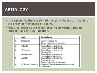
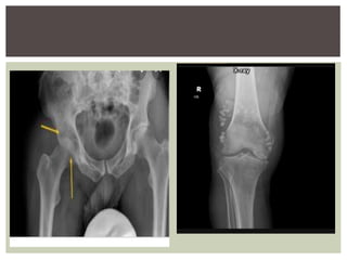
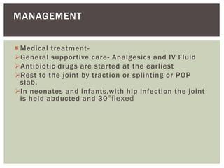
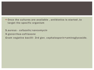
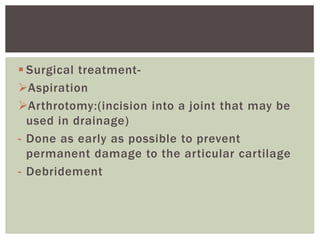
- Shaikh Yumna
SEPTIC ARTHRITIS
INTRODUCTION
 Septic arthritis is an inflammation of synovial
membrane with purulent effusion into the joint
capsule due to infection
 Also referred as Infectious arthritis
 It is commonly caused by Staphylococcus aureus,
N.gonorrhea and less commonly by streptococci,
pneumococci, gonococci etc.
 Seen in every age group ,M=F.
 Common sites-hip joint in childrens ,knee joint in
adults.
 It is caused by the invasion of bacteria ,viruses or fungi into
the synovial membrane of a joint.
 May also begin as the result of an open wound , trauma
,surgery, or unsterile injection.
AETIOLOGY
 Age>80 years old.
 Medical conditions-OA,RA,gout,diabetes HIV.
 Weak immune system.
 Recent joint surgery.
 IV drug use.
 Immunosuppressive drug therapy.
RISK FACTORS
HAEMATOGENOUS
DIRECT
IMPLANTATION
OF THE BACTERIA
IATROGENIC
SECONDARY TO
OSTEOMYELITIS TO
ADJACENT BONE
Inflammation of the synovial membrane
Bacteria enters the body
triggers inflammatory process
inflammation of the synovial membrane with excessive
production of joint fluid/pus
Fluid contains a large no. of cells, bacteria and fibrin
Formation of pus in the joint followed by erosion and
destruction of articular cartilage and the bone.
PATHOGENESIS
Pathologic dislocation may occur
The capsule may get perforated and pus may escape out
forming a sinus.
If untreated, joint may get disorganized and lead to fibrous or
bony ankylosis.
 H/O recent trauma /infection.
 High grade fever.
 Painful , swollen and inflamed joint.
 Warmth and marked local tenderness.
 Restricted movements of affected joint due to pain
and muscle spasm.
CLINICAL FEATURES
 Commonly seen in children younger than 1 year.
 occurs due to spread of infection from the neighbouring bone
lesion or from a septic umbilicus.
 Clinical features:-
Fever.
painful hip.
unable to move the limb
swelling and tenderness around the hip
occasionally in late presentations, the child may present with
a limp when he/she begins to walk
limb shortening (indicative of head destruction)
resembles the condition CDH.
ACUTE SEPTIC ARTHRITIS OF INFANCY
(TOM SMITH ARTHRITIS)
 Investigations
 Radiography
complete loss of head and neck of femur function with poorly
developed acetabulum
there is marked upriding of the trochanter with absence of
femoral head and neck
Laboratory testing includes WBC count,ESR
and CRP.These values are usually elevate
in those with septic arthritis.
INVESTIGATIONS
 X-ray –
Early radiographs may be normal or may show
increased joint space d/t fluid or pus collection.
Late radiographs show destruction of the cartilage ,
new bone formation ,and eventually bony ankylosis or
even pathological dislocation.
 Ultrasonography-
To detect collection of fluid in the joint.
INVESTIGATIONS
Synovial fluid analysis- technique is done by
aspiration of synovial fluid from the affected
area
 Medical treatment-
General supportive care- Analgesics and IV Fluid
Antibiotic drugs are started at the earliest
Rest to the joint by traction or splinting or POP
slab.
In neonates and infants,with hip infection the joint
is held abducted and 30°flexed
MANAGEMENT
 Once the cultures are available , antibiotics is started ,to
target the specific organism
S.aureus - cefazolin/vancomycin
N.gonorrhea-cefriaxone
Gram negative bacilli- 3rd gen. cephalosporin+aminoglycoside.
 Surgical treatment-
Aspiration
Arthrotomy:(incision into a joint that may be
used in drainage)
- Done as early as possible to prevent
permanent damage to the articular cartilage
- Debridement
Physiotherapy treatment-
Primary aims
Patient education about the exercise and the
management of their condition.
Reduce pain.
Restore and maintain physiological function.
Interventions
 Electrotherapy modatilies –Ice, hydrotherapy, heat, TENS
,IFT, Ultrasound.
 Cold- useful in the acute phase or active period of
inflammation. Application of cold produces local analgesia,
increase superficial circulation at the site of application and
decrease intra articular temperature.
10-20mins 1-2 times a day
 Heat-useful in chronic phase. Provides pain relief, reduces
muscle spasm and improves the elasticity of soft tissues.
20-30mins 1-2 times a day
Gentle assisted movements through the joint range
should be started to prevent contractures
Isometrics “static muscle contraction” exercises to
maintain muscle tone without increasing
inflammation
Strong movements of the adjacent joints .
Assisted functional training task and compensatory
methods to facilitate ADLs
Orthotic device
 Gait training with walker
Position the walker in front of the patient
Assist the patient to a standing position
patient leans forward ad pushes up with the arms from the
chair arm rest/ bed to come to stand
Instruct the patient to position his/her body within the frame of
the walker and ask the patient to grasp the hand rest
Instruct the patient to move the walker forward by lifting it
up,moving it forward and setting it down
Instruct the patient to take a step forward with the weak leg
Then ask he patient to move His/her strong leg forward
Instruct to take short steps ,keep head up and eyes looking
forward
Returning to sit
As the patient approaches the chair /bed , the patient turns in
small circles towards the stronger side
Ask the patient to move backwards until the chair /bed can be
felt against the patients leg
Then the patient lowers to the chair/bed in a controlled manner
THANKYOU

Septic Arthritis.pptx

  • 1.
  • 2.
    INTRODUCTION  Septic arthritisis an inflammation of synovial membrane with purulent effusion into the joint capsule due to infection  Also referred as Infectious arthritis  It is commonly caused by Staphylococcus aureus, N.gonorrhea and less commonly by streptococci, pneumococci, gonococci etc.  Seen in every age group ,M=F.  Common sites-hip joint in childrens ,knee joint in adults.
  • 3.
     It iscaused by the invasion of bacteria ,viruses or fungi into the synovial membrane of a joint.  May also begin as the result of an open wound , trauma ,surgery, or unsterile injection. AETIOLOGY
  • 4.
     Age>80 yearsold.  Medical conditions-OA,RA,gout,diabetes HIV.  Weak immune system.  Recent joint surgery.  IV drug use.  Immunosuppressive drug therapy. RISK FACTORS
  • 5.
  • 6.
    Inflammation of thesynovial membrane Bacteria enters the body triggers inflammatory process inflammation of the synovial membrane with excessive production of joint fluid/pus Fluid contains a large no. of cells, bacteria and fibrin Formation of pus in the joint followed by erosion and destruction of articular cartilage and the bone. PATHOGENESIS
  • 7.
    Pathologic dislocation mayoccur The capsule may get perforated and pus may escape out forming a sinus. If untreated, joint may get disorganized and lead to fibrous or bony ankylosis.
  • 8.
     H/O recenttrauma /infection.  High grade fever.  Painful , swollen and inflamed joint.  Warmth and marked local tenderness.  Restricted movements of affected joint due to pain and muscle spasm. CLINICAL FEATURES
  • 10.
     Commonly seenin children younger than 1 year.  occurs due to spread of infection from the neighbouring bone lesion or from a septic umbilicus.  Clinical features:- Fever. painful hip. unable to move the limb swelling and tenderness around the hip occasionally in late presentations, the child may present with a limp when he/she begins to walk limb shortening (indicative of head destruction) resembles the condition CDH. ACUTE SEPTIC ARTHRITIS OF INFANCY (TOM SMITH ARTHRITIS)
  • 11.
     Investigations  Radiography completeloss of head and neck of femur function with poorly developed acetabulum there is marked upriding of the trochanter with absence of femoral head and neck
  • 13.
    Laboratory testing includesWBC count,ESR and CRP.These values are usually elevate in those with septic arthritis. INVESTIGATIONS
  • 14.
     X-ray – Earlyradiographs may be normal or may show increased joint space d/t fluid or pus collection. Late radiographs show destruction of the cartilage , new bone formation ,and eventually bony ankylosis or even pathological dislocation.  Ultrasonography- To detect collection of fluid in the joint. INVESTIGATIONS
  • 15.
    Synovial fluid analysis-technique is done by aspiration of synovial fluid from the affected area
  • 18.
     Medical treatment- Generalsupportive care- Analgesics and IV Fluid Antibiotic drugs are started at the earliest Rest to the joint by traction or splinting or POP slab. In neonates and infants,with hip infection the joint is held abducted and 30°flexed MANAGEMENT
  • 19.
     Once thecultures are available , antibiotics is started ,to target the specific organism S.aureus - cefazolin/vancomycin N.gonorrhea-cefriaxone Gram negative bacilli- 3rd gen. cephalosporin+aminoglycoside.
  • 20.
     Surgical treatment- Aspiration Arthrotomy:(incisioninto a joint that may be used in drainage) - Done as early as possible to prevent permanent damage to the articular cartilage - Debridement
  • 21.
    Physiotherapy treatment- Primary aims Patienteducation about the exercise and the management of their condition. Reduce pain. Restore and maintain physiological function.
  • 22.
    Interventions  Electrotherapy modatilies–Ice, hydrotherapy, heat, TENS ,IFT, Ultrasound.  Cold- useful in the acute phase or active period of inflammation. Application of cold produces local analgesia, increase superficial circulation at the site of application and decrease intra articular temperature. 10-20mins 1-2 times a day  Heat-useful in chronic phase. Provides pain relief, reduces muscle spasm and improves the elasticity of soft tissues. 20-30mins 1-2 times a day
  • 23.
    Gentle assisted movementsthrough the joint range should be started to prevent contractures Isometrics “static muscle contraction” exercises to maintain muscle tone without increasing inflammation Strong movements of the adjacent joints . Assisted functional training task and compensatory methods to facilitate ADLs Orthotic device
  • 24.
     Gait trainingwith walker Position the walker in front of the patient Assist the patient to a standing position patient leans forward ad pushes up with the arms from the chair arm rest/ bed to come to stand Instruct the patient to position his/her body within the frame of the walker and ask the patient to grasp the hand rest Instruct the patient to move the walker forward by lifting it up,moving it forward and setting it down Instruct the patient to take a step forward with the weak leg Then ask he patient to move His/her strong leg forward Instruct to take short steps ,keep head up and eyes looking forward
  • 25.
    Returning to sit Asthe patient approaches the chair /bed , the patient turns in small circles towards the stronger side Ask the patient to move backwards until the chair /bed can be felt against the patients leg Then the patient lowers to the chair/bed in a controlled manner
  • 26.