This case report describes a 53-year-old woman with a large ulnar nerve schwannoma. Schwannomas are rare peripheral nerve tumors that are often misdiagnosed due to nonspecific symptoms. Key diagnostic features of this patient's schwannoma included a mobile mass with a positive Tinel's sign. MRI showed characteristic target sign morphology. Surgical removal of the encapsulated tumor was performed while preserving ulnar nerve function. Histological analysis confirmed the diagnosis of schwannoma through S100 protein staining.
In this presentation, several aspects about imaging, sentinel node and treatment of the N0 neck in head and neck cancer are discussed with emphasis on oral cancer. 2015
In this presentation, several aspects about imaging, sentinel node and treatment of the N0 neck in head and neck cancer are discussed with emphasis on oral cancer. 2015
Cervical Sympathetic chain ganglioneuroma : case report and review of literatureiosrphr_editor
The IOSR Journal of Pharmacy (IOSRPHR) is an open access online & offline peer reviewed international journal, which publishes innovative research papers, reviews, mini-reviews, short communications and notes dealing with Pharmaceutical Sciences( Pharmaceutical Technology, Pharmaceutics, Biopharmaceutics, Pharmacokinetics, Pharmaceutical/Medicinal Chemistry, Computational Chemistry and Molecular Drug Design, Pharmacognosy & Phytochemistry, Pharmacology, Pharmaceutical Analysis, Pharmacy Practice, Clinical and Hospital Pharmacy, Cell Biology, Genomics and Proteomics, Pharmacogenomics, Bioinformatics and Biotechnology of Pharmaceutical Interest........more details on Aim & Scope).
The temporal branch of the facial nerve is a commonly injured nerve during facial trauma due to its superficial course over the zygomatic arch, and is a commonly damaged nerve during facial surgery.1 We report a case of trauma to the left temporal fossa, and subsequent unilateral forehead paralysis. Early exploration revealed external suture compression as the origin of his paralysis. Removal of the suture led to complete resolution of the neurological deficit. The differential diagnosis did not include the possibility of the compression of the nerve by a suture, however the decision for early exploration led to a full recovery.
We report a case of acquired anterior thoracic lung herniation in a 63-year-old female. This painful herniation developed four years after uncomplicated video-assisted thoracic surgery for lung cancer resection and adjuvant radiation for concomitant breast cancer. The herniation site was remote from all prior incisions, and demonstrated intercostal muscle denervation and radiation fibrosis. The 8 cm x 10 cm chest wall defect was reconstructed with inlay PROCEED mesh and reinforced with a pedicled latissimus dorsi flap. Five months postoperatively the patient had complete resolution of symptoms, no evidence of herniation, and a stable wound.
This is initial data from the Figure 8 FlatWire Sternal Closure System. FlatWire is a simple, strong, and cost effective replacement for traditional steel wire for sternal cerclage.
This is a CME article that appears in Plastic and Reconstructive Surgery, the gold standard of publications within the field. Reconstructing the eyelid can be difficult and complicated. This article discusses the various approaches to defects caused by cancer.
This is a pilot study which examines the use of the fistbump instead of a traditional handshake in the hospital setting. In the hospital we use automatic doors, automatic sinks, and alcohol based hand sanitizer. However the rise of antibiotic resistant bacteria continues to increase. We propose ceasing handshaking within the hospital and opting instead for the fun fistbump will reduce the transmission of bacteria.
This is a paper that Dr. W. Thomas McClellan co-authored on the anatomy and reconstruction of the inframammary fold. This critical structure is often injured during breast augmentation and understanding of the anatomy is crucial to a good outcome in breast augmentation.
This is a powerpoint presentaiton given by W. Thomas McClellan, MD FACS, a Board Certified Plastic Surgeon who specializes in breast augmentation. This presentation is unique and critical because it gives patients detailed information about what is important regarding breast augmentation. For example: How to choose a surgeon, what is important in the operating room, postoperative care, how to pick a size, type of breast implant.
This is a paper which describes an innovative approach for skin sparing mastectomy. This incision tends to distract the eye and be less noticeable. Additionally it allows excellent access to the axilla for lymph node sampling and reduces the excessive retraction on the skin flaps.
micro teaching on communication m.sc nursing.pdfAnurag Sharma
Microteaching is a unique model of practice teaching. It is a viable instrument for the. desired change in the teaching behavior or the behavior potential which, in specified types of real. classroom situations, tends to facilitate the achievement of specified types of objectives.
Tom Selleck Health: A Comprehensive Look at the Iconic Actor’s Wellness Journeygreendigital
Tom Selleck, an enduring figure in Hollywood. has captivated audiences for decades with his rugged charm, iconic moustache. and memorable roles in television and film. From his breakout role as Thomas Magnum in Magnum P.I. to his current portrayal of Frank Reagan in Blue Bloods. Selleck's career has spanned over 50 years. But beyond his professional achievements. fans have often been curious about Tom Selleck Health. especially as he has aged in the public eye.
Follow us on: Pinterest
Introduction
Many have been interested in Tom Selleck health. not only because of his enduring presence on screen but also because of the challenges. and lifestyle choices he has faced and made over the years. This article delves into the various aspects of Tom Selleck health. exploring his fitness regimen, diet, mental health. and the challenges he has encountered as he ages. We'll look at how he maintains his well-being. the health issues he has faced, and his approach to ageing .
Early Life and Career
Childhood and Athletic Beginnings
Tom Selleck was born on January 29, 1945, in Detroit, Michigan, and grew up in Sherman Oaks, California. From an early age, he was involved in sports, particularly basketball. which played a significant role in his physical development. His athletic pursuits continued into college. where he attended the University of Southern California (USC) on a basketball scholarship. This early involvement in sports laid a strong foundation for his physical health and disciplined lifestyle.
Transition to Acting
Selleck's transition from an athlete to an actor came with its physical demands. His first significant role in "Magnum P.I." required him to perform various stunts and maintain a fit appearance. This role, which he played from 1980 to 1988. necessitated a rigorous fitness routine to meet the show's demands. setting the stage for his long-term commitment to health and wellness.
Fitness Regimen
Workout Routine
Tom Selleck health and fitness regimen has evolved. adapting to his changing roles and age. During his "Magnum, P.I." days. Selleck's workouts were intense and focused on building and maintaining muscle mass. His routine included weightlifting, cardiovascular exercises. and specific training for the stunts he performed on the show.
Selleck adjusted his fitness routine as he aged to suit his body's needs. Today, his workouts focus on maintaining flexibility, strength, and cardiovascular health. He incorporates low-impact exercises such as swimming, walking, and light weightlifting. This balanced approach helps him stay fit without putting undue strain on his joints and muscles.
Importance of Flexibility and Mobility
In recent years, Selleck has emphasized the importance of flexibility and mobility in his fitness regimen. Understanding the natural decline in muscle mass and joint flexibility with age. he includes stretching and yoga in his routine. These practices help prevent injuries, improve posture, and maintain mobilit
Report Back from SGO 2024: What’s the Latest in Cervical Cancer?bkling
Are you curious about what’s new in cervical cancer research or unsure what the findings mean? Join Dr. Emily Ko, a gynecologic oncologist at Penn Medicine, to learn about the latest updates from the Society of Gynecologic Oncology (SGO) 2024 Annual Meeting on Women’s Cancer. Dr. Ko will discuss what the research presented at the conference means for you and answer your questions about the new developments.
Ozempic: Preoperative Management of Patients on GLP-1 Receptor Agonists Saeid Safari
Preoperative Management of Patients on GLP-1 Receptor Agonists like Ozempic and Semiglutide
ASA GUIDELINE
NYSORA Guideline
2 Case Reports of Gastric Ultrasound
Anti ulcer drugs and their Advance pharmacology ||
Anti-ulcer drugs are medications used to prevent and treat ulcers in the stomach and upper part of the small intestine (duodenal ulcers). These ulcers are often caused by an imbalance between stomach acid and the mucosal lining, which protects the stomach lining.
||Scope: Overview of various classes of anti-ulcer drugs, their mechanisms of action, indications, side effects, and clinical considerations.
These simplified slides by Dr. Sidra Arshad present an overview of the non-respiratory functions of the respiratory tract.
Learning objectives:
1. Enlist the non-respiratory functions of the respiratory tract
2. Briefly explain how these functions are carried out
3. Discuss the significance of dead space
4. Differentiate between minute ventilation and alveolar ventilation
5. Describe the cough and sneeze reflexes
Study Resources:
1. Chapter 39, Guyton and Hall Textbook of Medical Physiology, 14th edition
2. Chapter 34, Ganong’s Review of Medical Physiology, 26th edition
3. Chapter 17, Human Physiology by Lauralee Sherwood, 9th edition
4. Non-respiratory functions of the lungs https://academic.oup.com/bjaed/article/13/3/98/278874
Ethanol (CH3CH2OH), or beverage alcohol, is a two-carbon alcohol
that is rapidly distributed in the body and brain. Ethanol alters many
neurochemical systems and has rewarding and addictive properties. It
is the oldest recreational drug and likely contributes to more morbidity,
mortality, and public health costs than all illicit drugs combined. The
5th edition of the Diagnostic and Statistical Manual of Mental Disorders
(DSM-5) integrates alcohol abuse and alcohol dependence into a single
disorder called alcohol use disorder (AUD), with mild, moderate,
and severe subclassifications (American Psychiatric Association, 2013).
In the DSM-5, all types of substance abuse and dependence have been
combined into a single substance use disorder (SUD) on a continuum
from mild to severe. A diagnosis of AUD requires that at least two of
the 11 DSM-5 behaviors be present within a 12-month period (mild
AUD: 2–3 criteria; moderate AUD: 4–5 criteria; severe AUD: 6–11 criteria).
The four main behavioral effects of AUD are impaired control over
drinking, negative social consequences, risky use, and altered physiological
effects (tolerance, withdrawal). This chapter presents an overview
of the prevalence and harmful consequences of AUD in the U.S.,
the systemic nature of the disease, neurocircuitry and stages of AUD,
comorbidities, fetal alcohol spectrum disorders, genetic risk factors, and
pharmacotherapies for AUD.
Prix Galien International 2024 Forum ProgramLevi Shapiro
June 20, 2024, Prix Galien International and Jerusalem Ethics Forum in ROME. Detailed agenda including panels:
- ADVANCES IN CARDIOLOGY: A NEW PARADIGM IS COMING
- WOMEN’S HEALTH: FERTILITY PRESERVATION
- WHAT’S NEW IN THE TREATMENT OF INFECTIOUS,
ONCOLOGICAL AND INFLAMMATORY SKIN DISEASES?
- ARTIFICIAL INTELLIGENCE AND ETHICS
- GENE THERAPY
- BEYOND BORDERS: GLOBAL INITIATIVES FOR DEMOCRATIZING LIFE SCIENCE TECHNOLOGIES AND PROMOTING ACCESS TO HEALTHCARE
- ETHICAL CHALLENGES IN LIFE SCIENCES
- Prix Galien International Awards Ceremony
Title: Sense of Smell
Presenter: Dr. Faiza, Assistant Professor of Physiology
Qualifications:
MBBS (Best Graduate, AIMC Lahore)
FCPS Physiology
ICMT, CHPE, DHPE (STMU)
MPH (GC University, Faisalabad)
MBA (Virtual University of Pakistan)
Learning Objectives:
Describe the primary categories of smells and the concept of odor blindness.
Explain the structure and location of the olfactory membrane and mucosa, including the types and roles of cells involved in olfaction.
Describe the pathway and mechanisms of olfactory signal transmission from the olfactory receptors to the brain.
Illustrate the biochemical cascade triggered by odorant binding to olfactory receptors, including the role of G-proteins and second messengers in generating an action potential.
Identify different types of olfactory disorders such as anosmia, hyposmia, hyperosmia, and dysosmia, including their potential causes.
Key Topics:
Olfactory Genes:
3% of the human genome accounts for olfactory genes.
400 genes for odorant receptors.
Olfactory Membrane:
Located in the superior part of the nasal cavity.
Medially: Folds downward along the superior septum.
Laterally: Folds over the superior turbinate and upper surface of the middle turbinate.
Total surface area: 5-10 square centimeters.
Olfactory Mucosa:
Olfactory Cells: Bipolar nerve cells derived from the CNS (100 million), with 4-25 olfactory cilia per cell.
Sustentacular Cells: Produce mucus and maintain ionic and molecular environment.
Basal Cells: Replace worn-out olfactory cells with an average lifespan of 1-2 months.
Bowman’s Gland: Secretes mucus.
Stimulation of Olfactory Cells:
Odorant dissolves in mucus and attaches to receptors on olfactory cilia.
Involves a cascade effect through G-proteins and second messengers, leading to depolarization and action potential generation in the olfactory nerve.
Quality of a Good Odorant:
Small (3-20 Carbon atoms), volatile, water-soluble, and lipid-soluble.
Facilitated by odorant-binding proteins in mucus.
Membrane Potential and Action Potential:
Resting membrane potential: -55mV.
Action potential frequency in the olfactory nerve increases with odorant strength.
Adaptation Towards the Sense of Smell:
Rapid adaptation within the first second, with further slow adaptation.
Psychological adaptation greater than receptor adaptation, involving feedback inhibition from the central nervous system.
Primary Sensations of Smell:
Camphoraceous, Musky, Floral, Pepperminty, Ethereal, Pungent, Putrid.
Odor Detection Threshold:
Examples: Hydrogen sulfide (0.0005 ppm), Methyl-mercaptan (0.002 ppm).
Some toxic substances are odorless at lethal concentrations.
Characteristics of Smell:
Odor blindness for single substances due to lack of appropriate receptor protein.
Behavioral and emotional influences of smell.
Transmission of Olfactory Signals:
From olfactory cells to glomeruli in the olfactory bulb, involving lateral inhibition.
Primitive, less old, and new olfactory systems with different path
Flu Vaccine Alert in Bangalore Karnatakaaddon Scans
As flu season approaches, health officials in Bangalore, Karnataka, are urging residents to get their flu vaccinations. The seasonal flu, while common, can lead to severe health complications, particularly for vulnerable populations such as young children, the elderly, and those with underlying health conditions.
Dr. Vidisha Kumari, a leading epidemiologist in Bangalore, emphasizes the importance of getting vaccinated. "The flu vaccine is our best defense against the influenza virus. It not only protects individuals but also helps prevent the spread of the virus in our communities," he says.
This year, the flu season is expected to coincide with a potential increase in other respiratory illnesses. The Karnataka Health Department has launched an awareness campaign highlighting the significance of flu vaccinations. They have set up multiple vaccination centers across Bangalore, making it convenient for residents to receive their shots.
To encourage widespread vaccination, the government is also collaborating with local schools, workplaces, and community centers to facilitate vaccination drives. Special attention is being given to ensuring that the vaccine is accessible to all, including marginalized communities who may have limited access to healthcare.
Residents are reminded that the flu vaccine is safe and effective. Common side effects are mild and may include soreness at the injection site, mild fever, or muscle aches. These side effects are generally short-lived and far less severe than the flu itself.
Healthcare providers are also stressing the importance of continuing COVID-19 precautions. Wearing masks, practicing good hand hygiene, and maintaining social distancing are still crucial, especially in crowded places.
Protect yourself and your loved ones by getting vaccinated. Together, we can help keep Bangalore healthy and safe this flu season. For more information on vaccination centers and schedules, residents can visit the Karnataka Health Department’s official website or follow their social media pages.
Stay informed, stay safe, and get your flu shot today!
Couples presenting to the infertility clinic- Do they really have infertility...Sujoy Dasgupta
Dr Sujoy Dasgupta presented the study on "Couples presenting to the infertility clinic- Do they really have infertility? – The unexplored stories of non-consummation" in the 13th Congress of the Asia Pacific Initiative on Reproduction (ASPIRE 2024) at Manila on 24 May, 2024.
Factory Supply Best Quality Pmk Oil CAS 28578–16–7 PMK Powder in Stockrebeccabio
Factory Supply Best Quality Pmk Oil CAS 28578–16–7 PMK Powder in Stock
Telegram: bmksupplier
signal: +85264872720
threema: TUD4A6YC
You can contact me on Telegram or Threema
Communicate promptly and reply
Free of customs clearance, Double Clearance 100% pass delivery to USA, Canada, Spain, Germany, Netherland, Poland, Italy, Sweden, UK, Czech Republic, Australia, Mexico, Russia, Ukraine, Kazakhstan.Door to door service
Hot Selling Organic intermediates
MANAGEMENT OF ATRIOVENTRICULAR CONDUCTION BLOCK.pdfJim Jacob Roy
Cardiac conduction defects can occur due to various causes.
Atrioventricular conduction blocks ( AV blocks ) are classified into 3 types.
This document describes the acute management of AV block.
MANAGEMENT OF ATRIOVENTRICULAR CONDUCTION BLOCK.pdf
Schwannoma of the ulnar nerve
1. Schwannoma of the Ulnar Nerve: A Case and Review of the Literature
Ashley Boustany, MS II
West Virginia University School of Medicine
W. Thomas McClellan, M.D.
Plastic Surgeon
Private Practice
Abstract
We report a case of a large ulnar nerve schwannoma, a rare type of soft tissue neoplasm.
Diagnostic pearls are described to facilitate a more accurate and timely diagnosis. These
characteristics include mobility, Tinel’s sign, MRI target sign, S100 histological staining, Antoni
patterns, and others. With a correct diagnosis, the tumor can be extirpated with preservation of
nerve function and a low risk of recurrence.
Introduction
Schwannomas are the most common type of tumor arising in peripheral nerves. (1)
However, peripheral nerve tumors are rare, representing less than 8% of soft tissue neoplasms.
Schwannomas are non-invasive tumors arising from peripheral nerve sheaths and are
encapsulated by epineurium. (2) There is a 2:1 occurrence of upper limb to lower limb
schwannomas, generally on the volar surface. There is no predisposition for sex or race, but they
usually develop in 30-60 year olds. (1) Schwannomas are often misdiagnosed due to their
indistinct signs and symptoms, which may lead to detrimental neurologic deficits if approached
incorrectly. It has been shown that less than a quarter of diagnostic tests provide an accurate
diagnosis of schwannoma. (3)
Case
We present a 53 year old right hand dominant female with a slowly enlarging mass
within her proximal volar right forearm (Figure 1). The mass had been present for a minimum of
two years and had become more painful and noticeable over time. Dull, intermittent pain was
reported at rest over the mass, and was moderately sharper during flexion of the fingers or
bumping of the forearm. The level of discomfort increasingly hindered her ability to perform
activities of daily living.
She was initially treated conservatively at an outside facility for a ruptured forearm
muscle belly. Subsequently, her symptoms worsened and a CT guided biopsy of the suspected
mass was performed. The patient reported the biopsy was extremely painful and caused an
exacerbation of her forearm pain. Histopathology demonstrated a paucicellular specimen with
very rare fragments of fibrous tissue that was insufficient for diagnosis.
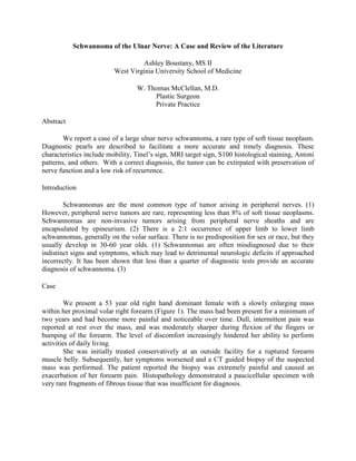
2. Figure 1. Preoperative: The dotted
line indicates the edge of the
palpable tumor. Additionally, the
olecranon, medial epicondyle, and
path of the ulnar nerve are
identified.
She was referred to our hand surgery service following the inconclusive biopsy. The 5 cm
firm mass was mobile perpendicular to the nerve axis and immobile along the parallel axis. A
very sensitive and positive Tinel’s sign radiated in the ulnar distribution, with dysthesias directly
over the mass and along the dorsal ulnar distribution of her hand. Semmes Weinstein testing
indicated diminished light touch and hypersensitivity to sharp touch in the same region.
However, her strength of the intrinsic muscles of the right hand was 5/5 without evidence of
weakness or loss of range of motion.
An MRI was performed and showed a 5.0 cm x 3.0 cm x 4.6 cm diameter mass
displacing the flexor digitorum superficialis and palmaris longus muscles (Figure 2). The mass
had smooth margins but displayed a complex heterogeneous signal. There was no evidence of
invasion of adjoining muscles or bones. With gadolinium injection, an intense irregular
enhancement was seen. A capsule surrounded the eccentrically placed mass. Images also
indicated the presence of a hemorrhagic center related to the needle biopsy. MRI diagnosis was
inconclusive, but the differential included, schwannoma, malignant histiocytoma,
neurofibrosarcoma, or soft tissue malignancy.
Figure 2. MRI: T1
weighted MRI of the
right upper extremity
showing a large well
demarcated but
heterogeneous mass.
The MRI lacked the
traditional “target
sign” that is
commonly associated
with schwannomas.
3. Figure 3. Intraoperative:
The proximal and distal
ulnar nerve are identified
with vascular loops. Note
how the fascicles of the
ulnar nerve are splayed
out over the tumor.
Identification of these
fascicles is crucial to
determine a safe
longitudinal entry point
through the epineurium.
She was taken to the operating room for exploration. Extirpation of the tumor was
performed via a longitudinal access incision through the epineurium and careful separation of the
nerve and tumor (Figure 3). The tumor measured 4.0 cm x 3.0 cm x 1.9 cm (Figure 4). It showed
a hypo-cellular tissue with Schwann cells strongly positive for S100 protein by immunostain
technique (Figure 5). Antoni B patterns predominated with few areas suggestive of Antoni A
patterns. Incision of the tumor also revealed hemorrhage and thrombosis consistent with prior
needle aspiration. Three months following surgery the patient has retained full ulnar motor and
sensory function as well as improving sensory paresthesias in her dorsal ulnar division.
Figure 4. Tumor: Gross
appearance of the Schwannoma
following removal from within
the ulnar nerve.
4. Figure 5. Histopathology:
A hypo-cellular tissue with
Schwann cells staining
strongly positive for S100
protein.
Discussion
Diagnosis of a schwannoma in the preoperative period is challenging because of the slow
growth and paucity of symptoms. Diagnostic accuracy is crucial to maintaining the integrity of
the nerve involved and to properly plan the appropriate surgical intervention. Often these tumors
present as palpable masses, tender to displacement without muscles weakness. Tinel’s sign is
positive in the majority of cases. The tumors are transversely mobile but immobile
longitudinally, likely due to their nested intraneural location. Schwannomas share many features
with other soft tissue tumors and are frequently misdiagnosed due to similarities. Differential
diagnosis should include neurofibroma, ganglion cysts, malignant tumors, lipomas, and
xanthomas. (1,4) Neurofibromas, in particular, cannot be distinguished from schwannomas on
physical examination. The symptoms appear to be nonspecific, which adds to difficulty in
diagnosis. (5) They may be differentiated on MRI or fine needle aspiration. Malignant masses
exhibit more distinct signs but are often mistaken for schwannomas in early stages of diagnosis.
Unlike schwannomas, malignant tumors often have immobility, firmness, constant pain at rest,
and motor weakness. (6) Weakness may occur if the benign tumor exceeds 2.5 cm and can be
location dependent. Kehoe et al (1995) analyzed 88 peripheral nerve tumors, where only one
was correctly diagnosed as a schwannoma preoperatively.
Gadolinium enhanced T1-weighted and T2-weighted MRIs are particularly useful in
diagnosing schwannomas. Koga et al (2007) found the presence of the target sign to be 100%
specific and 59% sensitive for the tumors. The target sign is the contrasting central and
peripheral intensities demonstrated on the images. Histological analysis credits the central
hyperintensities and hypointensities on T1 gadolinium and T2, respectively, to Antoni A cells.
Antoni A patterns are of low cellular concentration. Antoni B areas are of high cell
concentration and correspond to peripheral intensities on MRI and CT. (8)
Histological staining reveals a strongly positive S100 protein that is specific for
schwannomas and helps to rule out neurofibromas. (5,9) Imaging shows the tumors as round or
oval, eccentrically located in relation to the nerve, encapsulated, isolated, and non-invasive. In
comparison, neurofibromas are non-encapsulated and intimately surround the nerve. They cannot
be surgically removed without damaging the connected nerve, often necessitating nerve grafting
to repair functioning. (2,4,5) Schwannomas, on the other hand, can be separated surgically from
5. the nerve fascicles avoiding neurologic deficits. (4) This emphasizes the importance of a correct
preoperative diagnosis. Despite the structural differences of soft tissue tumors, they are difficult
to distinguish with imaging.
Fine needle aspiration tends to be extremely painful in cases of schwannomas, and
hemorrhaging may result with temporary worsening of symptoms. The results are frequently
inconclusive, but are helpful to exclude ganglion cysts. (3) Domanksi et al (2006) aspirated 116
different schwannomas, and results were not sufficient for diagnosis for about 44% of the cases.
Extirpation of the intraneural schwannoma can be challenging. Sterile tourniquet
dissection is recommended and assists in visualization. Loupe magnification and or use of the
operating microscope is highly recommended. Identification of the nerve proximal and distal to
the tumor is the first important step to reducing injury and traction neuropraxia. Identification of
the individual splayed nerve fascicles as they spread over of the tumor is critical in determining
the entry through epineurium. A longitudinal incision is created between the splayed fascicles
down to the tumor sheath. Once the outer layer of the tumor is identified, a plane can be
developed between the more superficial fascicles and the tumor wall. Slow, deliberate,
circumferential dissection with a “peanut” and Littler scissors facilitates delivery of the tumor.
Once the tumor is removed, the nerve is inspected for injury, the tourniquet is released, and
precise hemostasis is achieved. Repair of the epineurium is not required and the longitudinally
split muscle is repaired loosely over the nerve. Drains are optional, bulky dressing is preferred,
and immediate post operative hand therapy is instituted.
It is uncommon for schwannomas to recur in identical locations. (1) Das et al (2007)
found that surgical removal of schwannomas was successful in alleviating preoperative
symptoms while maintaining nerve functioning in 89% of their cases.
Conclusion
Schwannomas are rare peripheral nerve tumors that have important diagnostic and
radiographic features. These tumors are transversely mobile and longitudinally immobile, have a
positive Tinel’s sign, and exertional dysathesias or pain. MRI typically reveals the target sign of
biphasic contrast of peripheral and central regions and distinct encapsulation displacing the
intimately associated nerve fascicles. Surgical resection must be approached with caution to
protect nerve function and continuity. Surgical resection is associated with good outcomes. The
recurrence rate is low.
References
1. Ozdemir O, Kurt C, et.al. Schwannomas of the hand and wrist: long-term results and review of
literature. Journal of Orthopaedic Surgery. 2005;13(3):267-272.
2. Lin J, Martel W. Cross-sectional imaging of peripheral nerve sheath tumors: characteristic
signs on CT, MR imaging, and sonography. Amer Journ Roentgenology. 2001 Jan.;176:75-82.
3. Rockwell G, Achilleas T, et. al. Schwannoma of the hand and wrist. Plast Reconstr Surg. 2003
Mar.;111(3):1227-1232.
6. 4. Sandberg K, Nilsson J, et. al. Tumors of peripheral nerves in the upper extremity: A 22-year
epidemiological study. Scand J Plast Reconstr Hand Surg. 2009 Sep.;43:43-49.
5. Adani R, Baccarani A, et. al. Schwannomas of the upper extremity: diagnosis and treatment.
Chir Orani Mov. 2008 Sep.;92:85-88.
6. Ogose A, Hotta T, et. al. Tumors of peripheral nerves: correlation of symptoms, clinical signs,
imaging features, and histologic diagnosis. Skeletal Radiol. 1999 Jan.;28:183-188.
7. Kehoe N, Reid R, et. al. Solitary benign peripheral-nerve tumors: review of 32 years’
experience. J Bone Joint Surg. 1995 Oct.;77B:497-500.
8. Koga H, Matsumoto S, et. al. Definition of the target sign and its use for diagnosis of
schwannomas. Clinical Orthopaedics and Related Research. 2007 Aug.;464:224-229.
9. Das S, Ganju A, et. al. Tumors of the brachial plexus. Neurosurg. 2007June;22(6):E26.
10. Domanski H, Akerman M, et. al. Fine Needle Aspiration of Neurilemoma (Schwannoma). A
clinicocytopahologic study of 116 patients. Diagnostic Cytopathology. 2006;34:403-412.