1) Recurrent miscarriage is defined as 3 or more consecutive pregnancy losses at or before 20 weeks gestation. It affects approximately 1% of fertile couples.
2) The main causes of recurrent pregnancy loss are parental chromosomal abnormalities, uterine abnormalities, and antiphospholipid antibody syndrome. Other potential causes include endocrine, immunological, thrombophilic, and infectious factors.
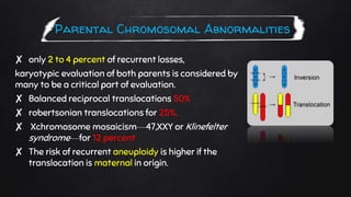
3) Evaluation of patients with recurrent miscarriage includes obtaining a thorough history, physical exam, testing for antiphospholipid antibodies and parental chromosome analysis, and evaluating the uterine cavity through procedures like hysterosalpingogram or hysteroscopy. No cause is identified in around 50% of couples.