Rna And Dna Editing Methods And Protocols 1st Edition H Ulrich Gringer
Rna And Dna Editing Methods And Protocols 1st Edition H Ulrich Gringer
Rna And Dna Editing Methods And Protocols 1st Edition H Ulrich Gringer
Rna And Dna Editing Methods And Protocols 1st Edition H Ulrich Gringer
Rna And Dna Editing Methods And Protocols 1st Edition H Ulrich Gringer
1.
Rna And DnaEditing Methods And Protocols 1st
Edition H Ulrich Gringer download
https://ebookbell.com/product/rna-and-dna-editing-methods-and-
protocols-1st-edition-h-ulrich-gringer-2108266
Explore and download more ebooks at ebookbell.com
2.
Here are somerecommended products that we believe you will be
interested in. You can click the link to download.
Rna And Dna Editing Molecular Mechanisms And Their Integration Into
Biological Systems 1st Edition Harold C Smith
https://ebookbell.com/product/rna-and-dna-editing-molecular-
mechanisms-and-their-integration-into-biological-systems-1st-edition-
harold-c-smith-1356238
Rna And Dna Diagnostics 1st Edition Volker A Erdmann Stefan Jurga
https://ebookbell.com/product/rna-and-dna-diagnostics-1st-edition-
volker-a-erdmann-stefan-jurga-5140964
Computational Studies Of Rna And Dna Bohdan Schneider Helen M Berman
Auth
https://ebookbell.com/product/computational-studies-of-rna-and-dna-
bohdan-schneider-helen-m-berman-auth-4390156
Rnadna And Cancer 1st Edition Joseph G Sinkovics Auth
https://ebookbell.com/product/rnadna-and-cancer-1st-edition-joseph-g-
sinkovics-auth-5482638
3.
Dna And RnaIsolation Techniques For Nonexperts Akash Gautam
https://ebookbell.com/product/dna-and-rna-isolation-techniques-for-
nonexperts-akash-gautam-46355016
Dna And Rna Origami Methods And Protocols Methods In Molecular Biology
2639 1st Ed 2023 Julin Valero Editor
https://ebookbell.com/product/dna-and-rna-origami-methods-and-
protocols-methods-in-molecular-biology-2639-1st-ed-2023-julin-valero-
editor-50124582
Dna And Rna Nanobiotechnologies In Medicine Diagnosis And Treatment Of
Diseases 1st Edition Dhiraj Bhatia
https://ebookbell.com/product/dna-and-rna-nanobiotechnologies-in-
medicine-diagnosis-and-treatment-of-diseases-1st-edition-dhiraj-
bhatia-4404980
Innate Dna And Rna Recognition Methods And Protocols 1st Edition
Hansjoachim Anders
https://ebookbell.com/product/innate-dna-and-rna-recognition-methods-
and-protocols-1st-edition-hansjoachim-anders-4705642
Synthetic Dna And Rna Programming Patrick Odonoghue Ilka Heinemann
https://ebookbell.com/product/synthetic-dna-and-rna-programming-
patrick-odonoghue-ilka-heinemann-10893916
6.
Me t ho d s i n Mo l e c u l a r Bi o l o g y ™
Series Editor
John M. Walker
School of Life Sciences
University of Hertfordshire
Hatfield, Hertfordshire, AL10 9AB, UK
For other titles published in this series, go to
www.springer.com/series/7651
8.
RNA and DNAEditing
Methods and Protocols
Edited by
Ruslan Aphasizhev
DepartmentofMicrobiologyandMolecularGenetics,
SchoolofMedicine,UniversityofCalifornia,Irvine,CA,USA
v
Foreword
RNA editing isa disparate field held together by history and friendships between the
researchers. The term was coined by Robb Benne in his 1986 Cell paper in which he
described the presence of four nonencoded uridylyl nucleotides in the middle of the
mRNA for cytochrome oxidase subunit II in the trypanosomatid protist, Crithidia
fasciculata. The striking thing was that these precise U-insertions at the RNA level pre-
cisely compensated for or “edited” an encoded −1 frameshift in the mitochondrial max-
icircle DNA encoded gene. In 1989, Janet Shaw and I tried to define RNA editing as “any
modification of the sequence of an mRNA molecule within coding regions, except
for splicing of introns.” This definition encompassed a single C to U substitution in
the mRNA for mammalian apolipoprotein B (apoB), specific A to I modifications in the
mRNA for the glutamate receptor, multiple C to U substitutions in plant chloroplast and
mitochondrial mRNAs, and even insertions of G residues within coding regions of nega-
tive strand RNA viruses. But it failed to cover specific nucleotide changes in tRNAs from
Acanthamoeba and marsupials, and the most diverse and striking phenomenon of all, spe-
cific insertions of multiple C and dinucleotide residues and other modifications in all
mitochondrial transcripts in Physarum, including rRNAs. The sexy term, RNA editing,
was hijacked (just joking) to describe all of these disparate genetic events, in spite of the
growing realization that quite different mechanisms were involved.
A book on RNA editing edited by Benne was published in 1994. The actual birth of
the RNA editing research field can probably be traced to a meeting in Albany organized
by Harold Smith the same year, which led to the establishment of a Gordon Conference
on RNA editing in 1989 and thereby made the field respectable. This field was the “flavor
of the week” for a while, but has had its ups and downs as is the case for any field of
research. Even as the realization grew that the various editing phenomena were only
related by the name, the field held together due to its scientific comaraderie and subse-
quent cross fertilization of ideas which, I believe, has led to many really exciting and
unexpected discoveries, such as for example cytidine deaminase-induced DNA editing
which is involved in the generation of antibody diversity. The field experienced some
intense soul searching when it debated encompassing the large well-established field of
nucleotide modifications in general as a type of “editing.” The decision to do this not only
provided the editing field with some very smart colleagues but also proved highly benefi-
cial to the mechanistic understanding of all types of editing.
In 1990, Beat Blum, Norbert Bakalara, and I uncovered the mechanism of the
U-insertion/deletion editing in trypanosome mitochondria by discovering a novel class of
small mitochondrial RNAs which we termed “guide RNAs” since they guided the editing
machinery to specific sites in the mRNAs. The solution to the precise site specificity of this
type of editing was simply base-pairing in trans. The site specificity of A to I editing in
neurons also proved to be due to base-pairing, except the complementary sequence is in
cis downstream of the editing site. This same base-pairing mechanism turned out to be
used by the small siRNAs which mediate the cleavage of RNAs in the Nobel Prize winning
RNA interference phenomenon, which was discovered in 1998, and also by the hitherto
mysterious yeast snoRNAs that determine the sites of pseudoU formation and
11.
vi Foreword
2¢-O-methylation inrRNAs. Alas, the latter two fields never directly acknowledged their
debt to the editing paradigm, but did so inadvertently by using the term, guide RNAs, to
describe the siRNAs and snoRNAs. But base-pairing site specificity was not the answer to
other types of editing and nucleotide modifications. The precise apoB mRNA C to U
change was due to the recognition of an upstream “mooring sequence” by a deamination
protein together with other factors, and the specificity of C to U changes in plant organel-
lar RNAs was also due to the recognition of specific sequences by proteins. The viral G
addition phenomenon turned out to be due to “stuttering” of a polymerase. And the
incredible Physarum C-insertion phenomenon is cotranscriptional also with some sequence
specificity.
A clear indication of the state of advancement and general acceptance of any field of
research is to have a “Methods” book published, which describes “cookbook” procedures
to repeat some of the most interesting discoveries in your own laboratory. Hence, the
publication of this Methods in Molecular Biology book (and a Methods in Enzymology book
in 2007 edited by Jonatha Gott) is to be celebrated. There are 16 chapters describing
state-of-the-art research in trypanosome U-insertion/deletion editing, A to I editing in
Drosophila, C to U RNA and DNA editing, C to U editing in plant chloroplasts and mito-
chondria and RNA modifications.
Larry Simpson
12.
vii
Preface
The term “RNAediting,” coined in 1986 to describe the insertion of four uridines into a
mitochondrial transcript in Trypanosoma brucei, has evolved into a collective definition of
processes that change RNA nucleotide sequence. Setting these pathways apart from
splicing,
5¢ capping, or 3¢ extensions is uncomplicated while drawing a clear distinction from RNA
modifications is less so. Spread throughout the Eukarya, editing creates genetic information
de novo, alters decoding capacity, influences structure, stability, export, and other aspects
of nucleic acids metabolism by inserting, deleting, adding, or modifying nucleotides.
Evolutionarily unrelated, although sometimes confined to the same organism or organelle,
editing events occur cotranscriptionally or posttranscriptionally. Mechanistically, RNA edit-
ing reactions include guide RNA-directed cascades of
nucleolytic and phosphoryl transfer
reactions, RNA polymerase stuttering, site-specific deamination, 3¢–5¢ polymerization, and
others. A more recent but most exciting development, DNA editing is emerging as the key
component of antibody gene diversification and antiviral defense.
Such diversity of organisms and editing types stimulated development of many unique
genetic, molecular, biochemical, and computational approaches. The purpose of this
volume is to introduce methods developed over the last few years to study the diversity of
editing substrates, mechanisms of specificity, and functions of RNA and DNA editing
enzymes and complexes.
I wish to express my sincere gratitude to the authors for their contributions and con-
tinued enthusiasm for our filed. This volume is dedicated to Rob Benne in appreciation of
his seminal discovery.
Ruslan Aphasizhev
14.
ix
Contents
Foreword . . . . . . . . . . . . . . . . . . . . . . . . . . . . . . . . . . . . . . . . . . . . . . . . . . . . . . . . . . . . . . . v
Preface . . . . . . . . . . . . . . . . . . . . . . . . . . . . . . . . . . . . . . . . . . . . . . . . . . . . . . . . . . . . vii
Contributors . . . . . . . . . . . . . . . . . . . . . . . . . . . . . . . . . . . . . . . . . . . . . . . . . . . . . . . . . . . . xi
Part I
Uracil Insertion/Deletion RNA Editing
in Mitochondrion of Trypanosoma brucei
1 Three-Dimensional Reconstruction of Trypanosoma brucei
Editosomes Using Single-Particle Electron Microscopy . . . . . . . . . . . . . . . . . . . . 3
H. Ulrich Göringer, Holger Stark, Cordula Böhm,
Bjoern Sander, and Monika M. Golas
2 iCODA: RNAi-Based Inducible Knock-In System
in Trypanosoma brucei . . . . . . . . . . . . . . . . . . . . . . . . . . . . . . . . . . . . . . . . . . . . . 23
Gene-Errol Ringpis, Richard H. Lathrop, and Ruslan Aphasizhev
Part II Adenosine to Inosine RNA Editing
3 Perturbing A-to-I RNA Editing Using Genetics
and Homologous Recombination . . . . . . . . . . . . . . . . . . . . . . . . . . . . . . . . . . . . 41
Cynthia J. Staber, Selena Gell, James E.C. Jepson, and Robert A. Reenan
4 Laser Microdissection and Pressure Catapulting of Single
Human Motor Neurons for RNA Editing Analysis . . . . . . . . . . . . . . . . . . . . . . . . 75
Hui Sun, Aruna Raja, Mary A. O’Connell, Valerie Mann, Brendon Noble,
and Liam P. Keegan
5 Biochemical Identification of A-to-I RNA Editing Sites
by the Inosine Chemical Erasing (ICE) Method . . . . . . . . . . . . . . . . . . . . . . . . . . 89
Masayuki Sakurai and Tsutomu Suzuki
Part III Cytidine to Uracil RNA and DNA Editing
6 Identifying mRNA Editing Deaminase Targets by RNA-Seq . . . . . . . . . . . . . . . . 103
Brad R. Rosenberg, Scott Dewell, and F. Nina Papavasiliou
7 Mouse and Other Rodent Models of C to U RNA Editing . . . . . . . . . . . . . . . . . . 121
Valerie Blanc and Nicholas O. Davidson
8 In Vivo Analysis of RNA Editing in Plastids . . . . . . . . . . . . . . . . . . . . . . . . . . . . . 137
Stephanie Ruf and Ralph Bock
9 Identifying Specific Trans-Factors of RNA Editing in Plant
Mitochondria by Multiplex Single Base Extension Typing . . . . . . . . . . . . . . . . . . 151
Mizuki Takenaka
15.
x Contents
10 Complementationof Mutants in Plant Mitochondrial RNA
Editing by Protoplast Transfection . . . . . . . . . . . . . . . . . . . . . . . . . . . . . . . . . . . . 163
Mizuki Takenaka and Anja Zehrmann
11 A High-Throughput Assay for DNA Deaminases . . . . . . . . . . . . . . . . . . . . . . . . . 171
Meng Wang, Cristina Rada, and Michael S. Neuberger
12 Biochemical Fractionation and Purification
of High-Molecular-Mass APOBEC3G Complexes . . . . . . . . . . . . . . . . . . . . . . . . 185
Ya-Lin Chiu
Part IV tRNA Editing and RNA Modifications
13 Analysis of tRNA Editing in Native and Synthetic Substrates . . . . . . . . . . . . . . . . 209
Jessica L. Spears, Kirk W. Gaston, and Juan D. Alfonzo
14 Post-transcriptional Modification of RNAs by Artificial Box
H/ACA and Box C/D RNPs . . . . . . . . . . . . . . . . . . . . . . . . . . . . . . . . . . . . . . . 227
Chao Huang, John Karijolich, and Yi-Tao Yu
15 Functional Analysis of Noncoding RNAs in Trypanosomes:
RNA Walk, a Novel Approach to Study RNA–RNA Interactions
Between Small RNA and Its Target . . . . . . . . . . . . . . . . . . . . . . . . . . . . . . . . . . . 245
Chaim Wachtel and Shulamit Michaeli
16 A Post-Labeling Approach for the Characterization
and Quantification of RNA Modifications Based on
Site-Directed Cleavage by DNAzymes . . . . . . . . . . . . . . . . . . . . . . . . . . . . . . . . . 259
Madeleine Meusburger, Martin Hengesbach, and Mark Helm
Index . . . . . . . . . . . . . . . . . . . . . . . . . . . . . . . . . . . . . . . . . . . . . . . . . . . . . . . . . . . . . . . . . . 271
16.
xi
Contributors
Juan D. Alfonzo• Department of Microbiology, The Ohio State University,
Columbus OH, USA
Ruslan Aphasizhev • Department of Microbiology Molecular Genetics,
School of Medicine, University of California, Irvine, CA, USA
Valerie Blanc • Department of Medicine, Washington University School
of Medicine, St. Louis, MO, USA
Ralph Bock • Max Planck Institute of Molecular Plant Physiology,
Potsdam-Golm, Germany
Cordula Böhm • Department of Genetics, Darmstadt University of Technology,
Darmstadt, Germany
Ya-Lin Chiu • Department of Medicine, Gladstone Institute of Virology
and Immunology, University of California, San Francisco, CA, USA
Nicholas O. Davidson • Department of Medicine, Washington University School
of Medicine, St. Louis, MO, USA
Scott Dewell • Genomics Resource Center, The Rockefeller University,
New York, NY, USA
Kirk W. Gaston • Department of Microbiology, The Ohio State Center
for RNA Biology, The Ohio State University, Columbus, OH, USA
Selena Gell • Molecular Biology Cellular Biology and Biochemistry,
Brown University, Providence, RI, USA
Monika M. Golas • The Water and Salt Research Center, Institute of Anatomy,
Aarhus University, Århus C, Denmark
H. Ulrich Göringer • Department of Genetics, Darmstadt University
of Technology, Darmstadt, Germany
Mark Helm • Department of Chemistry, Institute of Pharmacy and Molecular
Biotechnology, Ruprecht-Karls Universität Heidelberg, Heidelberg, Germany;
Institute of Pharmacy and Biochemistry, Johannes Gutenberg-University Mainz,
Mainz, Germany
Martin Hengesbach • Institute of Pharmacy and Molecular Biotechnology,
University of Heidelberg, Heidelberg, Germany
Chao Huang • Department of Biochemistry and Biophysics,
University of Rochester Medical Center, Rochester, NY, USA
James E.C. Jepson • Molecular Biology Cellular Biology and Biochemistry,
Brown University, Providence, RI, USA
John Karijolich • Department of Biochemistry and Biophysics, University
of Rochester Medical Center, Rochester, NY, USA
Liam P. Keegan • MRC Human Genetics Unit, Institute of Genetics and Molecular
Medicine, Western General Hospital, Edinburgh, UK
17.
xii Contributors
Richard H.Lathrop • Department of Computer Science, School of Information
and Computer Sciences, Institute for Genomics and Bioinformatics,
University of California, Irvine, CA, USA
Valerie Mann • MRC Centre for Regenerative Medicine, University of Edinburgh,
Edinburgh, UK
Madeleine Meusburger • Institute of Pharmacy and Molecular Biotechnology,
University of Heidelberg, Heidelberg, Germany
Shulamit Michaeli • The Mina and Everard Goodman Faculty of Life Sciences
and the Advanced Materials and Nanotechnology Institute, Bar-Ilan University,
Ramat-Gan, Israel
Michael S. Neuberger • Medical Research Council Laboratory of Molecular
Biology, Cambridge, UK
Brendon Noble • MRC Centre for Regenerative Medicine, University of Edinburgh,
Edinburgh, UK
Mary A. O’Connell • MRC Human Genetics Unit, Institute of Genetics
and Molecular Medicine, Western General Hospital, Edinburgh, UK
F. Nina Papavasiliou • Laboratory of Lymphocyte Biology, The Rockefeller
University, New York, NY, USA
Cristina Rada • Medical Research Council Laboratory of Molecular Biology,
Cambridge, UK
Aruna Raja • MRC Human Genetics Unit, Institute of Genetics and Molecular
Medicine, Western General Hospital, Edinburgh, UK
Robert A. Reenan • Molecular Biology Cellular Biology and Biochemistry,
Brown University, Providence, RI, USA
Gene-Errol Ringpis • Department of Microbiology and Molecular Genetics,
School of Medicine, University of California, Irvine, CA, USA
Brad R. Rosenberg • Laboratory of Lymphocyte Biology, The Rockefeller University,
New York, NY, USA
Stephanie Ruf • Max-Planck-Institut für Molekulare Pflanzenphysiologie,
Potsdam-Golm, Germany
Masayuki Sakurai • Department of Chemistry and Biotechnology, Graduate School
of Engineering, University of Tokyo, Tokyo, Japan
Bjoern Sander • Stereology and Electron Microscopy Research Laboratory,
Aarhus University, Århus C, Denmark
Jessica L. Spears • Department of Microbiology, The Ohio State Center
for RNA Biology, The Ohio State University, Columbus, OH, USA
Cynthia J. Staber • Molecular Biology Cellular Biology and Biochemistry,
Brown University, Providence, RI, USA
Holger Stark • Research Group of 3D Electron Cryomicroscopy, Max-Planck-
Institute for Biophysical Chemistry, Göttingen, Germany; Göttingen Centre
for Molecular Biology, University of Göttingen, Göttingen, Germany
Hui Sun • MRC Human Genetics Unit, Institute of Genetics and Molecular
Medicine, Western General Hospital, Edinburgh, UK
Tsutomu Suzuki • Department of Chemistry and Biotechnology, Graduate School
of Engineering, University of Tokyo, Bunkyo-ku, Tokyo, Japan
Mizuki Takenaka • Molekulare Botanik, Universität Ulm, Ulm, Germany
18.
xiii
Contributors
Chaim Wachtel •The Mina and Everard Goodman Faculty of Life Sciences
and the Advanced Materials and Nanotechnology Institute, Bar-Ilan University,
Ramat-Gan, Israel
Meng Wang • Medical Research Council Laboratory of Molecular Biology,
Cambridge, UK
Yi-Tao Yu • Department of Biochemistry and Biophysics, University of Rochester
Medical Center, Rochester, NY, USA
Anja Zehrmann • Molekulare Botanik, Universität Ulm, Ulm, Germany
4 Göringer etal.
complexes largely depends on the assembly step. One such example
is the RNA editing machinery in kinetoplastid protozoa such as
African trypanosomes and Leishmania. The RNA editing com-
plexes generate messenger ribonucleic acid (mRNA) molecules
from immature pre-messenger RNA (pre-mRNA) by the inser-
tion and/or deletion of exclusively uridylate residues (2, 3). In
trypanosomes, more than 20 proteins are involved in the process
and the catalytic complexes have been termed as editosomes.
Different RNA editing complexes sedimenting between about 5
and 40 Svedberg units (S) have been identified.
For the three-dimensional (3D) structural analysis of such
multicomponent complexes, single-particle transmission electron
microscopy (EM) represents the method of choice (4, 5). In con-
trast to X-ray crystallography and nuclear magnetic resonance
(NMR), single-particle EM can successfully deal with low sample
concentrations (in case of the editosomes typically below 10 mg/ml)
even in the case of transient and fragile assemblies (6). The basic
principle of single-particle EM is the computational merging of
many thousands of individual particles imaged in the electron
microscope to reconstruct their 3D structure (7, 8). Due to their
pronounced radiation sensitivity, biological samples have to be
imaged at low-dose conditions. As a consequence, the raw EM
images are noisy and signals need to be enhanced by computa-
tional averaging methods. In addition, the structural analysis of
macromolecular assemblies is often hampered by the disintegra-
tion and/or aggregation of the complexes during purification
and sample preparation, thereby limiting the applicability of single-
particle EM on a number of macromolecular machines. However,
a novel approach offers a promising protocol to tackle these limi-
tations (9). Recent developments in single-particle EM allow the
analysis of structural transitions and dynamics processes during
the assembly and catalytic cycle of macromolecular machines on
the 3D level.
1. SDM-79 medium (all media components from Invitrogen,
Karlsruhe, Germany) supplemented with 10% (v/v) fetal calf
serum (10).
2. pLEW100/TAP, a derivative of pLEW100 (11).
3. Primers used to amplify the coding region of TbMP42
(GenBankAF382335):TbMP42-5¢-primer:CCGCTCGAGAT
GAAGCGTGTTACTTCACATATTTCG;TbMP42-3¢-primer:
TGCTCTAGACACCCTCAACACTGACCCAAGCC.
2. Materials
2.1. TAP Purification
of RNA Editing
Complexes
6 Göringer etal.
1. Oxidation buffer: 50 mM NaOAc, pH 4.8, 10 mM MgCl2
,
100 mM NaCl.
2. Coupling buffer: 100 mM sodium phosphate, pH 7.2,
150 mM NaCl, 50 mM NaBH3
CN.
3. IAsys surface plasmon resonance (SPR) instrument
(NeoSensors, Sedgefield, UK).
4. Aminosilane-coated SPR reaction cuvette (NeoSensors,
Sedgefield, UK).
1. Gradient preparation device, e.g., GradientMaster (Biocomp,
Fredericton, NB, Canada).
2. Ultracentrifuge, rotors and centrifugation tubes: Sorvall or
Beckman ultracentrifuge with suitable rotor with a capacity of
about 13.2 ml per centrifugation tube (e.g., Sorvall TH-641).
3. Gradient fractionation device: custom-made needle/tube system
connected to a peristaltic pump and a UV spectrophotometer
(GE Healthcare, Buckinghamshire, UK) or tube piercing
device (Brandel Isco tube piercer, Isco, Lincoln, USA).
4. EM-grade 25% (v/v) glutaraldehyde solution (Electron
Microscopy Sciences, Hatfield, PA, USA). See Note 2.
1. Custom-made black plastic (polyoxymethylene) or Teflon
block with holes of about 25–30 and 100–200 ml volume.
2. Home-made carbon film indirectly coated on mica. Carbon
films can be prepared by evaporating carbon on freshly cleaved
mica using a carbon evaporating device.
3. Copper EM grids covered with a perforated carbon film,
either home-made or commercial, e.g., Quantifoil (Quantifoil
Micro Tools, Jena, Germany) or C-flat (Protochips, Raleigh,
NC, USA).
4. Liquid nitrogen storage container for cryogrids.
5. Negative staining solution, e.g., 2% (w/v) uranyl formate in
water (see Note 3).
6. Glycerol-free buffer: 20 mM HEPES-KOH pH 7.5, 30 mM
KCl, 10 mM Mg(OAc)2
, 0.5 mM DTT.
1. Freeze-plunging device (home-made or commercial, e.g.,
Vitrobot, FEI, Eindhoven, The Netherlands or Cryoplunge,
Gatan, Pleasanton, CA, USA). See Note 4.
2. Glow-discharging device (home-made or Leica Microsystems,
Wetzlar, Germany).
3. Buffer-exchange columns, e.g., Zeba spin columns (Pierce,
Rockford, IL, USA).
4. Liquid N2
. Gaseous ethane. See Note 4.
2.3. Surface Plasmon
Resonance
2.4. GraFix
Fractionation
2.5. Negative Staining
and Cryo-negative
Staining
2.6. Unstained
Cryopreparation
26.
7
Three-Dimensional Reconstruction ofTrypanosoma brucei Editosomes
5. Copper EM grids covered with a perforated carbon film,
either home-made or commercial, e.g., Quantifoil (Quantifoil
Micro Tools) or C-flat (Protochips).
6. Liquid nitrogen storage container for cryogrids.
1. Electron cryomicroscope equipped with LaB6
cathode or field
emission gun (FEG), suitable lens, cryostage, and low-dose
capabilities (e.g., FEI or JEOL, Tokyo, Japan).
2. For imaging at room temperature: Room temperature side-
entry holder or loading system (e.g., FEI or JEOL).
3. For imaging under cryogenic conditions: Side-entry cryohol-
der (Gatan) or cryoloading system (FEI).
4. For imaging on a charge-coupled device (CCD) camera:
Slow-scan CCD camera optimized for low-dose work (e.g.,
4k×4k CCD camera from Tietz, Gauting, Germany or Gatan,
or FEI).
5. For imaging on photographic film: Kodak SO163 photo-
graphic film.
6. For imaging on photographic film: High-resolution scanner,
e.g., Tango, Heidelberger Druckmaschinen, Heidelberg,
Germany or CoolScan, Nikon, Tokyo, Japan.
7. Computer cluster for image processing and graphical work-
station for particle selection, visualization of single-particle
image processing intermediates, and 3D maps.
8. Image processing software, e.g., IMAGIC (http://www.
imagescience.de), SPIDER (http://www.wadsworth.org/
spider_doc/spider/docs/spider.html), EMAN (http://blake.
bcm.tmc.edu/eman/eman1), XMIPP (http://xmipp.cnb.
csic.es), or FREALIGN (http://emlab.rose2.brandeis.edu/
grigorieff/downloads.html).
9. 3D visualization software, e.g., Amira (www.amiravis.com),
Chimera (http://www.cgl.ucsf.edu/chimera/download.
html), PyMOL (http://pymol.sourceforge.net).
Native RNA editing complexes were isolated by tandem-affinity
purification (TAP) (12). For that we generated a trypanosome
cell line that expresses a C-terminally TAP-tagged version of the
mitochondrial protein TbMP42, which is an integral component
of the Trypanosoma brucei editosome (13, 14). The open reading
frame was amplified from genomic DNA of T. brucei strain 427
(15). The resulting PCR fragment was cloned into pLEW100/TAP,
2.7. Electron
Microscopy
and Single-Particle
Image Processing
3. Methods
3.1. TAP Purification
of RNA Editing
Complexes
9
Three-Dimensional Reconstruction ofTrypanosoma brucei Editosomes
eluate on the protein level, protein bands were excised from
SDS-containing polyacrylamide gels and “in gel”-digested with
trypsin. Peptides were identified by mass spectrometry (MS). The
MS spectra were analyzed using MS-Fit (19).
Apparent sedimentation coefficients (S-values) of the purified
complexes were determined by isokinetic density centrifugation
in linear glycerol gradients. To increase the detection limit, RNA
editing complexes were radioactively labeled by protein iodina-
tion (20). In the presence of Chloramine-T, sodium iodide
(Na125
I) is oxidized to molecular I2
or ICl. Both molecules react
specifically with tyrosines and to a lesser degree with histidines to
form stable, covalent protein-125
I bonds.
1. Add 3.7 MBq Na125
I (74 TBq/mmol) to 0.5 mg TAP eluate
and transfer the sample to a fume hood (see Note 1). Start
the reaction by adding 10 ml Chloramine-T (2.5 mg/ml)
freshly dissolved in 50 mM sodium phosphate, pH 7.5.
Incubate for 2 min at room temperature.
2. Quench the reaction by adding 20 ml of 3 mg/ml Na2
S2
O5
freshly dissolved in 50 mM sodium phosphate, pH 7.5.
3. Separate radioactively labeled complexes in a 2.2 ml 0–40%
(v/v) glycerol gradient for 2 h at 100,000×g at 4°C (Beckman
TLS55 rotor).
4. Collect eleven 0.2 ml fractions from the top of the gradient.
5. Quantify gradient fractions by TCA precipitation followed by
scintillation counting.
6. Determine the glycerol concentration of the gradient frac-
tions by measuring the refractive index. To correlate a specific
refractive index with an apparent S-value, marker molecules
(5S rRNA, thyroglobin (19S), 23S rRNA, 30S and 50S ribo-
somal subunits) should be run on a separate gradient.
Quantitative data for the interaction of mRNA, gRNA, and
mRNA/gRNA hybrid molecules with ~20S editosomes can be
derived from SPR experiments.
For a covalent attachment of the RNA to the SPR-
microcuvette, 3¢-oxidized RNA was bound to the surface of an
amino silane microcuvette. In the presence of NaIO4
, RNA is
oxidized at the 2¢ and 3¢ position of the 3¢ terminal ribose ring by
conversion of the hydroxyl groups to aldehyde groups. The alde-
hyde groups form Schiff’s bases with the primary amines of the
aminosilane surface, which can be reduced to secondary amines.
1. For the 3¢ oxidation, incubate 10 mg of RNA with 20 mM
NaIO4
in oxidation buffer for 3 h at 4°C in the dark.
2. Purify oxidized RNA by gel filtration.
3.2. Surface Plasmon
Resonance
29.
10 Göringer etal.
3. Insert an aminosilan-coated microcuvette into the IAsys
instrument and add the oxidized RNA in 200 ml of coupling
buffer. Incubate for 3 h at 27°C without stirring. Monitor the
coupling reaction in real time.
4. Wash the cuvette 3× for 10 min with editing buffer.
The interaction of editing complexes with the RNA surface
can be monitored in real time by SPR. All steps should be per-
formed at 27°C with a stirrer frequency of 80 Hz. All buffers
must be prewarmed to 27°C to avoid fluctuations in the resonant
angle caused by a temperature shift.
1. Wash the cuvette with editing buffer until a stable baseline is
reached.
2. Add a known editosome concentration (2–40 nM). An inter-
action of the RNA editing complexes with the RNA surface
results in an increase of the resonant angle until saturation is
reached. Typically, binding is completed within 1–2 min.
3. Add 3×200 ml editing buffer. Dissociation of editosomes
from the RNA surface is achieved by dilution with excess of
binding buffer. Dissociation results in a decrease of the reso-
nant angle.
4. Regenerate the surface by adding 3×200 ml 6 M guanidinium-
HCl in editing buffer. This step is required to remove remaining
editosomes from the RNA surface.
5. Wash the cuvette 3× for 10 min with editing buffer and set
the baseline. The cuvette is now ready for the next binding
event.
6. Determine kdiss
and kass
values by plotting observed on rates
(kon(obs)
) as a function of the complex concentration
(kon(obs)
=kass
×[complex]+kdiss
). Equilibrium dissociation con-
stants (Kd
) are calculated as Kd
=kdiss
/kass
.
The RNA editing complexes isolated from the mitochondria of
trypanosomes using the TAP-tag approach are a mixture of differ-
ent assemblies (6). Thus, an additional purification step is favor-
able. To this end, we use gradient ultracentrifugation resulting in
a separation of complexes dependent on their sedimentation char-
acteristics. In addition, the complexes are further stabilized by a
mild chemical fixation gradient during ultracentrifugation using
the GraFix protocol (9). For visualization of the RNA editing
complexes by EM, this approach was necessary (see Note 5).
1. Prepare glycerol or sucrose density gradients in a suitable buf-
fer freshly (see Note 6). For RNA editing complexes, use low
and high percentage buffers (see Note 2). Use a gradient
forming device and keep the gradients at 4°C for about 1 h.
3.3. GraFix
Fractionation
30.
11
Three-Dimensional Reconstruction ofTrypanosoma brucei Editosomes
2. As an optional step for 13.2 ml gradient tubes, 0.5 ml of
cushion buffer can be added prior to sample loading (see
Note 7).
3. Carefully load the sample (up to 1.5 ml, depending on the
sample concentration) on top of the gradient/cushion. Load
at least 10–20 mg of protein on a 13.2 ml gradient. Overloading
of the gradient should be avoided to prevent inter-particle
crosslinking.
4. Spin, e.g., for 14 h at 247,000×g and 4°C using a Sorvall
TH-641 rotor.
5. Carefully fractionate the gradient from the bottom at 4°C
using a needle introduced from the top of the gradient or
alternatively use a piercing device (see Note 8). Fractions of
10 drops (corresponding to a volume of about 300 ml) are
well suited for a 13.2 ml gradient.
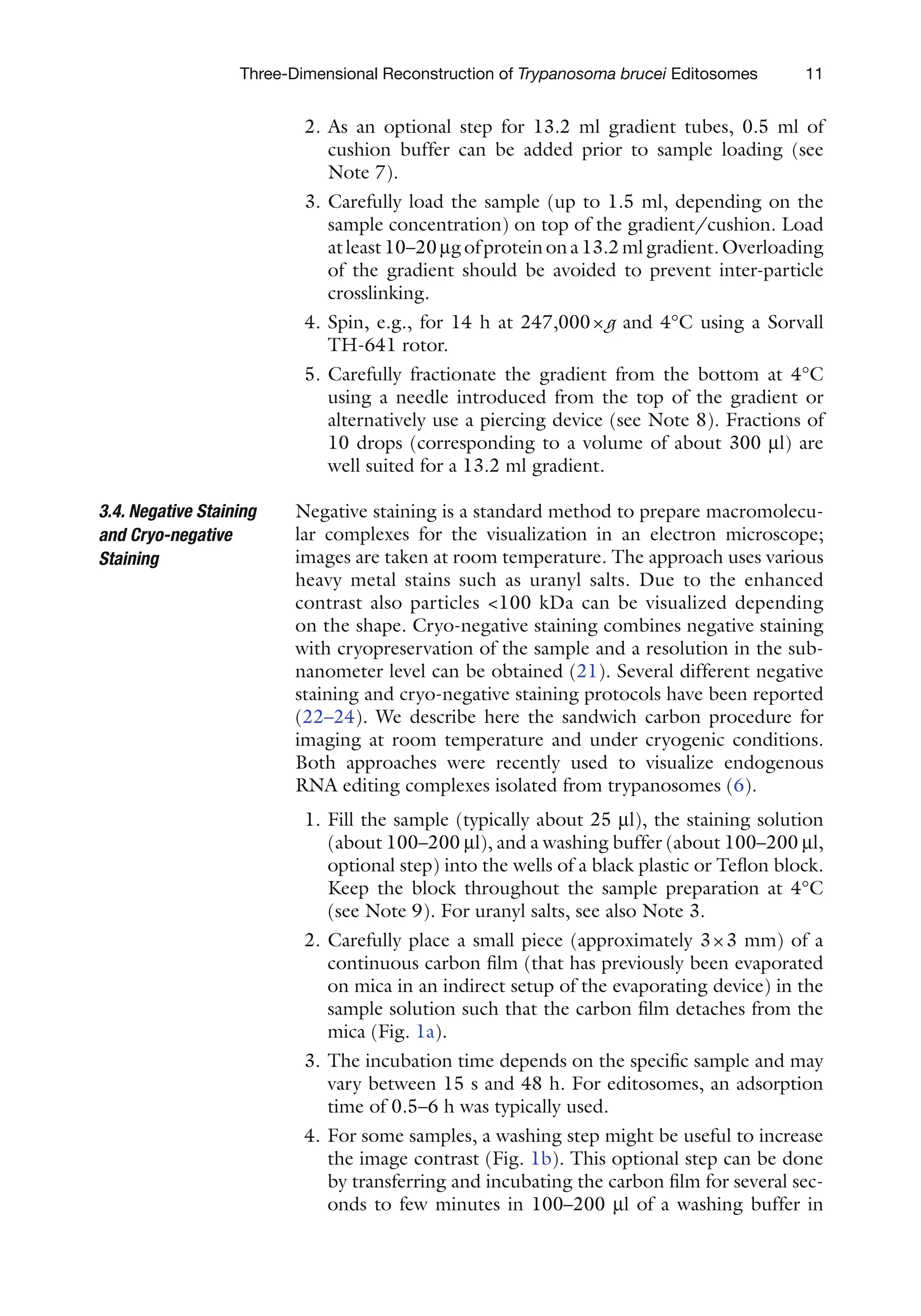
Negative staining is a standard method to prepare macromolecu-
lar complexes for the visualization in an electron microscope;
images are taken at room temperature. The approach uses various
heavy metal stains such as uranyl salts. Due to the enhanced
contrast also particles 100 kDa can be visualized depending
on the shape. Cryo-negative staining combines negative staining
with cryopreservation of the sample and a resolution in the sub-
nanometer level can be obtained (21). Several different negative
staining and cryo-negative staining protocols have been reported
(22–24). We describe here the sandwich carbon procedure for
imaging at room temperature and under cryogenic conditions.
Both approaches were recently used to visualize endogenous
RNA editing complexes isolated from trypanosomes (6).
1. Fill the sample (typically about 25 ml), the staining solution
(about 100–200 ml), and a washing buffer (about 100–200 ml,
optional step) into the wells of a black plastic or Teflon block.
Keep the block throughout the sample preparation at 4°C
(see Note 9). For uranyl salts, see also Note 3.
2. Carefully place a small piece (approximately 3×3 mm) of a
continuous carbon film (that has previously been evaporated
on mica in an indirect setup of the evaporating device) in the
sample solution such that the carbon film detaches from the
mica (Fig. 1a).
3. The incubation time depends on the specific sample and may
vary between 15 s and 48 h. For editosomes, an adsorption
time of 0.5–6 h was typically used.
4. For some samples, a washing step might be useful to increase
the image contrast (Fig. 1b). This optional step can be done
by transferring and incubating the carbon film for several sec-
onds to few minutes in 100–200 ml of a washing buffer in
3.4. Negative Staining
and Cryo-negative
Staining
31.
12 Göringer etal.
exactly the same way as carried out for the particle adsorption
step (see Subheading 3.4, step 2).
5. Lift the carbon/mica out of the particle or optional washing
solution, blot it carefully without touching the carbon film
and transfer it to the staining solution (Fig. 1c). The carbon
must completely detach from the mica. The particles face
down towards the staining solution.
6. After about 2 min, the carbon film is lifted out of the staining
solution by putting an EM grid covered with a perforated
Fig. 1. Cryo-negative staining and negative staining. (a) Initially, the sample (particles are depicted as gray spheres) and
the staining solution (two wells) are filled into wells of a black plastic or Teflon block. Optionally, wells can be filled with
a washing buffer, if necessary. Most of the steps are identical for both room temperature negative staining and cryo-
negative staining (a–d); the procedures differ only in the final step (e or f). A small piece of carbon film (dark gray) that
was indirectly evaporated on a piece of mica (light gray) is placed into the sample solution. Thereby, the carbon film
detaches from the mica except for a small area where the tweezers are touching it, and the macromolecules can adsorb
for a defined period of time. (b) Optionally, the carbon film to which the particles have adsorbed can be transferred to
a well filled with a washing buffer to improve the staining quality. This step can also be repeated, if necessary.
(c) Subsequent to particle adsorption or the optional washing step, the carbon film is transferred to a well filled with a
staining solution. Thereby, the mica completely detaches from the carbon film and falls down to the bottom of the well,
while the carbon film with the particles facing towards the staining solution is floating on the surface of the staining solu-
tion. A copper EM grid (top) covered with a perforated carbon film is placed on top of the floating carbon film and the EM
grid is carefully removed from the staining solution. Excess liquid is blotted from the side without destroying the carbon
film. (d) Another piece of carbon film is placed into a second well filled with staining solution so that the carbon film is
completely floating on the staining solution.The EM grid with the particles facing up is submerged underneath the second
carbon film and lifted out of the staining solution to form the sandwich. Again, excess liquid is blotted. (e) For room tem-
perature negative staining, the grid is dried and can then be stored at a dry place until imaging. (f) For cryo-negative
staining, the EM grid is frozen in liquid nitrogen. Cryo grids must be stored and imaged under cryogenic conditions.
32.
13
Three-Dimensional Reconstruction ofTrypanosoma brucei Editosomes
carbon film on top of the floating carbon (Fig. 1c). Submerge
the EM grid underneath the staining solution and carefully
remove the EM grid out of the staining solution. Blot excess
liquid from the side.
7. To form the carbon sandwich, float another piece of carbon
film onto the surface of a second well filled with the staining
solution so that the mica completely detaches from the car-
bon film (Fig. 1d). Submerge the EM grid with the particles
facing up underneath the floating carbon film and lift the EM
grid out of the solution to embed the particles in a layer of
staining solution between the two carbon films. Again, blot
the EM grid carefully from the side.
8. Let the EM grid air-dry for imaging at room temperature
(Fig. 1e) or freeze the grid in liquid N2
for cryo-negative
staining (Fig. 1f). Once frozen, the grid must be kept under
cryogenic conditions.
Unstained cryopreparations are obtained by freeze-plunging of
the sample into liquid ethane (25). This results in the vitrification
of the sample, i.e., the amorphous structure of the buffer is pre-
served and no crystalline ice is formed. Unstained cryoimaging
can be performed for all samples of sufficient concentration and
molecular mass (typically 250 kDa). In addition, the buffer must
be compatible with vitrification. In particular, glycerol, sucrose,
and high salt concentrations may interfere with the vitrification.
In all such cases (e.g., GraFix fractions), the sample needs to be
buffer exchanged to a suitable buffer.
1. Optional step in case substances with an adverse effect on the
vitrification are present in the particle buffer: Buffer exchange
the GraFix sample for a glycerol-free buffer using a Zeba spin
column (see Note 10).
2. Fill 25–30 ml of sample into a well of the black plastic or
Teflon block and proceed as described in Subheading 3.4,
step 2. Keep the sample at 4°C.
3. The adsorption time needs to be adjusted according to the
sample and may vary between 15 s and 48 h.
4. Place a copper EM grid covered with a perforated carbon film
on top of the floating carbon film, submerge it underneath
the surface, and carefully remove it out of the solution. Prior
to usage, the EM grid can be glow discharged.
5. Mount the tweezers holding the EM grid in the freeze-
plunger filled with liquid ethane (see also Note 4). Blot the
grid carefully and plunge it into the liquid ethane (see
Note 11). The EM grid is transferred and stored in liquid N2
until imaging.
3.5. Unstained
Cryopreparation
33.
14 Göringer etal.
The transmission electron microscope is used to image biological
samples in the form of two-dimensional (2D) projection views.
Due to the radiation sensitivity of biological material, imaging has
to be conducted at low-dose conditions, and a further protection
of the sample can be achieved by imaging at cryogenic tempera-
tures (i.e., liquid N2
- or He-cooling) as compared to room tem-
perature (26). To obtain the 3D information out of the data set
of 2D projections, the particle views have to be subjected to sin-
gle-particle image processing (7, 8). An overview of the steps is
given in Fig. 2 and the single-particle image processing of the
~20S and ~35–40S RNA editing complexes isolated from T. brucei
is summarized in Figs. 3 and 4, respectively.
1. Negatively stained grids can be transferred using standard
room temperature holders into the microscope. For cryoim-
aging, the grid has to be mounted in a specialized side-entry
cryoholder or a cryoloading system. During the mounting
and transfer of the cryogrids, the sample has to be kept at
cryogenic conditions (see Note 12).
2. For de novo structure determinations, a slow-scan CCD cam-
era offers significant advantages compared to conventional
photographic film; the latter usually is advantageous for high-
resolution work (27). See Note 13. The magnification of the
electron microscope has to be adjusted depending on, e.g.,
the size of the object, number of particles, pixel size of the
detector, and desired resolution of the images. For CCD
camera images, magnifications of about 50,000–275,000-
fold and for photographic films, magnifications of 27,000–
60,000-fold are typically used. Images can be taken untilted
or at a tilt angle supported by the cryostage/holder combina-
tion. In particular, for the random conical tilt (RCT) tech-
nique (28), images are first taken at the selected tilt angle
(e.g., 45°) and subsequently in an untilted mode at the same
position to reduce the beam damage of the tilted images (as
the tilted images are used for 3D calculation).
3. For photographic film only: Photographic film has to be digi-
tized with a scanner of appropriate quality (e.g., Tango or
Coolscan). Low quality images (e.g., poor contrast, drift,
charging) can be discarded prior to this step.
4. Select individual particles from the raw EM images by manual
or (semi-) automated procedures (see Note 14). Algorithms for
manual and/or (semi-) automated particle selection are imple-
mented in all major single-particle image processing software
packages and individual programs are also available (e.g.,
(29–32)). Automated procedures can be combined with a post-
selection step in which all positions that were found by the
software, but do not show a particle of desired quality are
removed.
3.6. Electron
Microscopy
and Single-Particle
Image Processing
34.
15
Three-Dimensional Reconstruction ofTrypanosoma brucei Editosomes
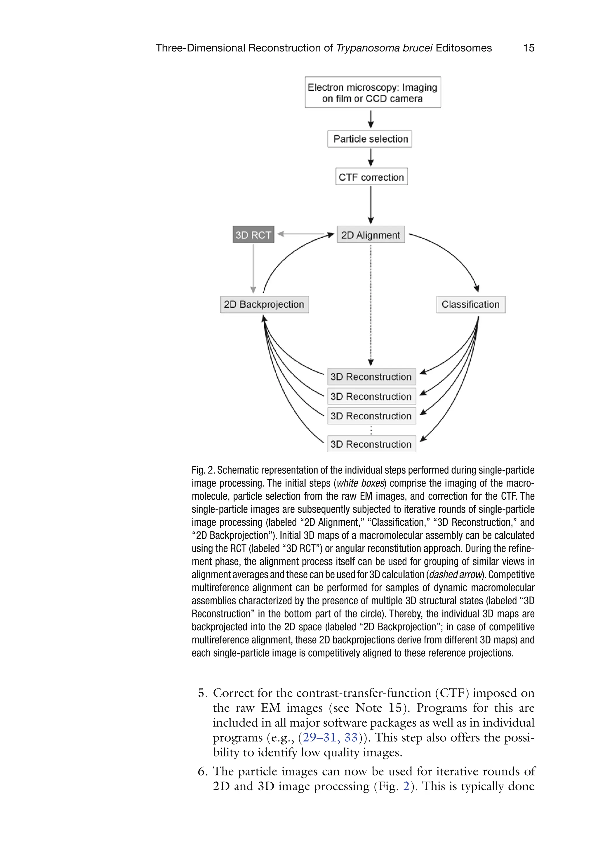
Fig. 2. Schematic representation of the individual steps performed during single-particle
image processing. The initial steps (white boxes) comprise the imaging of the macro-
molecule, particle selection from the raw EM images, and correction for the CTF. The
single-particle images are subsequently subjected to iterative rounds of single-particle
image processing (labeled “2D Alignment,” “Classification,” “3D Reconstruction,” and
“2D Backprojection”). Initial 3D maps of a macromolecular assembly can be calculated
using the RCT (labeled “3D RCT”) or angular reconstitution approach. During the refine-
ment phase, the alignment process itself can be used for grouping of similar views in
alignmentaveragesandthesecanbeusedfor3Dcalculation(dashedarrow).Competitive
multireference alignment can be performed for samples of dynamic macromolecular
assemblies characterized by the presence of multiple 3D structural states (labeled “3D
Reconstruction” in the bottom part of the circle). Thereby, the individual 3D maps are
backprojected into the 2D space (labeled “2D Backprojection”; in case of competitive
multireference alignment, these 2D backprojections derive from different 3D maps) and
each single-particle image is competitively aligned to these reference projections.
5. Correct for the contrast-transfer-function (CTF) imposed on
the raw EM images (see Note 15). Programs for this are
included in all major software packages as well as in individual
programs (e.g., (29–31, 33)). This step also offers the possi-
bility to identify low quality images.
6. The particle images can now be used for iterative rounds of
2D and 3D image processing (Fig. 2). This is typically done
35.
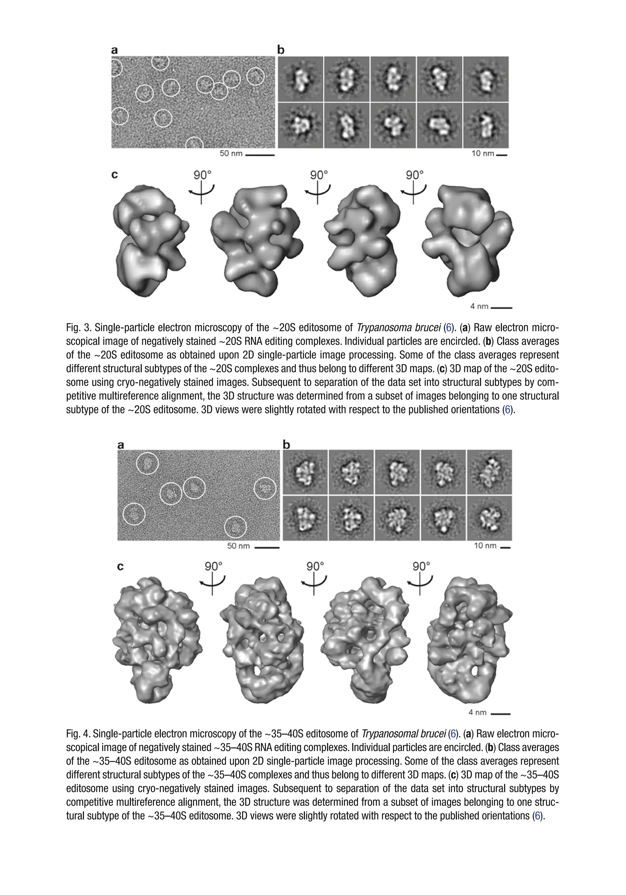
Fig. 3. Single-particleelectron microscopy of the ~20S editosome of Trypanosoma brucei (6). (a) Raw electron micro-
scopical image of negatively stained ~20S RNA editing complexes. Individual particles are encircled. (b) Class averages
of the ~20S editosome as obtained upon 2D single-particle image processing. Some of the class averages represent
different structural subtypes of the ~20S complexes and thus belong to different 3D maps. (c) 3D map of the ~20S edito-
some using cryo-negatively stained images. Subsequent to separation of the data set into structural subtypes by com-
petitive multireference alignment, the 3D structure was determined from a subset of images belonging to one structural
subtype of the ~20S editosome. 3D views were slightly rotated with respect to the published orientations (6).
Fig. 4. Single-particle electron microscopy of the ~35–40S editosome of Trypanosomal brucei (6). (a) Raw electron micro-
scopical image of negatively stained ~35–40S RNA editing complexes.Individual particles are encircled.(b) Class averages
of the ~35–40S editosome as obtained upon 2D single-particle image processing. Some of the class averages represent
different structural subtypes of the ~35–40S complexes and thus belong to different 3D maps. (c) 3D map of the ~35–40S
editosome using cryo-negatively stained images. Subsequent to separation of the data set into structural subtypes by
competitive multireference alignment, the 3D structure was determined from a subset of images belonging to one struc-
tural subtype of the ~35–40S editosome. 3D views were slightly rotated with respect to the published orientations (6).
36.
17
Three-Dimensional Reconstruction ofTrypanosoma brucei Editosomes
in one of the major software packages such as, e.g., IMAGIC
(29),SPIDER(30),EMAN(31),XMIPP(34),orFREALIGN
(35) (see Note 16). One round of 2D image processing typi-
cally comprises of an alignment step and a classification step.
During the alignment, the particle images are aligned towards
references to bring similar views into register (see Note 17).
These aligned images can subsequently be used to group sim-
ilar views into classes in order to increase the signal-to-noise-
ratio. Alignment and classification are iteratively repeated
until the result is stable.
7. The class or alignment averages are then used to calculate the
3D map of the macromolecular assembly. For this, all pro-
grams listed above can be used. Several different techniques
exist including, e.g., RCT (28), angular reconstitution (36),
and projection matching (37) (see Note 18).
8. For a refinement of the initial model (e.g., a low-resolution 3D
map determined using the RCT or angular reconstitution
approach), the Subheading 3.6, steps 6 and 7 are repeated until
the result is stable and no improvement of the structure is
observed. 2D backprojections of the 3D map are used as an
alignment reference during the refinement of the 3D map. For
dynamic macromolecular assemblies, several 3D structural
maps representing different compositional and/or conforma-
tional states can be determined using the RCT technique.
These different 3D maps can be used to generate sets of 2D
backprojections for a competitive multireference alignment.
Each single-particle image is thereby competitively aligned to
these reference projections, and the data set is separated into
subsets representing individual structural subtypes. These indi-
vidual structural subtypes are refined separately. Typically, dur-
ing the refinement steps, a large number of references is used to
cover all possible angular directions. This makes more advanced
schemes such as the corrims (38) necessary to speedup the cal-
culation process.
9. Finally, the 3D map is visualized as sections and/or surface
views in a 3D viewer and can be annotated by, e.g., fitting of
substructures, labeling, recognition of structural elements,
and other techniques.
1. 125
I emits gamma rays with a maximal energy of 0.035 MeV
and requires lead shielding. Unbound iodine is volatile and
must be handled under a fume hood.
4.
Notes
37.
18 Göringer etal.
2. We recommend using fresh EM-grade solutions of 25% (v/v)
glutaraldehyde since the activity of the crosslinker may
decrease over time. Glutaraldehyde and other crosslinkers are
highly toxic and appropriate safety precautions according to
the manufacturer should be followed.
3. Prepare the uranyl formate solution always freshly and keep
the solution cool and in the dark. Uranyl salts are radioactive
and toxic and thus appropriate safety precautions should be
followed.
4. Liquid N2
and liquid ethane can cause severe burns. Note the
asphyxiation hazard. Ethane is extremely flammable and forms
explosive mixtures with air. Appropriate protection is required.
5. Depending on the particle, GraFix can have a number of
advantages: (1) significant reduction of particle disintegration
and aggregation, (2) improved adsorption towards the car-
bon support film, (3) increased image contrast, and (4)
increase in angular diversity.
6. As the gradient contains a low amount of glutaraldehyde,
only buffer reagents compatible with the fixation reagent can
be used. Glutaraldehyde, for example, reacts with primary
amines (39, 40), and thus all gradient buffers must be free of
primary amines.
7. Some purification protocols result in the presence of primary
amine compounds in the sample in addition to the protein
or RNA/protein complex of interest. These include, e.g.,
elution peptides contaminating polypeptides at high concen-
tration and Tris-OH. As a direct contact of such a sample
with the crosslinker has to be avoided, a cushion free of pri-
mary amines and free of glutaraldehyde is used before loading
the sample.
8. We highly recommend fractionating the gradient from the
bottom since low molecular mass substances such as peptides
and detergents are enriched in the top fractions of the gradi-
ent. These low molecular mass substances may interfere with
the image contrast of the EM specimen and may cause stain-
ing artifacts. Using a needle/tube system connected to a peri-
staltic pump, mixing of the gradient, e.g., during the insertion
of the needle has to be avoided.
9. During long-term incubation, the block has to be kept at 4°C
and condensation of water vapor on the sample must be
avoided. This can be accomplished by placing the block into
a dry, precooled Petri dish, mounting the lid and storing the
block/Petri dish in a cold room. Even during short-term
incubation, the block must be kept at 4°C and condensation
of water vapor must be avoided. However, it is usually suffi-
cient to place the block in an ice-cooled Petri dish.
38.
19
Three-Dimensional Reconstruction ofTrypanosoma brucei Editosomes
10. As the recovery of the sample upon buffer exchange may vary
with the properties of the selected particle (for large multi-
megadalton assemblies in the range of about 20–95%), we
recommend to prepare a negative staining room temperature
EM grid to adapt the incubation time for the unstained cryo-
preparation accordingly.
11. The blotting properties (i.e., time, pressure, one-sided vs.
two-sided, frequency, etc.) as well as the environmental prop-
erties (temperature and humidity) have to be adapted to the
specific sample under investigation. Assessment of the vitrifi-
cation quality must be done by using the electron cryomicro-
scope under cryoconditions.
12. Mounting and transfer of room temperature EM grids is
straightforward. Significant warming and ice contamination
of cryo-EM grids during the mounting and transfer under
cryogenic conditions must be avoided.
13. Slow-scan CCD cameras offer an increased phase transmis-
sion and spectral signal-to-noise ratio (27), which are impor-
tant factors in the setup of a novel structure. Images can be
recorded in tile mode – i.e., with slight overlap of the images –
and stitched to larger images to compensate for the smaller
imaging area of the CCD camera. In contrast, photographic
film shows superior quality in the very high resolution range.
14. Manual selection of particles is more time-intensive compared
to (semi-) automated selection procedures, but offers the
opportunity to get an overview about the typical views and the
quality of the imaged particles (e.g., homogeneity, aggregation,
disintegration). In particular for particles with so-far unknown
structure, manual selection is advantageous. For the selection
of larger data sets of particles with known structure, automated
particle selection can be performed using “Boxer” (EMAN
software package (31)) or “Signature” (32). Manual postselec-
tion is often recommended to remove low quality images (e.g.,
aggregates, broken particles, ice contamination). In any of the
methods, the particles should be picked centrally, i.e., a dis-
placement of the particle in direction of the edges should be
avoided. Particles are extracted from the raw images in a pixel
frame larger than the actual maximum dimensions of the visual-
ized particles. In general, a pixel frame in which the particles
amount to about two thirds of the frame is recommended.
Depending on the particle dimensions and the pixel size, typi-
cal values of the pixel frame are 64×64 to 512×512 pixels.
15. Computationally, raw EM images should be corrected for the
defocus and twofold astigmatism, and can also be corrected
for the experimental B factor, if desired. Low quality images
due to drift or charging can be easily identified in the 2D
power spectra as truncations of the Thon rings.
39.
20 Göringer etal.
16. File format type and definition of the coordinate system
including 3D angles may vary between the individual pro-
grams and thus need to be checked carefully when switching
from one software package to the other.
17. There are several ways to generate reference images for the
alignment. In case of a particle with an unknown structure,
selected single-particle views or the sum of all images (i.e., a
featureless “blob”) can be used as a first reference. Also, views
from a related particle (e.g., with minor differences in com-
position) can be used in the first alignment. For subsequent
rounds, class or alignment averages can be used (see below).
Once a 3D model of the analyzed or a related (e.g., with
minor differences in composition) particle is available, this
can be used to generate 2D projections by backprojecting the
3D map into 2D projections. For averaging, either a statisti-
cal classification approach such as multivariate statistical clas-
sification or – in particular in the advanced steps of the
refinement – the alignment itself is used. The former tech-
nique generates so-called class averages, the latter alignment
averages.
18. All of the different 3D reconstruction techniques are imple-
mented in one or more of the software packages listed in
Subheading 2.7, item 8. The selection of the approach pri-
marily depends on the stage of the project. RCT (28) and
angular reconstitution (36) can be used to determine the
structure of a particle whose structure was so-far unknown,
i.e., in a de novo approach. For refinement of an available
model, angular reconstitution and projection matching (37)
can be used. RCT structures are typically limited to the low
resolution range and suffer from a missing cone, but in con-
trast to the other techniques can determine the handedness of
the complex. Refinement of an RCT structure is possible by
both angular reconstitution and projection matching. Angular
reconstitution and projection matching can reconstruct 3D
maps to the subnanometer level.
Acknowledgments
MMG and BS are supported by a grant from the Danish Center
for Scientific Computing (DCSC). HS is supported by a grant
of the Bundesministerium für Bildung und Forschung (BMBF)
and a European “3D Repertoire” grant. HUG is supported as
an International Scholar of the Howard Hughes Medical
Institute (HHMI) and by the German Research Foundation
(DFG).
40.
21
Three-Dimensional Reconstruction ofTrypanosoma brucei Editosomes
References
1. Alberts, B. (1998) The cell as a collection of
protein machines: preparing the next genera-
tion of molecular biologists Cell 92, 291–4.
2. Madison-Antenucci, S., Grams, J., and
Hajduk, S. L. (2002) Editing machines: the
complexities of trypanosome RNA editing
Cell 108, 435–8.
3. Stuart, K. D., Schnaufer, A., Ernst, N. L., and
Panigrahi, A. K. (2005) Complex manage-
ment: RNA editing in trypanosomes Trends
Biochem Sci 30, 97–105.
4. Stark, H., and Lührmann, R. (2006) Cryo-
electron microscopy of spliceosomal compo-
nents Annu Rev Biophys Biomol Struct 35,
435–57.
5. Leschziner, A. E., and Nogales, E. (2007)
Visualizing flexibility at molecular resolution:
analysis of heterogeneity in single-particle
electron microscopy reconstructions Annu
Rev Biophys Biomol Struct 36, 43–62.
6. Golas, M. M., Böhm, C., Sander, B.,
Effenberger, K., Brecht, M., Stark, H., and
Göringer, H. U. (2009) Snapshots of the
RNA editing machine in trypanosomes cap-
tured at different assembly stages in vivo
EMBO J 28, 766–78.
7. van Heel, M., Gowen, B., Matadeen, R.,
Orlova, E. V., Finn, R., Pape, T., Cohen, D.,
Stark, H., Schmidt, R., Schatz, M., and
Patwardhan, A. (2000) Single-particle elec-
tron cryo-microscopy: towards atomic resolu-
tion Q Rev Biophys 33, 307–69.
8. Frank, J. (2002) Single-particle imaging of
macromolecules by cryo-electron microscopy
Annu Rev Biophys Biomol Struct 31, 303–19.
9. Kastner, B., Fischer, N., Golas, M. M., Sander,
B., Dube, P., Boehringer, D., Hartmuth, K.,
Deckert, J., Hauer, F., Wolf, E., Uchtenhagen,
H., Urlaub, H., Herzog, F., Peters, J. M.,
Poerschke, D., Lührmann, R., and Stark, H.
(2008) GraFix: sample preparation for single-
particle electron cryomicroscopy Nat Methods
5, 53–5.
10. Brun, R., and Schönenberger, M. (1979)
Cultivation and in vitro cloning of procyclic
culture forms of Trypanosoma brucei in a semi-
defined medium Acta Trop 36, 289–92.
11. Wirtz, E., Leal, S., Ochatt, C., and Cross, G. A.
(1999) A tightly regulated inducible expres-
sion system for conditional gene knock-outs
and dominant-negative genetics in Trypano-
soma brucei Mol Biochem Parasitol 99,
89–101.
12. Rigaut, G., Shevchenko, A., Rutz, B., Wilm,
M., Mann, M., and Seraphin, B. (1999) A
generic protein purification method for pro-
tein complex characterization and proteome
exploration Nat Biotechnol 17, 1030–2.
13. Brecht, M., Niemann, M., Schlüter, E.,
Müller, U. F., Stuart, K., and Göringer, H. U.
(2005) TbMP42, a protein component of the
RNA editing complex in African trypano-
somes, has endo-exoribonuclease activity Mol
Cell 17, 621–30.
14. Panigrahi, A. K., Schnaufer, A., Carmean, N.,
Igo, R. P., Jr., Gygi, S. P., Ernst, N. L.,
Palazzo, S. S., Weston, D. S., Aebersold, R.,
Salavati, R., and Stuart, K. D. (2001) Four
related proteins of the Trypanosoma brucei
RNA editing complex Mol Cell Biol 21,
6833–40.
15. Cross, G. A. (1975) Identification, purifica-
tion and properties of clone-specific glycopro-
tein antigens constituting the surface coat of
Trypanosoma brucei Parasitology 71,
393–417.
16. Hauser, R., Pypaert, M., Hausler, T., Horn,
E. K., and Schneider, A. (1996) In vitro
import of proteins into mitochondria of
Trypanosoma brucei and Leishmania tarento-
lae J Cell Sci 109 (Pt 2), 517–23.
17. Igo, R. P., Jr., Palazzo, S. S., Burgess, M. L.,
Panigrahi, A. K., and Stuart, K. (2000)
Uridylate addition and RNA ligation contrib-
ute to the specificity of kinetoplastid insertion
RNA editing Mol Cell Biol 20, 8447–57.
18. Igo, R. P., Jr., Weston, D. S., Ernst, N. L.,
Panigrahi, A. K., Salavati, R., and Stuart, K.
(2002) Role of uridylate-specific exoribonu-
clease activity in Trypanosoma brucei RNA
editing Eukaryot Cell 1, 112–8.
19. Clauser, K. R., Baker, P., and Burlingame,
A. L. (1999) Role of accurate mass measure-
ment (+/− 10 ppm) in protein identification
strategies employing MS or MS/MS and
database searching Anal Chem 71, 2871–82.
20. Hunter, W. M., and Greenwood, F. C. (1962)
Preparation of iodine-131 labelled human
growth hormone of high specific activity
Nature 194, 495–6.
21. Golas, M. M., Sander, B., Will, C. L.,
Lührmann, R., and Stark, H. (2003) Molecu
lar architecture of the multiprotein splicing
factor SF3b Science 300, 980–4.
22. Harris, J. R. (2007) Negative staining of
thinly spread biological samples Methods Mol
Biol 369, 107–42.
23. Adrian, M., Dubochet, J., Fuller, S. D., and
Harris, J. R. (1998) Cryo-negative staining
Micron 29, 145–60.
41.
22 Göringer etal.
24. Golas, M. M., Sander, B., Will, C. L.,
Lührmann, R., and Stark, H. (2005) Major
conformational change in the complex SF3b
upon integration into the spliceosomal U11/
U12 di-snRNP as revealed by electron cryo-
microscopy Mol Cell 17, 869–83.
25. Adrian, M., Dubochet, J., Lepault, J., and
McDowall, A. W. (1984) Cryo-electron
microscopy of viruses Nature 308, 32–6.
26. Chiu, W., Downing, K. H., Dubochet, J.,
Glaeser, R. M., Heide, H. G., Knapek, E.,
Kopf, D. A., Lamvik, M. K., Lepault, J.,
Robertson, J. D., Zeitler, E., and Zemlin, F.
(1986) Cryoprotection in electron micros-
copy J Microsc 141, 385–91.
27. Sander, B., Golas, M. M., and Stark, H.
(2005) Advantages of CCD detectors for
de novo three-dimensional structure determi-
nation in single-particle electron microscopy
J Struct Biol 151, 92–105.
28. Radermacher, M. (1988) Three-dimensional
reconstruction of single particles from ran-
dom and nonrandom tilt series J Electron
Microsc Tech 9, 359–94.
29. van Heel, M., Harauz, G., Orlova, E. V.,
Schmidt, R., and Schatz, M. (1996) A new
generation of the IMAGIC image processing
system J Struct Biol 116, 17–24.
30. Shaikh, T. R., Gao, H., Baxter, W. T., Asturias,
F. J., Boisset, N., Leith, A., and Frank, J.
(2008) SPIDER image processing for single-
particle reconstruction of biological macro-
molecules from electron micrographs Nat
Protoc 3, 1941–74.
31. Tang, G., Peng, L., Baldwin, P. R., Mann, D.
S., Jiang, W., Rees, I., and Ludtke, S. J. (2007)
EMAN2: an extensible image processing suite
for electron microscopy J Struct Biol 157,
38–46.
32. Chen, J. Z., and Grigorieff, N. (2007)
SIGNATURE: a single-particle selection sys-
tem for molecular electron microscopy J Struct
Biol 157, 168–73.
33. Sander, B., Golas, M. M., and Stark, H.
(2003) Automatic CTF correction for single
particles based upon multivariate statistical
analysis of individual power spectra J Struct
Biol 142, 392–401.
34. Sorzano, C. O., Marabini, R., Velazquez-
Muriel, J., Bilbao-Castro, J. R., Scheres, S.
H., Carazo, J. M., and Pascual-Montano, A.
(2004) XMIPP: a new generation of an open-
source image processing package for electron
microscopy J Struct Biol 148, 194–204.
35. Grigorieff, N. (2007) FREALIGN: high-res-
olution refinement of single particle structures
J Struct Biol 157, 117–25.
36. van Heel, M. (1987) Angular reconstitution:
a posteriori assignment of projection direc-
tions for 3D reconstruction Ultramicroscopy
21, 111–23.
37. Penczek, P. A., Grassucci, R. A., and Frank, J.
(1994) The ribosome at improved resolution:
new techniques for merging and orientation
refinement in 3D cryo-electron microscopy of
biological particles Ultramicroscopy 53, 251–70.
38. Sander, B., Golas, M. M., and Stark, H.
(2003) Corrim-based alignment for improved
speed in single-particle image processing
J Struct Biol 143, 219–28.
39. Hopwood, D. (1972) Theoretical and practi-
calaspectsofglutaraldehydefixationHistochem
J 4, 267–303.
40. Migneault, I., Dartiguenave, C., Bertrand,
M.J.,andWaldron,K.C.(2004)Glutaraldehyde:
behavior in aqueous solution, reaction with
proteins, and application to enzyme crosslink-
ing Biotechniques 37, 790–6, 8–802.
24 Ringpis, Lathrop,and Aphasizhev
processing (2, 6). Because Trypanosoma brucei is a diploid asexually
reproducing organism, inducible knockouts of essential genes
require sequential generation of three clonal cell lines and as
many selective markers (7); a fourth marker is required for a
knock-in construction. For reasons that are not clear, successful
knockouts and knock-ins of essential mitochondrial genes (8, 9)
have been achieved only in blood stream (BF) parasites which
are deficient in oxidative phosphorylation and grow to a low cell
density (~106
cells/mL). These technical restrictions made RNA
interference (10) a method of choice for gene silencing in BF
and procyclic (PF) parasites, which have an actively respiring
mitochondrion and can be cultured at higher cell density (~107
cells/mL).
Transgenic cell lines have been generated for both BF and PF
T. brucei to allow tetracycline-regulated (11) ectopic expression
of double-stranded (ds) RNA driven by either T7 RNA poly-
merase or RNA polymerase I promoters (12). Typically, the RNAi
cassette is designed toward protein-coding regions, although
achieving sufficient knockdown may require empirical selection
among several gene fragments (13) or inclusion of untranslated
regions (UTRs) (14). Targeting unique UTRs for RNAi also has
been used as a knock-in method. In this strategy, RNAi targets
exclusively 5¢- or 3¢-UTR triggering degradation of the endoge-
nous mRNA while the gene of interest flanked by heterologous
UTRs is expressed (15). Limitations of this approach include a
requirement for precise UTR mapping to avoid knockdown of
closely spaced adjacent mRNAs and UTRs that are too short for
effective RNAi knockdown.
Although biochemical and structural analyses of recombinant
editing enzymes were instrumental in defining catalytic residues
and domain organization (16, 17), functional studies would
greatly benefit from an in vivo mutagenesis system which also
allows in vitro analysis of purified editing complexes. We have
developed an iCODA in vivo complementation approach for PF
parasites based on tetracycline-inducible co-expression of a
dsRNA and a synthetic gene which encodes the same polypeptide
as the one targeted by RNAi. A fragment corresponding to the
RNAi-targeted region is assembled from overlapping DNA oligo-
nucleotides using CODA technology (18) with at least one silent
mutation per 12 bp. These mutations are designed based on a
genome-wide analysis of codon bias and codon context to mini-
mize effects on translation. Transcription of the synthetic gene
produces an RNAi-resistant mRNA as the only source for the
protein of interest. Consequentially, the cell survives unless a
mutation disrupting catalysis or complex association is introduced
into the synthetic gene. Introduction of the TAP tag allows
for protein complex purification and downstream biochemical
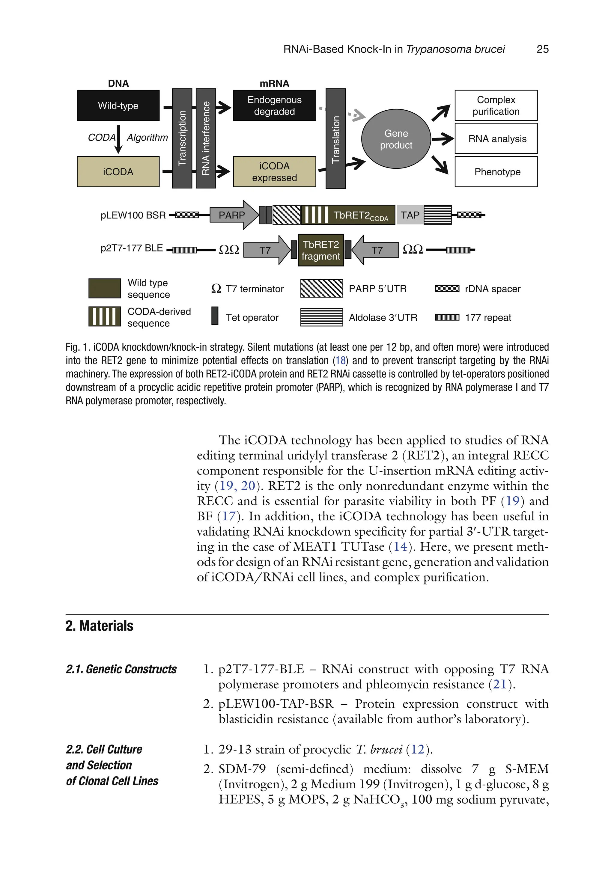
analysis (Fig. 1).
44.
25
RNAi-Based Knock-In inTrypanosoma brucei
The iCODA technology has been applied to studies of RNA
editing terminal uridylyl transferase 2 (RET2), an integral RECC
component responsible for the U-insertion mRNA editing activ-
ity (19, 20). RET2 is the only nonredundant enzyme within the
RECC and is essential for parasite viability in both PF (19) and
BF (17). In addition, the iCODA technology has been useful in
validating RNAi knockdown specificity for partial 3¢-UTR target-
ing in the case of MEAT1 TUTase (14). Here, we present meth-
ods for design of an RNAi resistant gene, generation and validation
of iCODA/RNAi cell lines, and complex purification.
1. p2T7-177-BLE – RNAi construct with opposing T7 RNA
polymerase promoters and phleomycin resistance (21).
2. pLEW100-TAP-BSR – Protein expression construct with
blasticidin resistance (available from author’s laboratory).
1. 29-13 strain of procyclic T. brucei (12).
2. SDM-79 (semi-defined) medium: dissolve 7 g S-MEM
(Invitrogen), 2 g Medium 199 (Invitrogen), 1 g d-glucose, 8 g
HEPES, 5 g MOPS, 2 g NaHCO3
, 100 mg sodium
pyruvate,
2.
Materials
2.1. Genetic Constructs
2.2. Cell Culture
and Selection
of Clonal Cell Lines
TbRET2CODA
iCODA
CODA Algorithm
Wild-type
Endogenous
degraded
Gene
product
Transcription
RNA
interference
Translation
PARP 5UTR
Aldolase 3UTR
Tet operator
T7 terminator
177 repeat
rDNA spacer
pLEW100 BSR
p2T7-177 BLE
PARP TAP
T7
iCODA
expressed
TbRET2
fragment
T7
ΩΩ ΩΩ
Ω
A
N
R
m
A
N
D
Complex
purification
Phenotype
RNA analysis
Wild type
sequence
CODA-derived
sequence
Fig. 1. iCODA knockdown/knock-in strategy. Silent mutations (at least one per 12 bp, and often more) were introduced
into the RET2 gene to minimize potential effects on translation (18) and to prevent transcript targeting by the RNAi
machinery.The expression of both RET2-iCODA protein and RET2 RNAi cassette is controlled by tet-operators positioned
downstream of a procyclic acidic repetitive protein promoter (PARP), which is recognized by RNA polymerase I and T7
RNA polymerase promoter, respectively.
45.
26 Ringpis, Lathrop,and Aphasizhev
200 mg l-alanine, 100 mg l-arginine, 300 mg l-glutamine,
70 mg l-methionine, 80 mg l-phenylalanine, 600 mg l-proline,
60 mg l-serine, 160 mg l-taurine, 350 mg l-threonine, 200 mg
l-tyrosine, 10 mg adenosine, 10 mg guanosine, 50 mg
glucosamine-HCl, 4 mg folic acid, 2 mg p-aminobenzoic acid,
200 mg biotin, 8 mL MEM amino acids (50×) (Invitrogen),
6 mL MEM nonessential amino acids (100×) (Invitrogen) in
850 mL of water. Adjust pH to 7.3 with 5 M NaOH and bring
volume to 900 mL. Sterilize by filtration and store at +4°C up
to 2 weeks.
3. 29-13 Medium: SDM-79 medium supplemented with 50 mg/
mL of Geneticin (G418), 50 mg/mL of Hygromycin, 10 mg/mL
of hemin (EMD Chemicals), and 10% heat inactivated fetal
bovine serum (FBS, see Note 1).
4. Limiting dilution (LD) medium: same as 29-13 medium with
appropriate additional drug(s) and 20% FBS.
5. Humidified incubator set at 27°C with 5% CO2
.
1. Not I restriction endonuclease.
2. Cytomix: 120 mM KCl, 0.15 mM CaCl2
, 10 mM potassium
phosphate, 25 mM HEPES, 2 mM EDTA, 5 mM MgCl2,
pH 7.6.
3. Phosphate sucrose buffer: 277 mM sucrose, 1 mM MgCl2
,
7 mM potassium phosphate, pH 7.4.
4. EM buffer: 3:1 mixture of Cytomix and phosphate sucrose
buffer.
5. Bio-Rad Gene Pulser.
6. 0.4-cm electroporation cuvettes.
1. Extraction buffer (EB): 50 mM Tris–HCl pH 7.6, 150 mM
KCl, 2 mM EDTA. Stock of Tris–HCl is prepared as 1 M
solution with pH 7.6 at 20°C.
2. IgG binding buffer (IBB): 25 mM Tris–HCl pH 7.6, 150 mM
KCl, 1 mM EDTA, 0.1% NP-40.
3. TEV cleavage buffer (TCB): IBB supplemented with 1 mM
DTT (see Note 2).
4. Calmodulin binding buffer (CBB): 25 mM Tris–HCl pH 7.6,
150 mM KCl, 0.1% NP-40, 10 mM b-mercaptoethanol,
1 mM magnesium acetate, 1 mM imidazole, 2 mM CaCl2
.
5. Calmodulin elution buffer 1 (CEB1) for activity assays:
25 mM Tris–HCl pH 7.6, 150 mM KCl, 3 mM EGTA, 1 mM
EDTA, 0.1% NP40, 10% glycerol.
6. Calmodulin elution buffer 2 (CEB2) for mass spectrometry
analysis of complexes: 25 mM Tris–HCl pH 7.6, 50 mM KCl,
3 mM EGTA, 1 mM EDTA, 0.1% NP40.
2.3.
Electroporation
2.4. Tandem Affinity
Purification
46.
27
RNAi-Based Knock-In inTrypanosoma brucei
7. Disposable polystyrene columns (Pierce, product # 29920).
8. TEV protease (Invitrogen).
9. Sypro Ruby gel stain (Invitrogen).
10. Complete protease inhibitor (Roche). Dissolve one tablet in
1 mL of water.
The choice of ~400 bp-long DNA fragment for the RNAi
cassette
is based on the RNAit algorithm (http://trypanofan.path.cam.
ac.uk/software/RNAit.html) that uses BLAST searches to mini-
mize off-targeting (22). The p2T7-177 genetic construct
(partially diagrammed in Fig. 1) is a standard vehicle for induc-
ible expression of dsRNA fragments in trypanosomes (21). The
dual opposing T7 promoter architecture of this vector allows a
one-step cloning of the target sequence or multiple gene knock-
downs by cloning several target sequences. Integration into the
mini-chromosome 177-bp repeat region provides for low base-
line transcription.
The pLEW100 vector (23), partially diagrammed in Fig. 1,
is widely used for stable expression in T. brucei via integration
into the rRNA spacer. Procyclic acidic repetitive protein (PARP)
promoter-driven expression regulated by tet-operators allows for
inducible expression in PF parasites. Alternatively, if work in BF
parasites is planned, the pLEW100v5-BSD vector (http://tryps.
rockefeller.edu) may be a better alternative. In this construct,
expression is driven by the rRNA promoter that is active in both
PF and BF T. brucei. While overexpression is often performed in
the pLEW79-based mhTAP vector (24), pLEW100 variants pro-
vide a balance of efficient integration and tightly regulated expres-
sion which is better suited for iCODA.
The synthetic gene replacement construct is designed to (a)
be identical in amino acid sequence to the wild-type gene except
for deliberately introduced mutations; (b) differ from the wild-
type gene DNA sequence by at least 1 bp in any contiguous
12 bp; (c) avoid off-target cross-hybridization, which is known to
be a source of mutagenesis failure (25); (d) respect known or
hypothesized codon usage (26) and codon pair (27–29) prefer-
ences; (e) avoid any undesired DNA sequences, e.g., restriction
sites used in cloning; and (f) be easily assembled from purchased
synthetic oligonucleotides by polymerase extension.
These goals are all accomplished by making silent (synony-
mous) codon substitutions in the designed synthetic gene replace-
ment construct. For this purpose, we used the computationally
optimized DNA assembly technology (CODA, (18)) to design
the gene assembly and mfold (30) to identify undesired RNA
3.
Methods
3.1. Genetic
Constructs Design
47.
28 Ringpis, Lathrop,and Aphasizhev
secondary structures. However, any modern gene design software
that accepts sequence constraints and removes undesired RNA
secondary structures may be used instead of CODA. Conceptually,
the CODA system works by beginning with (a) a desired amino
acid sequence, (b) a candidate DNA sequence, and (c) a set of
constraints to satisfy. It then iteratively introduces silent codon
substitutions into the candidate DNA sequence until all con-
straints are satisfied. Finally, it outputs (a) a list of DNA oligo-
nucleotide sequences to purchase and (b) a set of instructions for
combining the purchased oligonucleotides by polymerase exten-
sion. The actual CODA implementation involves more technical
details; see Larsen et al. for further explanation (18).
1. Define the desired synthetic replacement sequence. To mini-
mize the DNA synthesis and assembly burden, an approxi-
mately 400 bp gene fragment is recommended for iCODA.
2. Prepare an initial candidate DNA sequence by replacing every
codon in the sequence from step 1 by the most prevalent
(most highly used) codon for that amino acid in the host
organism (26), unless the wild-type DNA sequence also uses
the most prevalent codon there, in which case choose the
second most prevalent codon. The result of this step is a DNA
sequence that is identical in encoded amino acid sequence to
step 1 and differs from the wild-type DNA sequence at every
codon (except for methionine and tryptophan, which each
have only one codon).
3. Prepare a list of desired DNA sequence constraints, which
includes any required DNA subsequences while prohibiting
(a) any four consecutive codons from the wild-type gene; (b)
restriction sites used in cloning from occurring anywhere in
the coding region of the gene; (c) low-usage codons or unde-
sired codon pairs; and (d) any other DNA sequences to
exclude. Prohibiting any four consecutive codons from the
wild-type gene guarantees that the desired synthetic gene
replacement construct has no more, and often fewer, than
11 contiguous base pairs that are identical to the wild-type gene
DNA sequence.
4. Compare the current candidate DNA sequence to the list of
sequence constraints from step 3. If any constraint violations
are found, introduce silent (synonymous) codon substitu-
tions into the candidate DNA sequence to eliminate them
while avoiding low-usage codons (26) and go to step 4.
5. Analyze the current candidate DNA sequence for undesired
RNA secondary structures, such as hairpins, cross-hybridiza-
tions, etc. (18, 25). If any undesired RNA secondary structures
are found, introduce silent (synonymous) codon substitutions
48.
29
RNAi-Based Knock-In inTrypanosoma brucei
into the candidate DNA sequence to
eliminate them while
avoiding low-usage codons (26), and go to step 4.
6. Divide the DNA sequence of step 5 into DNA oligonucle-
otide sequences that can be purchased commercially and
assemble them by polymerase extension as described in
Larsen et al. (18).
The following protocol may be used for transfection with a single
plasmid or co-transfection with two plasmids (see Note 3). All
materials and solutions that will come in contact with cells should
be kept on ice. Sterile technique must be employed when han-
dling cells.
1. From a mid-logarithmic culture maintained at ~5×10⁶
cells/
mL in 29-13 medium, seed 1–2×10⁶
cells/mL to achieve a
cell density of 4–5×10⁶
cells/mL on the following day.
Assume that the division time is ~12 h and 1.4×10⁷
cells per
transfection are required. Incubate 25–30 mL of suspension
culture at 27°C with mild agitation (~100 rpm) in a 75 cm2
cell culture flask in vertical position.
2. Digest 10–12 mg of plasmid DNA purified with QIAprep kit
(Qiagen) with 5 units of Not I restriction endonuclease in a
200-mL reaction volume at 37°C overnight.
3. Precipitate the DNA with 500 mL of ethanol for 20 min in
dry ice. Do not add extra salt or extract with organic solvents.
Wash the pellet with 70% ethanol, air-dry in the laminar hood,
and resuspend in 100 mL of sterile water (50 mL for
co-transfection).
1. When the cell count reaches ~5×10⁶
/mL, transfer cells into
a conical tube.
2. Using a fixed-angled rotor, centrifuge the cells 2,000×g at
4°C for 5 min. Aspirate the supernatant completely.
3. Fill the tube to 50% volume with EM buffer and gently
resuspend cells with pipetting. Repeat centrifugation and
aspiration.
4. Resuspend cells in EM buffer at 3×10⁷
/mL.
5. Transfer the DNA solution into a chilled 4-mm electropora-
tion cuvette. Use sterile water for the control transfection.
6. Add 450 mL of cell solution to each cuvette with 1-mL pipette
tip. Gently pipette twice.
7. Set electroporator to 1,500 V and 25 F. Electroporate cells
with two pulses with a 10-s interval. Expected time constant
is 0.8–1 ms. Chill on ice for 2 min (see Note 4).
3.2. Transfection
of Genetic Constructs
into 29-13 T. brucei
by Electroporation
3.2.1. Preparation
of Plasmids and Cell Lines
3.2.2.
Electroporation
49.
30 Ringpis, Lathrop,and Aphasizhev
8. Using a sterile transfer pipette, transfer cells to a 25-cm2
culture
flask with 10-mL 29-13 medium prewarmed to 27°C and
incubate with mild agitation at 27°C for 24 h.
For single-plasmid transfection, add 10 mL of 2.5 mg/mL
phleomycin (2.5 mg/mL final) or 10 mL of 10 mg/mL blastici-
din (10 mg/mL final). For co-transfection, add both drugs.
Incubate cells in 25 cm2
culture flask with mild agitation at 27°C
for 24 h.
1. Add 100 mL of LD medium into wells 2–12 (all rows) of a
96-well flat-bottom plate.
2. Add 100 mL of LD medium into wells B1, D1, F1, and H1.
3. Add 300 mL of culture into wells A1, C1, E1, and G1.
4. Transfer 100 mL of culture from A1 to B1, C1 to D1, E1 to
F1, and G1 to H1.
5. Using a multichannel pipetman, perform 1:1 serial dilutions
(100 mL) from lane 1 through lane 12.
6. Starting from lane 12, add 100 mL of LD medium to each
well to achieve 200 mL of total volume.
7. Incubate the 96-well plate at 27°C in humidified incubator
with 5% CO2
.
8. Monitor growth under an inverted microscope. Expect to see
cell growth in 10–14 days. If no live cells are present after
2 weeks, repeat the transfection.
9. When the well with highest dilution becomes confluent and
the next dilution remains clear, transfer the entire 200-mL
culture into 3 mL of LD medium in a 25-cm2
flask and incu-
bate upright without agitation for 24 h (see Note 5).
10. Start agitation (100 rpm) and continue until cell density
reaches 5–10×106
cells/mL.
11. Add 3 mL of 29-13 medium with drugs and continue incuba-
tion for another 24 h.
12. Stabilize culture by diluting to 106
cells/mL two to fourfold
every 24 h until division time is consistent.
Successful concurrent knockdown of endogenous protein and
knock-in of exogenous protein can be verified at both the
transcript and protein level. If antibodies are available, the deple-
tion of endogenous protein and expression of longer TAP-tagged
polypeptide can be verified by Western blotting (Fig. 2a). Parasite
cells are collected by centrifugation at different induction time
points, washed with phosphate buffered saline (PBS) buffer, and
boiled in SDS loading buffer for 3 min. Cell extract is separated
by SDS gel (3–5×10⁶
cells/well) and transferred by standard
3.2.3. Add Drug at 24-h
Posttransfection
3.2.4.
Limiting Dilution
3.3. Validation
of iCODA/RNAi
Cell Lines
50.
31
RNAi-Based Knock-In inTrypanosoma brucei
techniques. If antibodies are not accessible, Western blotting
with commercially available PAP reagent (Sigma) which recognizes
the Protein A moiety of the TAP tag and quantitative RT-PCR
(qRT-PCR) can be used to confirm expression of
TAP-tagged
proteins and knockdown of the endogenous transcript, respec-
tively. For total RNA isolation, 20–50 mL cultures are sufficient.
Since transcripts derived from the iCODA-optimized sequence
will be present in iCODA/RNAi cell lines, both qRT-PCR
primers must selectively hybridize with the RNAi-targeted region
of the wild-type transcript, but not with the iCODA-derived
sequence. The knockdown of the endogenous transcript after
~48 h of RNAi should be normalized to mock-induced cells (see
Note 6).
RET2-TAP
RET2 endo
coomassie
Tet induction (days) 0 1 2 3 4 5 6 7 8
a
0
1
2
3
4
5
6
7
0 20 40 60 80 100 120 140 160 180 200
Mock
iCODA/RNAi
RNAi
Mock
iCODA/RNAi
RET2 wt
RET2 D97A
0 20 40 60 80 100 120 140 160 180 200
0
1
2
3
4
5
tetracycline induction, hr
tetracycline induction, hr
Cumulative
cell
count
(Log)
Cumulative
cell
count
(Log)
b
c
Fig. 2. Functional complementation of RET2 RNAi by co-expression of the RNAi-resistant
transcript. (a) Western blotting analysis of RET2-iCODA/RNAi cells. (b) Growth kinetics of
RET2-RNAi and RET2-iCODA/RNAi cell lines. (c) Growth kinetics of RET2-D97A-iCODA/
RNAi cell line.
51.
32 Ringpis, Lathrop,and Aphasizhev
The RET2-RNAi-mediated growth phenotype is successfully
rescued via co-expression of the iCODA-modified transcript
(Fig. 2b). To confirm that the functional complementation
requires RET2’s enzymatic activity, a single amino acid mutation
(D97A) has been introduced into the active site. As seen from
Fig. 2c, the expression of the inactive RET2 does not counteract
the RNAi growth inhibition phenotype.
The following protocol is used to purify complexes from either
iCODA/RNAi cells or from cells expressing the TAP-tagged pro-
tein. All purification steps are performed at 4°C.
1. Build up the 150 mL culture with cell density of ~107
cells/mL
(29-13 medium plus appropriate antibiotics). Avoid dilutions
larger than five to tenfold as this can kill cells. Seed 2×850 mL
cultures (1×10⁶
cells/mL) in roller bottles (110×475 mm,
Bellco) and add tetracycline to 1 mg/mL. Incubate in the roller
apparatus until cell density reaches 12–15×106
cells/mL,
typically 48–72 h.
2. Harvest cells in a fixed-angle rotor at 5,000×g for 10 min.
3. Resuspend pellet in 100 mL cold PBS pH 7.6 and repeat
centrifugations.
4. Resuspend pellet in 50 mL of PBS, transfer into a conical
centrifuge tube, collect cells at 3,000×g for 10 min, and
proceed with complex purification. Pellet can be frozen in
liquid nitrogen and stored at −80°C for later use.
5. Resuspend cells in 6-mL EB (final volume). Add NP-40 to
0.4% and 300 mL of Complete protease inhibitor (Roche).
6. Sonicate the extract three times at 9 W for 10 s.
7. Spin the extract at 100,000×g for 15 min at 4°C in a TLA
100 rotor and promptly transfer supernatant to a clean 15-mL
conical tube.
8. Re-extract the pellet in 6 mL of EB (no NP-40) with sonica-
tion and centrifugation. Pool the two extracts – target volume
is ~12 mL.
9. Prepare the IgG Sepharose resin. Transfer 0.3 mL of slurry (GE
Healthcare) to a 15-mL tube and add 15-mL IBB. Centrifuge
300×g for 2 min (no brakes). Aspirate supernatant.
10. Filter the extract from step 8 through a syringe-driven low-
protein binding membrane into the resin-containing tube
from step 9. Incubate the extract with resin for 1 h with gentle
agitation.
11. Transfer the material into a disposable column (Pierce).
Reload three times to maximize recovery of resin. Collect
flow-through.
3.4. Tandem Affinity
Purification
of Complexes from
iCODA/RNAi Whole
Cell Lysates
52.
33
RNAi-Based Knock-In inTrypanosoma brucei
12. Wash the column with 5–6 full volumes of IBB. Gently
resuspend the resin with each wash and rinse the rims of the
column well.
13. Wash IgG column with two column volumes of TCB.
14. Mix 150 units TEV protease, 20 mL Complete protease
inhibitor, and 1.5 mL TCB. Close the column outlet, add
TEV solution, seal both ends of the column with parafilm,
and incubate overnight with gentle agitation.
15. Transfer 0.3 mL calmodulin resin into a 15-mL conical tube
pretreated with 2% Tween 20 (see Note 7). Fill the tube with
CBB, centrifuge at 300×g for 2 min and then aspirate the
supernatant. Repeat wash once.
16. Drain the IgG column (~1.5 mL) directly onto the prewashed
calmodulin resin.
17. Wash the IgG column with 3-mL CBB and collect the wash
into the tube containing the calmodulin resin. Expect to
recover ~4.5 mL total.
18. Add 6 mL 1 M CaCl2
to the suspension and incubate the tube
with gentle agitation for 1 h.
19. Transfer the suspension to a disposable column pretreated
with 2% Tween 20. Reload flow through 3–5 times. Collect
the flow-through.
20. Wash the column with 5–6 full column volumes with CBB
with resin.
21. Close the bottom of the column. Apply 0.5 mL CEB to the
resin and incubate for 10 min at room temperature with peri-
odic gentle mixing to resuspend the resin. Collect the elution
in a low-protein binding microcentrifuge tube. Repeat thrice
(see Note 8).
22. Flash-freeze fractions in liquid nitrogen and store at −80°C.
23. To visualize the complexes, load ~10 mL of eluted fractions
onto an SDS-PAGE gel and stain the gel with Sypro
Ruby.
Protein profiles of RECCs purified from RET2-mhTAP,
RET2-iCODA-TAP, and RET2-iCODA/RNAi cells are indistin-
guishable (Fig. 3). Collect aliquots at all purification steps start-
ing from total cell lysate. Quantitative Western blotting with
anti-CBP antibody (GenScript) can be used to estimate purifica-
tion yield for the bait protein, which is expected to be 25–30%.
For mass spectrometry analysis, final fraction is precipitated by
adding trichloric acid to 20% and deoxycholate to 0.1%, washed
with acetone twice and digested with trypsin by standard
protocols.
53.
34 Ringpis, Lathrop,and Aphasizhev
1. Many commercial batches of FBS are contaminated with tet-
racycline. Some manufacturers (BD, Invitrogen) claim to
have a tetracycline-free serum which does not always guaran-
tee the leak-proof transcriptional control of RNAi expression.
Apparently, mild to severe growth inhibition phenotypes can
be induced in T. brucei RNAi cells by tetracycline present at
levels undetectable with current testing methods. It is advis-
able to collect several serum samples and test for growth
kinetics of an RNAi cell line in which an essential gene is tar-
geted (available from the author’s laboratory). Typically,
serum supporting the fastest growth would be most suited
for cloning procedures and maintenance of uninduced cells.
4.
Notes
Fig. 3. Protein profiles of complexes purified from RET2 overexpression (mhTAP), RET2-
iCODA-TAP, and RET2-iCODA/RNAi cell lines. (Top panel ) TAP-purified complexes were
separated on an 8–16% gradient SDS-PAGE gel and stained with Sypro Ruby staining.
Cell lines are listed above the gel. mhCBP – 6His plus calmodulin binding peptide which
remain on the tagged protein upon TEV protease release from the IgG resin. (Bottom
panel) TAP-purified complexes were analyzed by Western blotting with anti-RET2 anti-
bodies. Affinity purification of structural RECC subunit (MP63) was used to demonstrate
gel mobility of the endogenous RET2.
54.
35
RNAi-Based Knock-In inTrypanosoma brucei
2. When preparing TCB, DTT should be added immediately
before applying to the IgG beads.
3. Co-transfection of both TAP and RNAi constructs requires
less time and is a default protocol. If obtaining clones resis-
tant to all four drugs is unsuccessful, sequential transfection is
recommended. First, transfect 29-13 cells with the protein
expression construct (blasticidin resistance) and stabilize cells
in a 10 mL of culture without clonal selection (~3 weeks).
Second, transfect cells with the RNAi construct and proceed
with clonal selection.
4. Incubation of the electroporated cells on ice longer than
5 min is not recommended. When performing multiple trans-
fections, electroporations should be staggered to decrease
the time between electroporation and inoculation into
medium. If an electric arc occurs, expect to obtain fewer or
no clones.
5. The medium color changes with cell growth from red at low
density to yellow in the stationary culture. Cells can be ready
for passage at 3–4 weeks posttransfection. Care should be
taken to avoid oversaturated (yellow) cultures. Transfer at
least 3–5 clones, but continue incubating the 96-well plate
for a few more days to ensure lack of cell growth in wells
with higher dilutions. If this occurs, discard previously
collected clones and transfer new ones from the highest dilu-
tion wells.
6. For RNAi knockdown of essential genes, induced and mock-
induced cell should be harvested at the same cell density.
Typically, RNAi is induced at 106
cells/mL and the culture is
maintained for 24–48 h; cell density should not exceed
5–6×10⁶
cells/mL.
7. Pretreatment of plastic tubes and columns during the calm-
odulin binding step with 2% Tween 20 is recommended to
improve purification yields. Pretreated plastic should be rinsed
with large amounts of distilled water before use.
8. Fractions 1 and 2 should contain most of the eluted material.
The first 50–100 mL of the Fraction 1 is the void volume and
does not need to be collected.
Acknowledgments
We thank George Cross and Elisabetta Ullu for kind gifts of cell
lines and plasmids. This work was supported by the NIH grants
RO1AI064653 to RA and R01CA112560 to RHL.
afternoon the principalcame in to tell her that an urgent telephone
message had just asked Joan Befort's teacher to come to Beforts' as
soon as she possibly could.
I said you would be down at once, went on Miss Weir. Was Joan
at school to-day?
No, Rebecca Mary remembered that Joan hadn't been at school
either that morning or that afternoon.
Probably measles or mumps, prophesied Miss Weir, who had been
made wise by years of experience. Foreigners are so helpless at
times. You will have to explain that the quarantine laws must be
obeyed. What do you know about the Beforts?
Rebecca Mary blushed, for when Miss Weir asked her she discovered
that she knew very very little about the Beforts.
Joan's mother is dead, and she and her father live with an old
woman who keeps house for them. Rebecca Mary tried her best to
make a complete garment out of her very small pattern. Joan is
devoted to her father. He took her to the Waloo for tea the other
afternoon. It was Joan's birthday, and she gave me the violets her
father had given her. Rebecca Mary's chin tilted a bit as she told her
principal that she, too, had been at the popular Waloo for tea. Joan
is an odd child, different from the others. It isn't only that she is a
foreigner, you know she has only been in this country a short time,
and she has picked up a very American way of expressing herself,
but underneath—underneath— she floundered helplessly.
Yes? Miss Weir waited for her to explain that underneath, and
when Rebecca Mary just stammered on she said gently, but, oh, so
firmly: That is why I ask you to visit the homes, so that you can
understand the 'underneath.'
Yes, murmured Rebecca Mary meekly, but when Miss Weir had
gone with Disapproval shouting, Fie, fie, Rebecca Mary Wyman,
from her unbending back Rebecca Mary was anything but meek. She
stamped her foot and threw a book on the floor and murmured
rebelliously that the days would have to be three times as long as
57.
they were ifshe were to get underneath the forty children in her
room.
She found the house, a modest frame cottage, in a block which held
only one other house. Joan was sitting on the steps, and she looked
very small and very forlorn until she saw Rebecca Mary. She jumped
to her feet and stood waiting, her arms full of what Rebecca Mary
naturally thought were playthings. She wore her hat and had a suit
case on the steps beside her.
Oh dear Miss Wyman! she called joyously. I thought you'd never
come. Mrs. Lee, over there, she nodded toward the next house,
said you couldn't be here a minute before half-past three. She
looked at the small silver clock which was one of the things she held
and shook it for the clock said plainly that in its opinion it was a
quarter to four. This must be an ignorant clock, she decided with a
frown, for I know you wouldn't wait a minute when you knew I
wanted you. It doesn't matter now, and I'm to tell you that I'm to be
your little girl! She was quite enchanted by the prospect, and she
expected Rebecca Mary to be enchanted, too.
My goodness gracious! And Rebecca Mary frowned. Old habits are
hard to break. What do you mean, Joan?
Joan was only too ready to explain. You see my father has gone
away for a long long time, we don't know how long, and Mrs.
Muldoon, who keeps our house for us, has gone, too. She said I was
to stay with you until she came back because at Mrs. Lee's they
have scarlet fever upstairs and the mumps downstairs. Rebecca
Mary could see for herself that Mrs. Lee had scarlet fever. A card on
the house was actually red in the face with its efforts to tell her that
Mrs. Lee had scarlet fever. Mrs. Muldoon said she guessed my
teacher was an all right person to leave me with, and so she's
loaned me to you. Yes, she has! as Rebecca Mary seemed unable to
believe it. I'm loaned to you until my father or Mrs. Muldoon comes
home again. Aren't you glad? Her lip quivered for Rebecca Mary
looked anything but glad.
58.
Rebecca Mary couldn'tsay she was glad, either. She seemed to have
lost her tongue for she just stood there and looked down at black-
haired, black-eyed Joan and wondered what in the world she would
do if Joan's absurd story was true.
Are you Joan's teacher? called Mrs. Lee from next door. Mrs.
Muldoon was sure that you would look after Joan while she was
away. Her son in Kansas City is sick. She went as soon as she got
the telegram, and she said she didn't know a living soul who would
look after Joan until she thought of you. I'd be glad to take her in
here if the health officer would let me. If you can't look after her I
suppose the Associated Charities could find some one, she
suggested.
Oh, no! exclaimed Rebecca Mary. Joan did not seem at all like an
Associated Charities case. Bewildered as Rebecca Mary was she
could see that.
That's what I thought, and Mrs. Muldoon thought so, too. Mr. Befort
is away on business she said. They're nice people, used to much
better days, I'd say. You won't have a mite of trouble with Joan.
Not a mite! promised Joan, winking fast to keep the tears in her
black eyes. It wasn't pleasant to be loaned to a teacher who didn't
want to borrow. I'll be so good you'll never know I'm there!
Shan't I? Rebecca Mary visualized the tiny apartment she had
shared with a fellow teacher until Miss Stimson had been called
home by the illness of her mother. At first Rebecca Mary had liked to
be alone, but even before Cousin Susan talked to her as only a
relative can talk to one, she had wished for a companion, not an
eight-year-old companion she thought quickly as she looked at Joan.
Goodness knows, she had enough of children during school hours.
But what could she do? Plainly Mrs. Lee and Joan expected her to
take Joan home and keep her indefinitely. It was absurd. But if she
didn't take her there was only the Associated Charities.
A little hand clutched her arm. You aren't h-happy because I-I'm
loaned to you, faltered a trembling little voice.
59.
Rebecca Mary wasalmost unkind enough to say she wasn't and to
ask how she could be, but the sob in Joan's voice made her
ashamed of herself and her frown. She dropped down on the top
step and put her arms around Joan and her clock and a framed
picture and a potato masher which she discovered made the odd
collection in Joan's arms. The potato masher hit her nose and she
frowned again.
Joan leaned against her with a tired sigh. It's—it's very hard when
no one wants you, she hiccoughed.
Rebecca Mary knew just how hard it was, but she didn't say so. Her
back was toward the street so that she did not see a limousine
coming toward them. It stopped in front of the cottage, and if it
hadn't been for the four-leaf clover in her pocket Rebecca Mary
would have been very much surprised to hear Mrs. Peter Simmons'
voice.
Does Mr. Frederick Befort live here? Upon my word! as Rebecca
Mary jumped up and faced her. I wondered if we should meet
again. Mr. Befort is one of the men at the factory so I have come to
get acquainted with his family, she explained with a friendly smile.
That's me! Joan was on her toes with importance. I'm all the
family Mr. Frederick Befort has, but I'm loaned to Miss Wyman!
60.
CHAPTER III
Fifteen minuteslater Rebecca Mary and Joan with Joan's suit case
and the picture and the clock and the potato masher were driving
away with Mrs. Simmons, while Mrs. Lee waved her apron and
promised to let them know the very first minute that Mr. Befort or
Mrs. Muldoon returned.
This is the picture of my very own father and my very own mother,
Joan explained as she showed Mrs. Simmons and Rebecca Mary the
photograph of a man in a very gorgeous uniform and with an order
on his breast standing beside a beautiful young woman in a smart
evening gown, a long string of pearls about her neck. There was a
coat of arms emblazoned on the silver frame, and Mrs. Simmons
touched it with her fingers to call Rebecca Mary's attention to the
splendor of it.
This clock was my mother's, too, Joan chattered on. And I've
wound it myself every night since she went away so I had to bring it
with me, and this, she looked at the potato masher doubtfully. I
don't know why I like it, but I do.
Then I'm glad you brought it with you. Mrs. Simmons patted the
small fingers which clutched the wooden potato masher and
wondered if the pictured father was dressed for a costume ball or if
his every-day clothes were so gorgeous. Did you ever see her
father? she asked Rebecca Mary.
Rebecca Mary quite forgot the brief glimpse she had had of Mr.
Befort's back as he was leaving the Viking room with Joan. Never!
she exclaimed with an emphasis which made Mrs. Simmons laugh. It
sounded so fierce, as though if Rebecca Mary ever had seen Mr.
Befort she would have told him a thing or two.
61.
He has onlybeen at the factory for a few months, Mrs. Simmons
explained. We'll stop at my house and telephone to the office. It will
be interesting to hear where he has gone and why he has gone.
But when they stopped at Mrs. Simmons' house, a big sprawling
mansion of brick and plaster and brown timbers, and telephoned to
the office all they learned was that Frederick Befort had gone away
on special business and could not be reached by any one—not by
any one at all.
Well, upon my word! Mrs. Simmons was quite taken aback by the
decisive answer from the office. I've half a mind to show that man
that I can reach Frederick Befort if I want to. It's ridiculous, perfectly
ridiculous, to think that any business is more important than his
child. What will you do? she asked Rebecca Mary.
I suppose I shall have to keep her until her father comes back,
sighed Rebecca Mary. I really can't turn her over to the Associated
Charities, but it seems to me that a good deal is expected of a
teacher.
She might stay here, suggested Mrs. Simmons. One of my maids
could look after her. How would you like that? she asked Joan, who
stood beside her.
It would be like home. Joan looked about the big spacious rooms
with their rich rugs and hangings, the attractive furnishings and
beautiful pictures. Our old home, I mean. But I wasn't loaned to
you. I was—I was loaned to Miss Wyman. Her lips quivered and
tears hung perilously near the edge of each black eye.
So you were, honey. Suddenly Rebecca Mary realized that a great
deal was being expected of Joan, too, and she hugged her. She felt
almost as sorry for Joan as she did for herself. It couldn't be
pleasant to be left on the door step with a picture and a clock and a
potato masher. It's ever so kind of you, Mrs. Simmons, but we'll
manage some way.
I'm sure she wouldn't bother me as much as she will you, and I
have an obligation toward her as long as her father works for my
62.
husband. Don't goyet, as Rebecca Mary rose and took Joan's hand.
We'll have a cup of tea, and then I'll take you home in the car.
I like to ride in cars, dimpled Joan, all smiles again. I always used
to.
Over her head Mrs. Simmons looked at Rebecca Mary and raised her
eyebrows questioningly, but Rebecca Mary could only shake her
head. Rebecca Mary began to see that there might be something in
her principal's wish to have her teachers know more of their pupils
than their ability to read and cipher. There was such a lot more
about Joan that Rebecca Mary would like to have known that very
minute.
Where was your old home, my dear? Mrs. Simmons did not
hesitate to ask for any information she wished to have.
Over the sea—at Echternach. Joan turned an eager face toward
her, quite willing to talk of that old home where she had lived with
her daddy and her mother until she had come to the United States
with her mother. Her mother had died suddenly, leaving Joan with a
grandmother who had lived only long enough to give the little girl
back to her father when he came a year later. And as she chattered
Mrs. Simmons and Rebecca Mary looked at the coat of arms on the
silver frame and at the photograph of the gorgeously uniformed man
and the beautiful woman.
Tell me about your father? Mrs. Simmons asked as soon as she
could slip a word in edgeways.
Joan looked up, a trifle puzzled by the question. Daddy? she
repeated. Why, he's just—daddy. He's like—well, his eyes always
look at me so lovingly and his mouth talks to me so sweetly and his
ears hear everything I say and his hands work for me and his feet
bring him to me. She kept her eyes on the photograph to make
sure she left nothing out. That's my daddy! she finished
triumphantly, and she looked up as if she dared them to find fault
with such a daddy.
Mrs. Simmons patted her shoulder, and Rebecca Mary hugged her.
63.
That's a verygood working description of a daddy, smiled Mrs.
Simmons. And here is Sako with the tea.
When the Japanese butler had placed the tray on the low table
beside Mrs. Simmons, Joan handed cups and passed sandwiches
quite as if she were accustomed to that pleasant task.
I'm consumed with curiosity, Mrs. Simmons whispered to Rebecca
Mary. She is a most unusual child. You must tell me anything you
learn about her. Echternach sounds German, doesn't it? And
although the war is over and we're told we are to forgive our
enemies, I can't quite forgive the Germans for all the dreadful things
they did. Nor the Turks. Of course the children aren't to be blamed,
but—That's my grandson, she told Joan, who was looking at a large
framed photograph on the table. Young Peter Simmons, and I'm
sinfully proud of him. He was my first grandchild, and even when he
was a fat bald-headed baby I knew that some day he would do
wonderful things. I suppose all grandmothers think that, just as all
mothers do. But I really didn't think Peter would do as wonderful
things as he has, she went on more to Rebecca Mary than to Joan.
You know he has a croix de guerre? She drew a quick breath and
looked at Rebecca Mary with a smile which was not at all a laughing
smile. I'm apt to be a bit foolish when I talk of young Peter
Simmons, she admitted as she wiped her eyes.
I don't wonder! Rebecca Mary drew a quick breath, too. I should
think you would be proud! She knew she should be proud if young
Peter Simmons belonged to her. She didn't care if he had scowled at
her.
My daddy has one of those. Joan's pink finger pointed to the cross
on young Peter Simmons' tunic. Only his is an eagle. She showed it
to them on her pictured father. He doesn't wear it every day.
Neither does my Peter, complained Peter's grandmother. Listen!
Doesn't that sound like Peter now? For a car had stopped before
the house, and there was a rush of young feet and a chatter of
young tongues. Don't you hope it is?
64.
Rebecca Mary musthave hoped it was for she turned a deep
crimson, and when young Peter Simmons did actually come in she
gazed at him as if he were the most wonderful, the most amazing,
man in the world. Rebecca Mary had never met a hero before and
although Peter looked like any young man of twenty-three, big and
brave and jolly, she knew that he was a hero and that the French
government had given him a cross to prove that he was a hero. No
wonder she drew a quick breath and that her eyes were full of awe
as she looked at him. She quite forgot that once he had scowled at
her, and she had scowled at him.
Peter was not alone, and Rebecca Mary and Joan were introduced to
Doris Kilbourne and Martha Farnsworth and Stanley Cabot. The girls
rushed across the room to kiss Granny Simmons and tell her about
their golf at the Country Club and to ask her if Peter wasn't a perfect
brute to beat them.
And Peter chuckled. You must expect to be beaten, he told them in
a lordly manner. Golf is no game for a girl, is it, Miss Wyman?
Rebecca Mary colored to have him appeal to her, and she
stammered a bit as she answered. I thought it was a game for
men, fat bald-headed old men.
The girls shrieked at that. There, Peter Simmons! I reckon that will
hold you for a while!
May we have some tea, Granny? drawled Doris in her soft rich
voice. Or is it all gone? She would have peeped into the tea pot to
see but Granny kept her brown fingers in her soft white hands.
Is it, Miss Wyman? Do you think you can find any tea for these
thirsty children?
Rebecca Mary was glad to pour tea. It gave her something to do
while the others laughed and chattered of golf and tennis and the
Country Club dances and a hundred other things about which she
knew nothing. Doris and Martha wore smartly cut skirts of heavy
white piqué. Doris had a green sweater and a soft green hat and
green stockings while Martha wore purple. Rebecca Mary could
65.
scarcely decide whichshe liked the best as she sat back in her low
chair, her hands loosely clasped on her knee. She wore a white skirt
herself and a white blouse but they were a little rumpled from
spending the day in school. But in her white hat and clothes and
with a red rose in each cheek she had only a faint family
resemblance to the girl in the shabby blue serge who had scowled at
Peter that day in the Viking room. Peter looked at her curiously.
There was something familiar about the rosy little face, but he could
not remember where he had seen it as he refused tea and lounged
back in a chair to smoke a cigarette.
Hello, who's the chap in the Prussian uniform? he asked suddenly,
and he lifted the photograph of Joan's father and mother from the
table where it lay beside the clock and the potato masher.
That's my father! Joan ran across to look at the picture with him.
And he has a medal, too. She pointed to it as she nodded at Peter.
So he has, a real German eagle. Peter was as astonished as she
could wish, and he lifted his eyebrows inquiringly at Granny as if he
would ask where the German eagle came from.
He showed it to me, Joan hinted delicately, and when Peter only
grinned, she went on not quite so delicately; I love to see medals.
Joan! Rebecca Mary was mortified to death. What would Peter
think?
You'd like to see it, too. You told the grandmother you would,
insisted Joan.
Would you? teased Peter, who had already discovered how easy it
was to make Rebecca Mary blush, and what fun it was, also.
She blushed then, all the way from the brim of her hat to the V of
her blouse, but she had to say, Yes, thank you. Goodness, if she
had imagined half the embarrassment her promise to Cousin Susan
would cause her she never would have made it.
All right, I'll show it to you, but it will be no treat to you, young
woman, he pinched Joan's cheek, if you have a German eagle in
66.
your family. Whereis your father now?
He's gone. Her eyes filled with tears, and Peter imagined that he
knew what she meant, that her father was dead, and he patted her
shoulder sympathetically. And I'm loaned to Miss Wyman! The
tears disappeared as she jubilantly announced what had happened.
I hope Miss Wyman is as pleased as you are. Peter grinned at
Rebecca Mary.
Rebecca Mary laughed softly and said that Miss Wyman was, and
she only told the truth, for if it had not been for Joan she knew very
well that she never would be in Mrs. Peter Simmons' lovely room
with young Peter Simmons laughing at her.
Joan had to ask him again before young Peter pulled a small box
from his pocket and showed her and Rebecca Mary the croix de
guerre. Rebecca Mary had never seen anything which brought such
a lump into her throat as that bronze cross on the red and green
ribbon. She could not keep her voice steady as she said:
How proud you must be of it!
Huh, grunted young Peter, closing the box with a snap and
thrusting it back into his pocket. It makes me feel like a sweep.
Why, every man in the section deserved a cross more than I did!
The French general didn't think so! Granny was indignant.
It's true! insisted Peter, red and embarrassed.
Oh! breathed Rebecca Mary. She liked to see Peter red and
embarrassed. She hadn't supposed that heroes ever were that way,
but she knew that school teachers were.
Stanley Cabot watched her face brighten. Stanley had been an artist
before the war and now that the war was over he was an artist
again, and the vivid expression of her face held his attention.
She looks as if she had just wakened up, he said to himself.
67.
But suddenly thebright color faded from Rebecca Mary's cheeks.
We must go home, she said quickly. Come, Joan.
Not yet, begged Granny. You can't stay? Peter, will you see if Karl
is waiting? He will drive them home. Yes, my dear, as Rebecca Mary
protested that it was not necessary, they could go home in the street
car. You have too much luggage, she laughed as Joan gathered her
photograph and her clock and her potato masher. The suit case is in
the car, isn't it? I hope you will come very soon again, she said
cordially, as she went into the hall with them. I want to see more of
you and of Joan. I love young people, and I love to have them with
me. It makes me feel young. I hate to be old, but I am old, and the
only way I can cheat myself is to have young people with me. You
and Joan must come to dinner some night. Come Thursday. Perhaps
we shall have heard something from Mr. Befort by then.
Joan, struggling with the potato masher and the clock, heard her.
My father's name, she said quickly, isn't Mr. Befort. It's Count
Ernach de Befort.
What! exclaimed Granny, who had no idea that she had been
entertaining a young countess.
Joan! cried Rebecca Mary very much surprised, indeed, to learn
that a young countess was in the third grade of the Lincoln school.
They were so amazed that Joan flushed and her fingers flew to her
guilty lips. Oh, she cried, I forgot! I wasn't to tell. They don't have
counts in this country.
Ernach de Befort, murmured Granny in Rebecca Mary's ear. That
sounds like a queer Franco-German combination. I'd like it better if it
were one thing or another, if it were French. Never mind, Joan, as
Joan began to whimper that she had forgotten that she wasn't to
tell. We'll keep the secret, won't we, Miss Wyman? Do you believe
her? she whispered to Rebecca Mary.
Rebecca Mary shook her head. Not for a second did she believe that
Joan's father was Count Ernach de Befort. She had met the active
imagination of a child too often, and she whispered that Joan was
68.
only playing alittle game of let's pretend before she said good-by
to Granny and promised to come Thursday to dinner.
Peter was waiting beside the luxurious limousine.
I hope I shall see you again soon, Miss Wyman, he said pleasantly,
and Rebecca Mary devoutly hoped he would, too. Good-by, Miss
Loan Child. He grinned at Joan as she sat with her arms full of her
treasures.
Good-by. Joan released one hand to wave it at him as they drove
away. He's very nice, don't you think so, Miss Wyman? And awfully
brave or he wouldn't have that cross. My father is as brave as a lion,
too. And she held the photograph up so that Rebecca Mary could
see how brave her father looked.
After Joan was tucked into Miss Stimson's abandoned bed Rebecca
Mary sat by the window in the soft darkness and recalled the
astonishing events of the day. How amazing they had been! And
how jolly! She hoped she would see Peter Simmons again, but there
wasn't much chance. He didn't go to the Lincoln school.
She laughed softly and jumped up and went to her desk to take out
the insurance policy which was such a bugbear to her now and
which was to be such a comfort to the old age that always had
loomed so blackly before her. She read it over and then giggled as
she took a sheet of paper and wrote across the top in large letters
—The Memory Insurance Company. And below in smaller letters
she copied and adapted the form of her old policy—by this policy of
insurance agrees to pay on demand to Rebecca Mary Wyman such
memories as she may have paid into the said company. And below
that she wrote in large letters again just one word—Payments.
She pressed her fountain pen against her lips and studied that one
word before she chuckled and began to enter her payments.
Kitchen curtains.
A four-leaf clover, origin unknown.
One loan child of mysterious parentage.
69.
A hero andhis croix de guerre.
What a lot there were! Why, it was only ten days since she had
promised to take out a memory insurance policy. Cousin Susan
would be pleased at the number of payments she had made on it
already. Her whole face twinkled as she read the list. A hero and a
croix de guerre! H-m! And that four-leaf clover! Where had it come
from? That list—why, that list represented securities that she
couldn't lose and which no one could take from her. So long as she
could remember anything she would remember Cousin Susan's
kitchen curtains which never would be bought now. She could
scarcely wait to make another payment, and she felt in each of her
two hundred and eight bones that there would be other payments,—
many of them.
70.
CHAPTER IV
The verynext day was Saturday so that Rebecca Mary was at home
when the postman made his first round. He brought her a letter
from her mother, and Rebecca Mary never suspected what a
wonderful surprise was packed in the square envelope.
Mrs. Wyman's favorite aunt, a woman of some wealth and many
years, had decided to give a few of her friends the legacies she had
meant to leave them at her death so that she could hear how they
were enjoyed. She had sent Mrs. Wyman a check for five thousand
dollars and a check for a thousand dollars to each of the Wyman
girls. Rebecca Mary's eyes fairly popped from her head when she
saw her check and read the letter. She couldn't believe that it was
her check.
I want you to spend at least a part of it on yourself, wrote
Mrs. Wyman. You have been so splendid and unselfish in
sharing everything with us that you have earned the right to be
a little foolish with some of this money. You never expected to
have it and so we never planned to use any of it for a new roof
or a kitchen stove. Take a little trip in your vacation, dear, or buy
some other pleasure. If you put it in the bank the interest would
pay your insurance premium, but you have sacrificed so much
to the future. Perhaps I have been wrong in making so much of
it for after all you are young but once. I do want my girls to
have some good times to remember. Write Aunt Ellen a little
note, and tell her that you are going to buy a lot of pleasure
which you will remember all of your life with her generous gift.
Rebecca Mary had to read that letter twice before she could quite
understand it, and then she looked at her loan child.
71.
Joan, she exclaimedbreathlessly, let us give three rousing cheers
for a four-leaf clover!
And after they had given three of the rousingest sort of cheers they
put on their hats and went down to the First National Bank, where
Rebecca Mary deposited the most beautiful check that she ever
hoped to see. And there they met Stanley Cabot, who was very
much pleased to see Rebecca Mary again and who introduced her to
his older brother, Richard Cabot, who was the youngest bank vice-
president that Waloo had ever had. Rebecca Mary had never
expected to know a vice-president of the First National Bank, and as
soon as she saw him her eyes changed from saucer size to service
plates, for she recognized him at once. He was the man who had
been with old Mrs. Peter Simmons that afternoon at the Waloo, the
man who had looked as if he could do things, the man who had
made her cheeks burn and her heart thump. She had never thought
that already he had done enough to make him a bank vice-
president. He looked too young. Rebecca Mary had always thought
of a banker, vice-president or president, as an old man with gray hair
and plenty of figure. Richard Cabot hadn't a gray hair in his head
and he was as slim and straight as an athlete. He seemed wonderful
to Rebecca Mary, who gazed at him with a surprise and interest
which amused and flattered him. He did not recognize her at all for
she had changed her face. At the Waloo tea room she had worn a
yellow brown scowl and at the bank she had on a pink smile. It was
not strange that Richard did not recognize her until she had agreed
that it was a gorgeous day and that Mrs. Simmons was a perfect old
dear. Then it was Richard who opened his eyes wide.
That's it! he exclaimed, and the puzzled look in his face was
chased away by a slight flush, which seemed rather strange to be on
the face of a banker. I thought I had seen you before, Miss Wyman.
And it was at the Waloo the afternoon Granny took me there for tea.
She would accept no refusal although I told her that bankers had no
time and little use for tea. But I was glad I went.
72.
He liked RebeccaMary's pink smile and self-conscious manner.
Richard knew any number of girls, all of those with whom he had
grown up and all the relatives and friends of the older men with
whom he was associated and who regarded him as Waloo's most
promising young man, and those girls had always met him
considerably more than half way. It was refreshing to meet a girl
who blushed and hesitated over the first steps to his acquaintance.
It made him feel big and mannish and important, which is exactly
the way you like to feel if you are a man. That is why when he met
Rebecca Mary at the bank door, after she had loaned that most
beautiful check in the world to the cashier, that he said more
impulsively than he usually spoke to a girl:
If you have finished your banking, may I walk up the avenue with
you?
My banking never takes long. Rebecca Mary was all in a flutter at
the thought of walking up the avenue with Mr. Richard Cabot. Why,
it would be like taking a stroll with the ten story bank building. I
just put a little in, and it seems to come out by itself, she explained
sadly.
The walk up the avenue was a royal progress for Richard seemed to
know every one. His hat was never on his head. Rebecca Mary was
rather tongue-tied, but Joan's tongue was not tied. Before they were
out of the bank she had told Richard that she had been loaned to
Rebecca Mary and that they were going to dinner at Mrs. Simmons'
house on Thursday evening.
I've never been to a party dinner in all my life, she finished with
great importance, so I hope nothing will happen.
What could happen? asked Richard with a smile for Rebecca Mary,
who gave him a shy smile in exchange.
Lots of things. Scarlet fever or mumps or——
My goodness gracious, Joan! I hope you haven't been neighborly
enough to take mumps or scarlet fever! The mere hint that Joan
might have been that neighborly was startling to Rebecca Mary.
73.
But I'm notgoing to think of them because they aren't going to
happen, and there isn't any good in thinking of what never will
happen, is there? went on Joan.
Not a bit, agreed Richard. Are you going in here? For Rebecca
Mary had stopped before the very smartest shop in Waloo.
We're going to buy clothes for the dinner, Joan whispered
confidentially. My father said that ladies, even as little ladies as I
am, can't ever go anywhere without buying new clothes. He thinks
it's very strange.
So it is. No wonder their money won't stay in the bank. I am very
glad to have met you, Miss Wyman, and I hope to see those new
clothes some time soon. He looked straight into Rebecca Mary's
gray eyes as he told her what he hoped to do before he said good-
by and went on up the avenue.
Joan, you are an awful chatterbox, rebuked Rebecca Mary.
I only talk because my head is so full of words that they just tumble
off my tongue. Don't the words want to tumble from your tongue?
Joan asked curiously as they went into the smartest shop.
Rebecca Mary looked at the beautiful frocks about her. Oh, Cousin
Susan was right, and her clothes were a disgrace. They weren't
clothes at all, they were only covering. She sent a little thank you
message to Aunt Ellen by telepathy before she began that easiest of
all tasks for a woman, to spend money.
She had an odd feeling that she was not herself as she went up Park
Terrace with Joan on Thursday evening, and she surely did not look
like her old shabby self. How could she when she wore a smart white
Georgette crepe frock under a smart beige cape and her big black
hat had been designed by a real milliner and not copied by a make
over person? Rebecca Mary had spent an hour with a hair dresser
that afternoon after school so that from the wave in her yellow
brown hair to the sole of her white pumps she was absolutely new.
She felt as new as she looked, for there is nothing which will take
the tired discouraged feeling from a woman, or a man either, quicker
74.
or more effectivelythan new clothes. Festal garments had been
found for Joan in the suit case which Mrs. Muldoon had packed so
that any one who saw Rebecca Mary and Joan walk up Park Terrace
knew at once that they were going out to dine.
They were early, and Rebecca Mary was dreadfully mortified. It
looked so eager, so hungry, she told herself crossly, to be early. Joan
was not mortified at all for in her small mind a guest could not go to
a party too early. Mrs. Simmons joined them in a very few minutes.
Joan curtsied prettily and kissed Granny's wrinkled white hand.
Did you teach her to do that in the Lincoln school? Granny asked
Rebecca Mary after Joan had gone into the sun room to see the gold
fish in their crystal globe. Have you heard anything from her father
yet? If Mr. Simmons were here we would soon know all about Mr.
Frederick Befort, Count Ernach de Befort, she corrected herself with
a chuckle of amusement. But he isn't here, and I don't like to make
trouble at the office. I hope Mr. Befort comes back soon for your
sake. Here is Richard Cabot. He asked himself, she explained as
Richard came toward them. He called me up and asked if I would
give him some dinner. He often drops in when Mr. Simmons is away
to keep me from being lonesome. I'm glad he came to-night.
Richard looked a trifle conscious himself as he took Rebecca Mary's
hand and told her that he was very glad to see her again.
And her new clothes, Mr. Cabot, whispered an anxious little voice
at his elbow. Joan was desperately afraid that Richard would not see
Rebecca Mary's new frock. You said you wanted to see her new
clothes soon, and here they are. Aren't they beautiful? And they
were marked down from sixty-nine fifty! Doesn't she look like a
princess?
I've never seen a princess, laughed Richard, his eyes telling
Rebecca Mary more than his lips how very much he liked her marked
down frock.
Haven't you? Joan looked quite surprised and sorry. I have. I've
seen the Belgian princess and some of the English ones and, of
75.
course, all ofthe German ones.
Rebecca Mary and Granny looked at each other as Joan spoke of the
many princesses she had seen. They couldn't help it. And Rebecca
Mary began to think that perhaps Joan had too much imagination.
It was a very gay little dinner, and before they had finished their
coffee young Peter Simmons and his mother ran in to ask what
Granny had heard from grandfather. They were followed almost at
once by Sallie Cabot and her husband, young Joshua Cabot, and
close on their heels came young Mrs. Hiram Bingham with her
adoring father-in-law. Richard drew Rebecca Mary to the other side
of the grand piano and told her how Sallie Cabot had eloped with
her great aunt and found a husband and of the jam rivalries which
had threatened the romance of Hiram and Judith Bingham. It was
like reading two volumes from the public library to hear Richard, and
Rebecca Mary's eyes sparkled. So there really was some romance in
the world. She had been afraid there wasn't any left. She had
thought it must all be shut up in books.
You ask Sallie, advised Richard, when she said that. She'll tell you
that there will be romance in the world as long as there are people
in it. I used to laugh at her but, by George, I'm beginning to think
that she is right!
Of course, I'm right, declared Sallie, who had strolled near enough
to hear herself quoted. Wherever did you find that child? she
asked Rebecca Mary with a nod toward Joan. Granny said she was
a mystery, but she is also a darling. She talks like an American
kiddie, but she doesn't act like an American. She acts more like a—
like a French child, she decided. Sallie Cabot had been at a French
convent so she thought she knew what French children were like.
Her mother was an American, from New Orleans. Rebecca Mary
didn't know what Joan's father was so she couldn't tell Sallie. She is
a dear, isn't she? When she told me she had been loaned to me I
was scared to death and furious, too, but she really is fun. I expect I
76.
was in arut, she confessed with a shamed little face and voice
which quite enchanted Richard.
A rut? What an unpleasant place for a pretty girl to be. May I tell
you that I love your frock?
Rebecca Mary glowed with pleasure to hear young Mrs. Joshua
Cabot admire her marked down frock. Every one in Waloo knew that
Mrs. Joshua Cabot could have a new frock every day and two for
Sunday if she wanted them.
I like it, Rebecca Mary admitted with adorable shyness.
So do I! Richard did not speak at all shyly but very emphatically.
Sallie smiled as she moved away. Any new fox trots, Granny? she
asked. I depend upon you to keep me up to the minute. Put on a
record, Peter, and let us jig a bit. You like to trot, don't you, Miss
Wyman?
Rebecca Mary admitted that she did, and Richard asked her to have
one with him as if he were afraid that some one would claim her
before he could. He was a perfect partner for he extended just far
enough above her five feet and three inches to hold her right, and
their steps suited perfectly. Rebecca Mary had never enjoyed a
dance more, she thought breathlessly, when at last they stopped
because the music stopped.
Here's your next partner, announced Peter, when he had changed
the record and another fox trot called them to dance.
If Rebecca Mary had been thrilled to dance with Waloo's youngest
bank vice-president you may imagine how bubbly she was inside to
fox trot with Waloo's hero. Peter smiled as he looked at the flushed
face so near his own. Lordy, but he hadn't realized what a jolly little
thing Granny had found. Nothing school marmish about her with her
shining gray eyes, which were almost black now, and her yellow-
brown hair and her pink cheeks and her smart new frock. Absolutely
nothing.
77.
Looking up tomake a little remark about the call of the fox trot,
Rebecca Mary caught the admiration in Peter's face, and she was so
astonished that she lost the step. That made her furious, and she
frowned impatiently.
By thunder! exclaimed Peter in quick surprise, and he stopped
dancing to look at her. Now I know where I saw you before! It was
at the Waloo, and you scowled at me like a pirate. I was scared to
death for fear you didn't like me.
You scowled at me first! Rebecca Mary's defense of her scowl was
more emphatic than logical.
Oh, come now! Peter wouldn't believe that he had been that
culpable. I couldn't scowl at you. My old Granny was quite broken
hearted to see you frown. She said if you were her daughter she'd
lock you up until you had learned to smile. Granny's strong for the
grins. Give one and you'll get one is her motto. You can see for
yourself how it works. You scowled at me,—sure it was that way!—
and I scowled at you, although I don't see now how I ever did it.
It's a very bad habit, Rebecca Mary told him severely. Her mouth
was as sober as a judge's mouth ever was, but her eyes crinkled
joyously. You should break yourself of it.
I shall, Peter told her promptly. Just how should I go to work? You
seem to have broken yourself of it. His eyes were full of boyish
admiration.
Not entirely. Rebecca Mary sighed, I wish I could. A frowning face
is horrid. If you ever see me scowl again I wish you would shout
'Pirate' at me as loud as you can. I'm afraid I do it unconsciously.
And sure enough her eyebrows did begin to bend together
unconsciously.
Pirate! shouted Peter instantly. I can see it's going to be some
work to be monitor of your eyebrows, he chuckled.
Rebecca Mary was sorry when the dance with Peter was over
although she turned politely to Joshua Cabot when he spoke to her.
78.
Peter's a luckychap, he said as he swung her out into the room.
All girls love a hero, and he's a hero all right. I'd like a decoration
myself, but I don't know as I'd care to be kissed on both cheeks by a
hairy French general. That duty should have been delegated to fat
Madame General or better still to pretty Mademoiselle General. Peter
is a good old scout, and modest. He blushes like a girl when any one
speaks of what he has done.
Rebecca Mary nodded. She had seen him blush. She colored
delicately herself, and Joshua looked wisely over her head to his
wife. Hello, another victim for old Peter, his glance seemed to tell
Sallie Cabot.
Joan danced, too, with old Mr. Bingham, who was not as light on his
feet as he had been once.
I do it for exercise, he explained to Granny. Judy thinks it's good
for me.
You needn't make any excuse to me, Hiram Bingham. I take
exercise myself, don't I, Peter? And if old Peter Simmons comes
home in time we shall dance nothing but fox trots at our golden
wedding.
A golden wedding! Joan had never heard of such a thing. What
does that mean, dear Granny Simmons? Would I like one?
Granny patted her rosy cheeks. If you have any kind of a wedding I
hope you will have a golden one, too. It stands, Joan, for fifty years
of self-control and unselfishness and forbearance and——
And love, interrupted Sallie Cabot quickly. Don't leave out the
love, Granny. No man and woman could live together for fifty years
without love.
I reckon you're right, Sallie, agreed Granny meekly.
I've never been to a golden wedding, ventured Joan, playing with
the black ribbon which kept Granny's glasses from losing
themselves. I've never been invited to one!
79.
You are invitedto mine this minute, Granny told her with beautiful
promptness.
Oh! Joan balanced herself on her toes and exclaimed rapturously:
A golden wedding! What good times I've had since I was loaned!
I suppose you young people think you are having good times,
murmured Granny wistfully, but they aren't a patch on the good
times we had, are they, Hiram? I like to take my memories out and
gloat over them when I hear you young people talk. I have a lot of
them, too. Why, Joan, if I should take all my memories out and put
them end to end I expect they would reach around the world, and if
they were piled one on top of the other they would be higher than
the Waloo water tower. She named the highest point in Waloo.
Joan was not the only one impressed by the vast number of
Granny's memories.
Imagine, Rebecca Mary turned to Richard, who was at her elbow,
having so many things you want to remember. Most of my
experiences I want to forget. And she shivered.
Have they been so unpleasant? Richard had never imagined he
could be so sympathetic. But I've heard that the hard experiences
are the very ones that people like best to remember.
Rebecca Mary shook her head. How can they? She didn't see how
any one would want to remember unpleasant experiences.
But you aren't going to have any more disagreeable times,
promised Richard confidently, as if he knew exactly what the future
had in store for her. You are going to walk on Pleasant Avenue from
now on.
I hope so. But Rebecca Mary was not so confident, although she
looked up and smiled at him. I surely have been on Pleasant
Avenue this evening, but now I must run back to Worry Street. I'm
like Cinderella, only out on leave. And she laughed at his prophecy
before she went over to tell Granny that she had never had such a
good time.
80.
Must you go?Granny held her hand in a warm friendly clasp and
thought that the child looked as if she had had a good time. Wait a
minute. Peter——
Rebecca Mary's heart thumped. Was Granny going to ask Peter to
take her home? But if Granny was she didn't for Richard interrupted
her.
Let me take Miss Wyman home. I have my car.
I have mine, too, grinned Peter.
But you have your mother. I'm alone.
Beggars cannot be choosers and although she would far rather have
gone with Peter it was pleasant to ride with Richard in his big car,
Joan tucked between them. Richard bent forward.
Tired? he asked gently.
I'm glad to be tired to-night. Rebecca Mary spoke almost fiercely.
I've been dead tired from work and from disappointment, but it
hasn't been often that I've been tired from pleasure. And then she
amazed herself and charmed Richard by telling him something of her
life, which had been so full of work and disappointment and so
empty of pleasure. She even told him of Cousin Susan and the price
she had paid for their tea at the Waloo, and Richard, banker though
he was, had never heard of kitchen curtains buying tea for two.
You were there that afternoon, she reminded him after she had
decided that she would not tell him about the four-leaf clover. It
would sound too foolish to a bank vice-president.
I know, Richard said hastily before he went on in his usual matter-
of-fact voice. You modern girls are wonderful. You are as brave as a
man, braver than lots of men I know.
That's because we have to be brave, Rebecca Mary explained. I
don't know why I've bored you with my stupid past, she said, rather
ashamed of her outburst. I've never spilled all my troubles on any
one before.
81.
I'm mighty flatteredthat you told them to me. It means that we are
going to be friends, doesn't it? He bent forward to see as well as to
hear that she would be friends with him. It was not often that
Richard had asked for a girl's friendship.
Rebecca Mary felt that in some occult feminine fashion, and she
offered him a warm little hand and said indeed she should be glad to
be friends with him. If her voice shook a trifle when she said that it
must have been because Richard was such a very important young
man in Waloo.
Before she went to bed Rebecca Mary took out her memory
insurance policy and entered another payment.
A fox trot with the hero of Waloo.
So far as her memory insurance went the most promising young
man in Waloo did not seem to exist although she liked him very very
much. But Rebecca Mary was like everybody else, she would rather
have what she wanted than what she could get.
82.
Welcome to ourwebsite – the perfect destination for book lovers and
knowledge seekers. We believe that every book holds a new world,
offering opportunities for learning, discovery, and personal growth.
That’s why we are dedicated to bringing you a diverse collection of
books, ranging from classic literature and specialized publications to
self-development guides and children's books.
More than just a book-buying platform, we strive to be a bridge
connecting you with timeless cultural and intellectual values. With an
elegant, user-friendly interface and a smart search system, you can
quickly find the books that best suit your interests. Additionally,
our special promotions and home delivery services help you save time
and fully enjoy the joy of reading.
Join us on a journey of knowledge exploration, passion nurturing, and
personal growth every day!
ebookbell.com


![5
Three-Dimensional Reconstruction of Trypanosoma brucei Editosomes
4. Editing buffer: 20 mM HEPES-KOH, pH 7.5, 30 mM KCl,
10 mM Mg(OAc)2
, 0.5 mM DTT.
5. Lysis buffer: 20 mM HEPES-KOH pH 7.5, 30 mM KCl,
10 mM Mg(OAc)2
, 0.5 mM DTT, 0.6% (v/v) Nonidet-P40.
6. IPP150: 10 mM Tris–HCl, pH 8, 150 mM NaCl, 1% (v/v)
Nonidet-P40.
7. IgG Sepharose (Amersham Biosciences, Freiburg, Germany)
equilibrated in editing buffer.
8. TEV cleavage buffer: 10 mM Tris–HCl, pH 8, 150 mM
NaCl, 1% (v/v) Nonidet-P40, 0.5 mM Na2
EDTA, 1 mM
DTT.
9. TEV protease (BioPioneer Inc., San Diego, CA, USA).
10. Calmodulin binding buffer: 10 mM Tris–HCl, pH 8, 150 mM
NaCl, 1% (v/v) Nonidet-P40, 10 mM 2-mercaptoethanol,
1 mM Mg(OAc)2
, 1 mM imidazol, 2 mM CaCl2
.
11. Calmodulin affinity resin (Stratagene, La Jolla, USA) equili-
brated in editing buffer.
12. Calmodulin elution buffer: 10 mM Tris–HCl, pH 8, 150 mM
NaCl, 1% (v/v) Nonidet-P40, 10 mM 2-mercaptoethanol,
1 mM Mg(OAc)2
, 1 mM imidazol, 5 mM EGTA.
13. RNasin©.
1. Calf intestine phosphatase.
2. Guanylyl transferase (Ambion Inc., Austin, TX, USA).
3. Polynucleotide kinase.
4. g-[32
P]-ATP.
5. a-[32
P]-GTP.
6. Na [125
I] (Hartmann Analytic). See Note 1.
7. Chloramine-T (Acros Organics, Geel, Belgium) dissolved in
50 mM sodium phosphate, pH 7.5.
8. Na2
S2
O5
(Acros Organics, Geel, Belgium) dissolved in 50 mM
sodium phosphate, pH 7.5.
9. Beckman TLS55 rotor.
10. Low percentage buffer: 10% (v/v) glycerol, 20 mM HEPES-
KOH pH 7.5, 30 mM KCl, 10 mM Mg(OAc)2
, 0.5 mM
DTT.
11. High percentage buffer: 40% (v/v) glycerol, 20 mM HEPES-
KOH, pH 7.5, 30 mM KCl, 10 mM Mg(OAc)2
, 0.5 mM
DTT, 0.1% (v/v) glutaraldehyde.
12. Cushion buffer: 20 mM HEPES-KOH pH 7.5, 30 mM KCl,
10 mM Mg(OAc)2
, 0.5 mM DTT, 10% (v/v) glycerol.
2.2. Biochemical
Analysis](https://image.slidesharecdn.com/1054133-250526164040-2b7845a3/75/Rna-And-Dna-Editing-Methods-And-Protocols-1st-Edition-H-Ulrich-Gringer-24-2048.jpg)
![10 Göringer et al.
3. Insert an aminosilan-coated microcuvette into the IAsys
instrument and add the oxidized RNA in 200 ml of coupling
buffer. Incubate for 3 h at 27°C without stirring. Monitor the
coupling reaction in real time.
4. Wash the cuvette 3× for 10 min with editing buffer.
The interaction of editing complexes with the RNA surface
can be monitored in real time by SPR. All steps should be per-
formed at 27°C with a stirrer frequency of 80 Hz. All buffers
must be prewarmed to 27°C to avoid fluctuations in the resonant
angle caused by a temperature shift.
1. Wash the cuvette with editing buffer until a stable baseline is
reached.
2. Add a known editosome concentration (2–40 nM). An inter-
action of the RNA editing complexes with the RNA surface
results in an increase of the resonant angle until saturation is
reached. Typically, binding is completed within 1–2 min.
3. Add 3×200 ml editing buffer. Dissociation of editosomes
from the RNA surface is achieved by dilution with excess of
binding buffer. Dissociation results in a decrease of the reso-
nant angle.
4. Regenerate the surface by adding 3×200 ml 6 M guanidinium-
HCl in editing buffer. This step is required to remove remaining
editosomes from the RNA surface.
5. Wash the cuvette 3× for 10 min with editing buffer and set
the baseline. The cuvette is now ready for the next binding
event.
6. Determine kdiss
and kass
values by plotting observed on rates
(kon(obs)
) as a function of the complex concentration
(kon(obs)
=kass
×[complex]+kdiss
). Equilibrium dissociation con-
stants (Kd
) are calculated as Kd
=kdiss
/kass
.
The RNA editing complexes isolated from the mitochondria of
trypanosomes using the TAP-tag approach are a mixture of differ-
ent assemblies (6). Thus, an additional purification step is favor-
able. To this end, we use gradient ultracentrifugation resulting in
a separation of complexes dependent on their sedimentation char-
acteristics. In addition, the complexes are further stabilized by a
mild chemical fixation gradient during ultracentrifugation using
the GraFix protocol (9). For visualization of the RNA editing
complexes by EM, this approach was necessary (see Note 5).
1. Prepare glycerol or sucrose density gradients in a suitable buf-
fer freshly (see Note 6). For RNA editing complexes, use low
and high percentage buffers (see Note 2). Use a gradient
forming device and keep the gradients at 4°C for about 1 h.
3.3. GraFix
Fractionation](https://image.slidesharecdn.com/1054133-250526164040-2b7845a3/75/Rna-And-Dna-Editing-Methods-And-Protocols-1st-Edition-H-Ulrich-Gringer-29-2048.jpg)