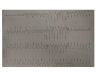
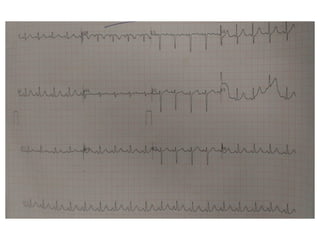
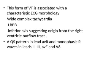
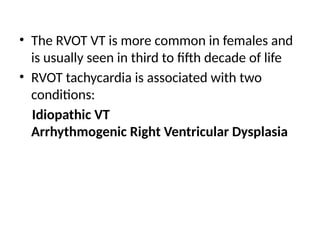
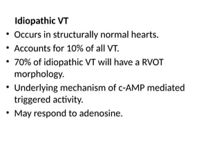
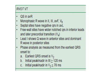
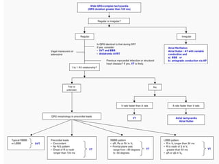
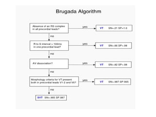
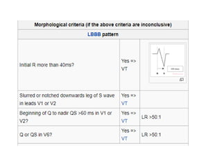
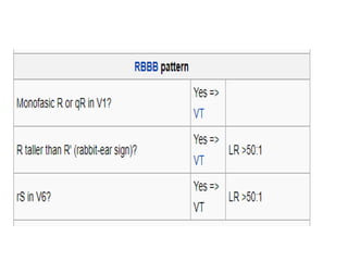
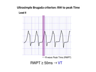
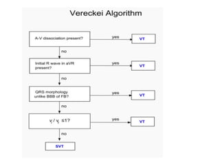
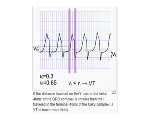
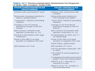
ECG PRESENTATION- RVOT VT
By Dr Kishor
• 60 YR OLD FEMALE PRESENTED WITH C/O
• SOB X 3 DAYS
NYHA class II
• Assosciated with PALPITATION
• No H/O CAD in past
• k/c/o SHTN since 10yrs
• H/O documented VT 1yr back reverted to sinus rhythm by
injection amoidarone.
• Since then patient on Tab amiodarone and Tab metoprolol
and patient stop drug byself for past 1 month
• 2D ECHO 1 YR BACK
LVID- 4.6/2.9/55%
NORMAL LVSF
GRADE I LVDD
NO MS/MR
NO AS/AR
NO PHTN
NO PE, NO CLOT
• CONSCIOUS, ORIENTED
• BP-130/80
• Inj Amiodarone 150 mg iv bolus given…….VT
not reverted
• Synchronized 100 J DC shock given…….VT
reverted
• This form of VT is associated with a
characteristic ECG morphology
Wide complex tachycardia
LBBB
inferior axis suggesting origin from the right
ventricle outflow tract .
• A QS pattern in lead avR and monophasic R
waves in leads II, III, avF and V6.
• The RVOT VT is more common in females and
is usually seen in third to fifth decade of life
• RVOT tachycardia is associated with two
conditions:
Idiopathic VT
Arrhythmogenic Right Ventricular Dysplasia
Idiopathic VT
• Occurs in structurally normal hearts.
• Accounts for 10% of all VT.
• 70% of idiopathic VT will have a RVOT
morphology.
• Underlying mechanism of c-AMP mediated
triggered activity.
• May respond to adenosine.
Arrhythmogenic Right Ventricular Dysplasia
• An inherited myocardial disease associated
with paroxysmal ventricular arrhythmias and
sudden cardiac death.
• Characterized pathologically by fibro-fatty
replacement of the right ventricular
myocardium.
• ARVD is associated with characteristic ECG
abnormalities:
Epsilon wave (most specific finding, seen in 30% of
patients)
T wave inversions in V1-3 (85% of patients)
Prolonged S-wave upstroke of 55ms in V1-3 (95% of
patients)
Localised QRS widening of 110ms in V1-3
Paroxysmal episodes of ventricular tachycardia with LBBB
morphology
TREATMENT
• Usually benign course
• Good prognosis
1. May respond acutely to carotid sinus massage,
Valsalva maneuvers or intravenous adenosine or
verapamil
2. Long-term oral therapy with either BB or CCB
3. Non-responders (33%)- class I or III
antiarrhythmic agents
RFA
• When medical therapy is ineffective or not
tolerated
• High success rate (>80%)
Ultrasimple Brugada criterion: RW to peak Time
• Brugada algorithm is the most commonly used
algorithm. SN 89%, SP 59.2%.
• The lead II R-wave-peak-time .SN 60%, SP
82.7%.
• The aVR algorithm .SN 87.1%, SP 48%.
THANK YOU

Right ventricular outflow tract vt.pptx

  • 1.
    ECG PRESENTATION- RVOTVT By Dr Kishor
  • 2.
    • 60 YROLD FEMALE PRESENTED WITH C/O • SOB X 3 DAYS NYHA class II • Assosciated with PALPITATION • No H/O CAD in past • k/c/o SHTN since 10yrs
  • 3.
    • H/O documentedVT 1yr back reverted to sinus rhythm by injection amoidarone. • Since then patient on Tab amiodarone and Tab metoprolol and patient stop drug byself for past 1 month • 2D ECHO 1 YR BACK LVID- 4.6/2.9/55% NORMAL LVSF GRADE I LVDD NO MS/MR NO AS/AR NO PHTN NO PE, NO CLOT
  • 5.
    • CONSCIOUS, ORIENTED •BP-130/80 • Inj Amiodarone 150 mg iv bolus given…….VT not reverted • Synchronized 100 J DC shock given…….VT reverted
  • 7.
    • This formof VT is associated with a characteristic ECG morphology Wide complex tachycardia LBBB inferior axis suggesting origin from the right ventricle outflow tract . • A QS pattern in lead avR and monophasic R waves in leads II, III, avF and V6.
  • 8.
    • The RVOTVT is more common in females and is usually seen in third to fifth decade of life • RVOT tachycardia is associated with two conditions: Idiopathic VT Arrhythmogenic Right Ventricular Dysplasia
  • 9.
    Idiopathic VT • Occursin structurally normal hearts. • Accounts for 10% of all VT. • 70% of idiopathic VT will have a RVOT morphology. • Underlying mechanism of c-AMP mediated triggered activity. • May respond to adenosine.
  • 10.
    Arrhythmogenic Right VentricularDysplasia • An inherited myocardial disease associated with paroxysmal ventricular arrhythmias and sudden cardiac death. • Characterized pathologically by fibro-fatty replacement of the right ventricular myocardium.
  • 11.
    • ARVD isassociated with characteristic ECG abnormalities: Epsilon wave (most specific finding, seen in 30% of patients) T wave inversions in V1-3 (85% of patients) Prolonged S-wave upstroke of 55ms in V1-3 (95% of patients) Localised QRS widening of 110ms in V1-3 Paroxysmal episodes of ventricular tachycardia with LBBB morphology
  • 14.
    TREATMENT • Usually benigncourse • Good prognosis 1. May respond acutely to carotid sinus massage, Valsalva maneuvers or intravenous adenosine or verapamil 2. Long-term oral therapy with either BB or CCB 3. Non-responders (33%)- class I or III antiarrhythmic agents
  • 15.
    RFA • When medicaltherapy is ineffective or not tolerated • High success rate (>80%)
  • 20.
  • 24.
    • Brugada algorithmis the most commonly used algorithm. SN 89%, SP 59.2%. • The lead II R-wave-peak-time .SN 60%, SP 82.7%. • The aVR algorithm .SN 87.1%, SP 48%.
  • 26.