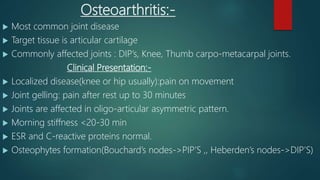
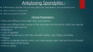
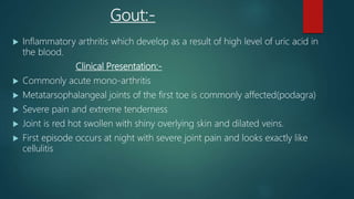
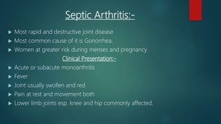
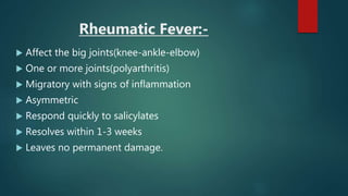
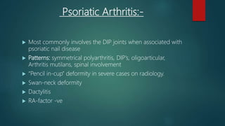
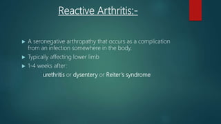
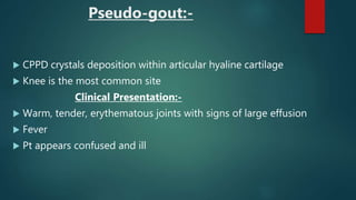
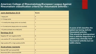
Rheumatology
A BRIEF REVISION OF RHEUMATOLOGY TOPIC
Dr. Sami Ur Rehman
Final Year MBBS
Quaid-e-Azam Medical College, Bahawalpur
#1:-Most common causes:-
 Rheumatoid Arthritis
 Osteoarthritis
 Ankylosing spondylitis
 Gout
 Rheumatic fever
 Psoriatic arthritis
 SLE
 Septic Arthritis
 Reactive Arthritis
 Enteropathic Arthropathy
 Pseudo-gout
 Reactive Arthritis
#2:-Five Clinical Features of each of the
above disease:-
Rheumatoid Arthritis:-
 Chronic inflammatory multisystemic disease with the main target
being the synovium.
Clinical Presentation:-
 Morning stiffness(>1hr) for 6 weeks
 Swelling of wrists , MCPs , PIPs for 6 weeks
 Swelling of 3 joints for 6 weeks
 RF +ve or anti-CCP
 CRP or ESR
 Some joints never involved in RA: DIPs ,, Joints of lower back
Osteoarthritis:-
 Most common joint disease
 Target tissue is articular cartilage
 Commonly affected joints : DIP’s, Knee, Thumb carpo-metacarpal joints.
Clinical Presentation:-
 Localized disease(knee or hip usually):pain on movement
 Joint gelling: pain after rest up to 30 minutes
 Joints are affected in oligo-articular asymmetric pattern.
 Morning stiffness <20-30 min
 ESR and C-reactive proteins normal.
 Osteophytes formation(Bouchard’s nodes->PIP’S ,, Heberden’s nodes->DIP’S)
Ankylosing Spondylitis:-
 Inflammatory disorder that primarily affects the axial skeleton and peripheral joints
 More common in young men
 90% are positive for HLAB27
Clinical Presentation:-
• Pain and stiffness in the lower back and buttocks
• Pain and stiffness that is worse in the mornings and during the night, but may be
improved by light exercise.
• Mild fever.
• Loss of appetite.
• Pain and tenderness in the ribs, shoulder blades, hips, thighs and heels.
• Fatigue.
• Mild to moderate anemia, which can make people pale, tired and short of breath.
• Inflammation of the bowel.
• Iritis or uveitis
Gout:-
 Inflammatory arthritis which develop as a result of high level of uric acid in
the blood.
Clinical Presentation:-
 Commonly acute mono-arthritis
 Metatarsophalangeal joints of the first toe is commonly affected(podagra)
 Severe pain and extreme tenderness
 Joint is red hot swollen with shiny overlying skin and dilated veins.
 First episode occurs at night with severe joint pain and looks exactly like
cellulitis
SLE:-
 Systemic disease in which tissues and multiple organs are damaged by pathogenic
autoantibodies and immune complexes.
Clinical Presentation:-
 Malar Rash
 Discoid rash
 Photosensitivity
 Oral ulcers
 Arthritis
 Serositis
 Renal involvement
 Neurological disorder(seizures or psychosis)
 Hematological disorder(hemolytic anemia,leukopenia,thrombocytopenia)
 Immunological disorder(anti-dsDNA , anti-SM, and other ANA’s
Septic Arthritis:-
 Most rapid and destructive joint disease
 Most common cause of it is Gonorrhea.
 Women at greater risk during menses and pregnancy
Clinical Presentation:-
 Acute or subacute monoarthritis
 Fever
 Joint usually swollen and red
 Pain at rest and movement both
 Lower limb joints esp. knee and hip commonly affected.
Rheumatic Fever:-
 Affect the big joints(knee-ankle-elbow)
 One or more joints(polyarthritis)
 Migratory with signs of inflammation
 Asymmetric
 Respond quickly to salicylates
 Resolves within 1-3 weeks
 Leaves no permanent damage.
Psoriatic Arthritis:-
 Most commonly involves the DIP joints when associated with
psoriatic nail disease
 Patterns: symmetrical polyarthritis, DIP’s, oligoarticular,
Arthritis mutilans, spinal involvement
 “Pencil in-cup” deformity in severe cases on radiology.
 Swan-neck deformity
 Dactylitis
 RA-factor -ve
Reactive Arthritis:-
 A seronegative arthropathy that occurs as a complication
from an infection somewhere in the body.
 Typically affecting lower limb
 1-4 weeks after:
urethritis or dysentery or Reiter’s syndrome
Pseudo-gout:-
 CPPD crystals deposition within articular hyaline cartilage
 Knee is the most common site
Clinical Presentation:-
 Warm, tender, erythematous joints with signs of large effusion
 Fever
 Pt appears confused and ill
Enteropathic Arthropathy:-
 a form of chronic, inflammatory arthritis associated with the
occurrence of an inflammatory bowel disease (IBD)=Ulcerative
colitis and Crohn’s disease.
Systemic Sclerosis:-
 Chronic multisysytem disease characterized by the thickening of the skin caused
by accumulation of the conn tissue and by involvement of the visceral
organs(lungs,git,kidneys)
Clinical Presentation:-
 Skin Thickening
 Raynaud’s phenomenon
 GIT: esophageal dysmotility, hypomotility of small intestine, dilation of large
intestine
 Lungs: Pulmonary fibrosis an Cor-pulmonale
 Kidneys: scleroderma renal crisis in which malignant HT develops and is
associated with acute renal failure.
Behcet Disease:-
#3:-Symptomatology of Rheumatology:-
 Pain
 Stiffness
 Swelling
 Erythema (redness) and warmth
 Weakness
 Locking and triggering
 Extra-articular features(like uveitis,3rd degree heart block,
aortic insufficiencies, rash, Raynaud phenomenon, etc.)
#4:-D/D of Polyarthritis with nodules:-
TYPE EXAMPLE
INFECTIVE
Bacterial: Lyme disease, subacute bacterial
endocarditis
Viral: rubella, mumps, glandular fever, chickenpox,
hepatitis B and C, human immunodeficiency virus
(HIV)
Post-infective Rheumatic fever
Degenerative Osteoarthritis: nodal with Heberden’s/Bouchard’s
nodes
Metabolic Haemochromatosis, gout
Inflammatory Rheumatoid arthritis, SLE, psoriatic arthritis
Other Hypertrophic pulmonary osteoarthropathy
#5:-Causes of false +ve RA factor:-
 SLE(Associated with positive Coomb's test)
 Sjogren syndrome
 Chronic liver disease
 Sarcoidosis
 Interstitial pulmonary fibrosis
 Infectious mononucleosis
 Hep B(Associated with HBsAg)
 TB
 Leprosy
 Syphilis(associated with +ve VDRL)
 Subacute bacterial endocarditis
 Visceral leishmaniasis
 Schistosomiasis
 malaria
Joint distribution (0–5) Score
1 large joint 0
2–10 large joints 1
1–3 small joints (large joints not counted) 2
4–10 small joints (large joints not counted) 3
>10 joints (at least one small joint) 5
Serology (0–3)
Negative RF and negative ACPA 0
Low positive RF or low positive ACPA 2
High positive RF or high positive ACPA 3
Acute-phase reactants
Normal CRP and normal ESR 0
Abnormal CRP or abnormal ESR 1
A score of ≥6 classifies the
patient as having definite
rheumatoid arthritis.
This should be distinguished
from a definite diagnosis as a
patient may clinically have
rheumatoid arthritis but not
fulfill criteria.
American College of Rheumatology/European League Against
Rheumatism classification criteria for rheumatoid arthritis:-

Rheumatology Rapid Review

  • 1.
    Rheumatology A BRIEF REVISIONOF RHEUMATOLOGY TOPIC Dr. Sami Ur Rehman Final Year MBBS Quaid-e-Azam Medical College, Bahawalpur
  • 2.
    #1:-Most common causes:- Rheumatoid Arthritis  Osteoarthritis  Ankylosing spondylitis  Gout  Rheumatic fever  Psoriatic arthritis  SLE  Septic Arthritis  Reactive Arthritis  Enteropathic Arthropathy  Pseudo-gout  Reactive Arthritis
  • 3.
    #2:-Five Clinical Featuresof each of the above disease:-
  • 4.
    Rheumatoid Arthritis:-  Chronicinflammatory multisystemic disease with the main target being the synovium. Clinical Presentation:-  Morning stiffness(>1hr) for 6 weeks  Swelling of wrists , MCPs , PIPs for 6 weeks  Swelling of 3 joints for 6 weeks  RF +ve or anti-CCP  CRP or ESR  Some joints never involved in RA: DIPs ,, Joints of lower back
  • 5.
    Osteoarthritis:-  Most commonjoint disease  Target tissue is articular cartilage  Commonly affected joints : DIP’s, Knee, Thumb carpo-metacarpal joints. Clinical Presentation:-  Localized disease(knee or hip usually):pain on movement  Joint gelling: pain after rest up to 30 minutes  Joints are affected in oligo-articular asymmetric pattern.  Morning stiffness <20-30 min  ESR and C-reactive proteins normal.  Osteophytes formation(Bouchard’s nodes->PIP’S ,, Heberden’s nodes->DIP’S)
  • 6.
    Ankylosing Spondylitis:-  Inflammatorydisorder that primarily affects the axial skeleton and peripheral joints  More common in young men  90% are positive for HLAB27 Clinical Presentation:- • Pain and stiffness in the lower back and buttocks • Pain and stiffness that is worse in the mornings and during the night, but may be improved by light exercise. • Mild fever. • Loss of appetite. • Pain and tenderness in the ribs, shoulder blades, hips, thighs and heels. • Fatigue. • Mild to moderate anemia, which can make people pale, tired and short of breath. • Inflammation of the bowel. • Iritis or uveitis
  • 7.
    Gout:-  Inflammatory arthritiswhich develop as a result of high level of uric acid in the blood. Clinical Presentation:-  Commonly acute mono-arthritis  Metatarsophalangeal joints of the first toe is commonly affected(podagra)  Severe pain and extreme tenderness  Joint is red hot swollen with shiny overlying skin and dilated veins.  First episode occurs at night with severe joint pain and looks exactly like cellulitis
  • 8.
    SLE:-  Systemic diseasein which tissues and multiple organs are damaged by pathogenic autoantibodies and immune complexes. Clinical Presentation:-  Malar Rash  Discoid rash  Photosensitivity  Oral ulcers  Arthritis  Serositis  Renal involvement  Neurological disorder(seizures or psychosis)  Hematological disorder(hemolytic anemia,leukopenia,thrombocytopenia)  Immunological disorder(anti-dsDNA , anti-SM, and other ANA’s
  • 9.
    Septic Arthritis:-  Mostrapid and destructive joint disease  Most common cause of it is Gonorrhea.  Women at greater risk during menses and pregnancy Clinical Presentation:-  Acute or subacute monoarthritis  Fever  Joint usually swollen and red  Pain at rest and movement both  Lower limb joints esp. knee and hip commonly affected.
  • 10.
    Rheumatic Fever:-  Affectthe big joints(knee-ankle-elbow)  One or more joints(polyarthritis)  Migratory with signs of inflammation  Asymmetric  Respond quickly to salicylates  Resolves within 1-3 weeks  Leaves no permanent damage.
  • 11.
    Psoriatic Arthritis:-  Mostcommonly involves the DIP joints when associated with psoriatic nail disease  Patterns: symmetrical polyarthritis, DIP’s, oligoarticular, Arthritis mutilans, spinal involvement  “Pencil in-cup” deformity in severe cases on radiology.  Swan-neck deformity  Dactylitis  RA-factor -ve
  • 12.
    Reactive Arthritis:-  Aseronegative arthropathy that occurs as a complication from an infection somewhere in the body.  Typically affecting lower limb  1-4 weeks after: urethritis or dysentery or Reiter’s syndrome
  • 13.
    Pseudo-gout:-  CPPD crystalsdeposition within articular hyaline cartilage  Knee is the most common site Clinical Presentation:-  Warm, tender, erythematous joints with signs of large effusion  Fever  Pt appears confused and ill
  • 14.
    Enteropathic Arthropathy:-  aform of chronic, inflammatory arthritis associated with the occurrence of an inflammatory bowel disease (IBD)=Ulcerative colitis and Crohn’s disease.
  • 15.
    Systemic Sclerosis:-  Chronicmultisysytem disease characterized by the thickening of the skin caused by accumulation of the conn tissue and by involvement of the visceral organs(lungs,git,kidneys) Clinical Presentation:-  Skin Thickening  Raynaud’s phenomenon  GIT: esophageal dysmotility, hypomotility of small intestine, dilation of large intestine  Lungs: Pulmonary fibrosis an Cor-pulmonale  Kidneys: scleroderma renal crisis in which malignant HT develops and is associated with acute renal failure.
  • 16.
  • 17.
    #3:-Symptomatology of Rheumatology:- Pain  Stiffness  Swelling  Erythema (redness) and warmth  Weakness  Locking and triggering  Extra-articular features(like uveitis,3rd degree heart block, aortic insufficiencies, rash, Raynaud phenomenon, etc.)
  • 18.
    #4:-D/D of Polyarthritiswith nodules:- TYPE EXAMPLE INFECTIVE Bacterial: Lyme disease, subacute bacterial endocarditis Viral: rubella, mumps, glandular fever, chickenpox, hepatitis B and C, human immunodeficiency virus (HIV) Post-infective Rheumatic fever Degenerative Osteoarthritis: nodal with Heberden’s/Bouchard’s nodes Metabolic Haemochromatosis, gout Inflammatory Rheumatoid arthritis, SLE, psoriatic arthritis Other Hypertrophic pulmonary osteoarthropathy
  • 19.
    #5:-Causes of false+ve RA factor:-  SLE(Associated with positive Coomb's test)  Sjogren syndrome  Chronic liver disease  Sarcoidosis  Interstitial pulmonary fibrosis  Infectious mononucleosis  Hep B(Associated with HBsAg)  TB  Leprosy  Syphilis(associated with +ve VDRL)  Subacute bacterial endocarditis  Visceral leishmaniasis  Schistosomiasis  malaria
  • 20.
    Joint distribution (0–5)Score 1 large joint 0 2–10 large joints 1 1–3 small joints (large joints not counted) 2 4–10 small joints (large joints not counted) 3 >10 joints (at least one small joint) 5 Serology (0–3) Negative RF and negative ACPA 0 Low positive RF or low positive ACPA 2 High positive RF or high positive ACPA 3 Acute-phase reactants Normal CRP and normal ESR 0 Abnormal CRP or abnormal ESR 1 A score of ≥6 classifies the patient as having definite rheumatoid arthritis. This should be distinguished from a definite diagnosis as a patient may clinically have rheumatoid arthritis but not fulfill criteria. American College of Rheumatology/European League Against Rheumatism classification criteria for rheumatoid arthritis:-