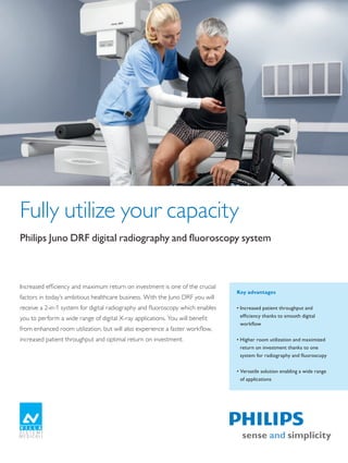
SlideShare a Scribd company logo
1 of 4
Fully utilize your capacity
Philips	Juno	DRF	digital	radiography	and	fluoroscopy	system


Increased efficiency and maximum return on investment is one of the crucial
                                                                                    Key advantages
factors in today’s ambitious healthcare business. With the Juno DRF you will
receive a 2-in-1 system for digital radiography and fluoroscopy which enables       •	Increased	patient	throughput	and	
you to perform a wide range of digital X-ray applications. You will benefit           efficiency	thanks	to	smooth	digital	
                                                                                      workflow
from enhanced room utilization, but will also experience a faster workflow,
increased patient throughput and optimal return on investment.                      •	Higher	room	utilization	and	maximized	
                                                                                      return	on	investment	thanks	to	one	
                                                                                      system	for	radiography	and	fluoroscopy


                                                                                    •	Versatile	solution	enabling	a	wide	range	
                                                                                      of	applications


The print quality of this copy is not an accurate representation of the original.
Juno DRF highlights


             The Juno DRF digital system for both radiography and fluoroscopy examinations enables you to increase
             your workflow and patient throughput while at the same time enhancing your room utilization thanks to
             a 2-in-1 system and a wide range of applications.

             Enhanced efficiency and patient throughput                   Higher room utilization and maximized return
             The	Juno	DRF	helps	you	to	accelerate	your	workflow	          on investment with 2-in-1 system
             and	patient	throughput	thanks	to	a	single	dynamic	           Idle	room	capacity	is	a	common	dilemma	with	dedicated	
             flat	panel	detector	for	all	your	high	resolution	digital	    fluoroscopy	systems.	They	are	usually	operated	at	full	
             radiography	and	high	frame	rate	fluoroscopy	applications.	   capacity	in	the	morning	and	then	see	utilization	drop	off	
             Performing	both	imaging	techniques	with	one	single	          in	the	afternoon.	With	the	Juno	DRF	2-in-1	system	you	
             digital	system	saves	your	time	and	your	money.               can	increase	utilization	rates	throughout	the	whole	day.	
                                                                          Higher	capacity	in	fluoroscopy	paired	with	an	enhanced	
             Smooth digital workflow, improved image quality              application	spectrum	in	digital	radiography	helps	you	
             Your	digital	images	are	instantly	available	with	no	need	    to	increase	room	utilization	and	further	maximize	your	
             to	handle	cassettes	or	wait	for	film	processing.	Benefit	    return	on	investment.	You	will	also	benefit	from	a	better	
             from	a	digital	environment	with	a	smooth,	accelerated	       space	allocation	as	well	as	lower	cost	for	maintenance	
             workflow,	higher	throughput	and	less	waiting	time	for	       and	personnel	training.
             your	patients.
                                                                          Versatile system, wide range of applications
             Specific	digital	processing	algorithms	for	individual	       The	digital	flat	panel	detector	of	the	Juno	DRF	delivers	
             anatomical	regions	recover	image	visualization	in	           the	same	high	spatial-resolution	image	quality	as	other	
             under-	and	overexposed	regions.	As	a	result,	you	will	       state-of-the-art	DR	systems	(148	micron	pixel)	and	the	
             experience	improved	image	quality	and	fewer	retakes.         DRF	images	are	not	affected	by	the	geometric	distortion	
                                                                          typical	with	Image	Intensifiers.	With	a	high	temporal	
                                                                          resolution	frame	rate	of	up	to	30	frames	per	second,	it	
                                                                          allows	you	to	perform	all	radiography	and	fluoroscopy	
                                                                          procedures	in	high	image	quality,	with	a	single	system.	
                                                                          The	Juno	DRF	offers	you	a	wide	range	of	applications,	
Significant reduction of preparation time due                             including	the	most	common	general	radiographic	
to automatic geometry positioning according to
preselected exam parameters.                                              procedures,		 astro-intestinal	studies,	tomography,	and	
                                                                                        g
                                                                          vascular	studies.	The	single	detector	even	enables	you	to	
                                                                          take	exams	on	the	table	with	source-to-image	distance	
                                                                          of	180	cm	(71").


                                                                          Experienced, reliable partner
                                                                          With	more	than	100	years	of	medical	X-ray	experience,	
                                                                          Philips	is	a	pioneer	and	leader	in	X-ray	imaging,	digital	
                                                                          radiography,	and	fluoroscopy.	As	a	global	partner	for	
                                                                          leading	healthcare	facilities	worldwide,	we	offer	a	
                                                                          comprehensive,	multi-modality	set	of	solutions	for	the	
                                                                          entire	radiology	department.	Our	customers	benefit	right	
                                                                          from	the	start	from	the	Philips	global	service	network.	
                                                                          Experienced	staff	will	support	you	at	every	level	to	
                                                                          ensure	you	get	the	highest	possible	uptime	and	return	on	
                                                                          investment	as	well	as	maximum	utility	from	your	system.

2

The print quality of this copy is not an accurate representation of the original.
Optional stitching function enables spine and       With SID up to 180 cm (71"), an extensive range of
                                               extremity reconstruction in distortion-free image   radiography exams can be taken, for example chest
                                               quality.                                            studies in 90° upright table position.


System specifications
Source	to	Image	Distance	(SID)            110	cm	to	180	cm	(43"	to	71")	provides	enhanced	radiographic	capabilities
Motorized	tilting                         -	90°/+90°	for	maximum	flexibility	in	fluoroscopy	applications	and	room	planning
                                          62	cm	(23")	facilitates	easy	transfer	and	positioning	of	patients	especially	with	limited	
Minimum	table	top	height	
                                          mobility
                                          Up	to	284	kg	(626	lbs)	in	all	movements	combined	with	a	large	table	top	size	allows	
Patient	capacity	
                                          for	bariatric	examinations
                                          203	cm	(79.9")	enables	examinations	without	repositioning	for	increased	patient	
Scan	range	without	table-top	movement
                                          comfort
Convenient functions
Auto	Grid	Selection	(AGS)	for	automatic	selection	of	two	grids	and	grid	parking
Automatic	exam-based	pre-positioning	of	the	table	geometry	for	reduced	exam	preparation	time
Optional	stitching	function	for	spine	and	extremity	reconstruction
The	tilting	tube	column	mechanism	(without	mechanical	bar)	enables	tomography	and	oblique	projections	in	any	table	position
Optional	vascular	imaging	e.	g.	for	Digital	Subtraction	Angiography	(DSA)	benefits	from	the	large	patient	coverage	range
Optional	reference	image	display	on	second	monitor
Detector specifications
                                          Electronic	flat	detector	based	on	CsI	(Cesium	Iodide)	scintillator	technology	delivers	
Type
                                          high	quality	digital	images
                                          43	cm	x	43	cm	(17"	x	17")	ensures	optimal	patient	coverage	for	different	exam	types	
Detector	size
                                          including	lungs	and	pelvis
Pixel	size                                148	μm	delivers	high	spatial-resolution	image	quality	for	radiographic	exams
Image	matrix	size                         2,880	pixel	x	2,881	pixel
Dynamic	frame	rate	                       Up	to	30	frames	per	second	enables	full	range	of	fluoroscopy	procedures
Pulsed	fluoroscopy                        Up	to	15	pulses	per	second	for	reduced	X-ray	dose


                                                                                                                                                    3

The print quality of this copy is not an accurate representation of the original.
Fluoroscopy procedures benefit
                                                                     from the large active area of the
                                                                     detector that allows visibility
                                                                     of large anatomic regions without
                                                                     repositioning


                                                                                    The flat panel detector
                                                                             delivers high spatial-resolution
                                                                                   image quality for all your
                                                                                 radiographic examinations


Site Planning
                                                                                                                 6000 (236.2“)

                                                                                             3000 (118.1“)                               3000 (118.1“)


                                                                  lavatory


                                                                                                                                               7         8       9
                                                                  cubicle


                                                                                                                                                                     5700 (224.4“)
                                                                  cubicle                                                                            3

                                                                                                                       1
                                                                                                                                     2
                    1	 	 emote	Controlled	Table
                       R
                    2	 	 ynamic	DR	Control	Room	Monitor	
                       D
                    3	 	 xamination	Room	Monitor		
                       E                                                                                                                                     6
                       with	Trolley	(optional)
                    4	 	 enerator	Control	Console
                       G
                    5	 	 able	Control	Console
                       T
                    6	 	 ynamic	DR	Workstation	Keyboard	
                       D                                                                                                         4                       5
                       and	Mouse
                    7	 	 ynamic	DR	Workstation	Cabinet
                       D
                    8	 	 enerator	Electronic	Cabinet
                       G
                    9	 	 lectronic	Cabinet
                       E


                    The	state-of-the-art	Juno	DRF	is	manufactured	by	Villa	Sistemi	Medicali	(VSM)	and	distributed	by	Philips	Healthcare.	


Please visit www.philips.com/fluoroscopy


                    ©	2011	Koninklijke	Philips	Electronics	N.V.
                    All	rights	are	reserved.

                    Philips	Healthcare	reserves	the	right	to	make	changes	in	specifications	and/or	to	discontinue	any	product	at	any	time	without	notice	or	
                    obligation	and	will	not	be	liable	for	any	consequences	resulting	from	the	use	of	this	publication.

                    Printed	in	The	Netherlands.
                    4522	962	75581	*	JUL	2011


The print quality of this copy is not an accurate representation of the original.

More Related Content

What's hot

Dental Digital Radiography in Easy to Understand Steps
Dental Digital  Radiography in Easy to Understand StepsDental Digital  Radiography in Easy to Understand Steps
Dental Digital Radiography in Easy to Understand StepsTheresa Nielsen
 
Jaminan mutu imaging
Jaminan mutu imagingJaminan mutu imaging
Jaminan mutu imagingHeri Kuswoyo
 
Anjali (qa qc on cr dr)
Anjali (qa qc on cr dr)Anjali (qa qc on cr dr)
Anjali (qa qc on cr dr)globalsoin
 
Introduction to digital radiography and pacs
Introduction to digital radiography and pacs Introduction to digital radiography and pacs
Introduction to digital radiography and pacs Irshad Basheer
 
Conventional versus digital/ dental implant courses
Conventional versus digital/ dental implant coursesConventional versus digital/ dental implant courses
Conventional versus digital/ dental implant coursesIndian dental academy
 
Exactitude Teleradiology Solutions - Canada
Exactitude Teleradiology Solutions - CanadaExactitude Teleradiology Solutions - Canada
Exactitude Teleradiology Solutions - CanadaIsmail Siddiqui
 
Information Technology in Radiology
Information Technology in RadiologyInformation Technology in Radiology
Information Technology in RadiologyIsmail Adegbenga
 
7 140319140043-phpapp01
7 140319140043-phpapp017 140319140043-phpapp01
7 140319140043-phpapp01Onikaroy
 
The Role of Computers in Medical Physics
The Role of Computers in Medical PhysicsThe Role of Computers in Medical Physics
The Role of Computers in Medical PhysicsVictor Ekpo
 
In Vivo Optical Imaging from the Whole Animal to the Cellular Level
In Vivo Optical Imaging from the Whole Animal to the Cellular LevelIn Vivo Optical Imaging from the Whole Animal to the Cellular Level
In Vivo Optical Imaging from the Whole Animal to the Cellular Levelabhitris
 
CBCT in Periodontology
CBCT in PeriodontologyCBCT in Periodontology
CBCT in PeriodontologyRinisha Sinha
 
Health Valley Event Presentatie John Lapré
Health Valley Event Presentatie John LapréHealth Valley Event Presentatie John Lapré
Health Valley Event Presentatie John LapréHealth Valley
 
Senthil kumar cmch
Senthil kumar cmchSenthil kumar cmch
Senthil kumar cmchrajisym
 
Biomedical Imaging In Vivo 2009
Biomedical Imaging In Vivo 2009Biomedical Imaging In Vivo 2009
Biomedical Imaging In Vivo 2009Servadio
 

What's hot (20)

Dental Digital Radiography in Easy to Understand Steps
Dental Digital  Radiography in Easy to Understand StepsDental Digital  Radiography in Easy to Understand Steps
Dental Digital Radiography in Easy to Understand Steps
 
Jaminan mutu imaging
Jaminan mutu imagingJaminan mutu imaging
Jaminan mutu imaging
 
Anjali (qa qc on cr dr)
Anjali (qa qc on cr dr)Anjali (qa qc on cr dr)
Anjali (qa qc on cr dr)
 
Introduction to digital radiography and pacs
Introduction to digital radiography and pacs Introduction to digital radiography and pacs
Introduction to digital radiography and pacs
 
ARP Products and services description
ARP Products and services descriptionARP Products and services description
ARP Products and services description
 
Conventional versus digital/ dental implant courses
Conventional versus digital/ dental implant coursesConventional versus digital/ dental implant courses
Conventional versus digital/ dental implant courses
 
Exactitude Teleradiology Solutions - Canada
Exactitude Teleradiology Solutions - CanadaExactitude Teleradiology Solutions - Canada
Exactitude Teleradiology Solutions - Canada
 
3d Imaging
3d Imaging3d Imaging
3d Imaging
 
Information Technology in Radiology
Information Technology in RadiologyInformation Technology in Radiology
Information Technology in Radiology
 
7 140319140043-phpapp01
7 140319140043-phpapp017 140319140043-phpapp01
7 140319140043-phpapp01
 
The Role of Computers in Medical Physics
The Role of Computers in Medical PhysicsThe Role of Computers in Medical Physics
The Role of Computers in Medical Physics
 
Specialized radiographic techniques
Specialized radiographic techniquesSpecialized radiographic techniques
Specialized radiographic techniques
 
In Vivo Optical Imaging from the Whole Animal to the Cellular Level
In Vivo Optical Imaging from the Whole Animal to the Cellular LevelIn Vivo Optical Imaging from the Whole Animal to the Cellular Level
In Vivo Optical Imaging from the Whole Animal to the Cellular Level
 
Linkedin
LinkedinLinkedin
Linkedin
 
CBCT in Periodontology
CBCT in PeriodontologyCBCT in Periodontology
CBCT in Periodontology
 
Health Valley Event Presentatie John Lapré
Health Valley Event Presentatie John LapréHealth Valley Event Presentatie John Lapré
Health Valley Event Presentatie John Lapré
 
Senthil kumar cmch
Senthil kumar cmchSenthil kumar cmch
Senthil kumar cmch
 
Small Animal Optical Imaging
Small Animal Optical ImagingSmall Animal Optical Imaging
Small Animal Optical Imaging
 
Fona cdr elite print
Fona cdr elite printFona cdr elite print
Fona cdr elite print
 
Biomedical Imaging In Vivo 2009
Biomedical Imaging In Vivo 2009Biomedical Imaging In Vivo 2009
Biomedical Imaging In Vivo 2009
 

Similar to Rf juno drf_product_overview

Logiq e9 xd clear 2.0
Logiq e9 xd clear 2.0 Logiq e9 xd clear 2.0
Logiq e9 xd clear 2.0 Madhuka Perera
 
COMPUTER APPLICATIONS IN DENTISTRY.pptx
COMPUTER APPLICATIONS IN DENTISTRY.pptxCOMPUTER APPLICATIONS IN DENTISTRY.pptx
COMPUTER APPLICATIONS IN DENTISTRY.pptxParkhiBhatngar
 
Pixium 4343F Detector - PhiGEM Parts
Pixium 4343F Detector - PhiGEM PartsPixium 4343F Detector - PhiGEM Parts
Pixium 4343F Detector - PhiGEM PartsMildred Galella
 
Go 2 d3d ceph_cataloge
Go 2 d3d ceph_catalogeGo 2 d3d ceph_cataloge
Go 2 d3d ceph_catalogeYenDoan19
 
Freedom Ultrasound RIS PACS
Freedom Ultrasound RIS PACS Freedom Ultrasound RIS PACS
Freedom Ultrasound RIS PACS Vivek Mehrotra
 
whitepaper-advanced-ultrasound-imaging-201507
whitepaper-advanced-ultrasound-imaging-201507whitepaper-advanced-ultrasound-imaging-201507
whitepaper-advanced-ultrasound-imaging-201507Ajay Anand
 
Carestream Whitepaper Advanced Ultrasound Imaging
Carestream Whitepaper Advanced Ultrasound ImagingCarestream Whitepaper Advanced Ultrasound Imaging
Carestream Whitepaper Advanced Ultrasound ImagingCarestream
 
Digital fluoro
Digital fluoroDigital fluoro
Digital fluoroRad Tech
 
Magnetom C¡ 0.35 T - Siemens- OPEN
Magnetom C¡ 0.35 T - Siemens- OPENMagnetom C¡ 0.35 T - Siemens- OPEN
Magnetom C¡ 0.35 T - Siemens- OPENJhon Arriaga Cordova
 
Mxr mobile diagnost_wdr_product_brochure
Mxr mobile diagnost_wdr_product_brochureMxr mobile diagnost_wdr_product_brochure
Mxr mobile diagnost_wdr_product_brochureJhon Arriaga Cordova
 
Mxr mobile diagnost_wdr_product_brochure
Mxr mobile diagnost_wdr_product_brochureMxr mobile diagnost_wdr_product_brochure
Mxr mobile diagnost_wdr_product_brochureJhon Arriaga Cordova
 
Mri d stream brochure_final_16 nov 12
Mri d stream brochure_final_16 nov 12Mri d stream brochure_final_16 nov 12
Mri d stream brochure_final_16 nov 12Jhon Arriaga Cordova
 
ctfluoroscopy11-230414043252-3728b13a (1).pdf
ctfluoroscopy11-230414043252-3728b13a (1).pdfctfluoroscopy11-230414043252-3728b13a (1).pdf
ctfluoroscopy11-230414043252-3728b13a (1).pdfNiteshChandraDas1
 

Similar to Rf juno drf_product_overview (20)

Logiq e9 xd clear 2.0
Logiq e9 xd clear 2.0 Logiq e9 xd clear 2.0
Logiq e9 xd clear 2.0
 
COMPUTER APPLICATIONS IN DENTISTRY.pptx
COMPUTER APPLICATIONS IN DENTISTRY.pptxCOMPUTER APPLICATIONS IN DENTISTRY.pptx
COMPUTER APPLICATIONS IN DENTISTRY.pptx
 
Pixium 4343F Detector - PhiGEM Parts
Pixium 4343F Detector - PhiGEM PartsPixium 4343F Detector - PhiGEM Parts
Pixium 4343F Detector - PhiGEM Parts
 
Symbia T Spect-CT
Symbia T  Spect-CTSymbia T  Spect-CT
Symbia T Spect-CT
 
Go 2 d3d ceph_cataloge
Go 2 d3d ceph_catalogeGo 2 d3d ceph_cataloge
Go 2 d3d ceph_cataloge
 
Freedom Ultrasound RIS PACS
Freedom Ultrasound RIS PACS Freedom Ultrasound RIS PACS
Freedom Ultrasound RIS PACS
 
whitepaper-advanced-ultrasound-imaging-201507
whitepaper-advanced-ultrasound-imaging-201507whitepaper-advanced-ultrasound-imaging-201507
whitepaper-advanced-ultrasound-imaging-201507
 
Carestream Whitepaper Advanced Ultrasound Imaging
Carestream Whitepaper Advanced Ultrasound ImagingCarestream Whitepaper Advanced Ultrasound Imaging
Carestream Whitepaper Advanced Ultrasound Imaging
 
Digital fluoro
Digital fluoroDigital fluoro
Digital fluoro
 
Magnetom C¡ 0.35 T - Siemens- OPEN
Magnetom C¡ 0.35 T - Siemens- OPENMagnetom C¡ 0.35 T - Siemens- OPEN
Magnetom C¡ 0.35 T - Siemens- OPEN
 
FieldStrength 50 advanced neuro
FieldStrength 50 advanced neuroFieldStrength 50 advanced neuro
FieldStrength 50 advanced neuro
 
Fona xdg brochure
Fona xdg brochureFona xdg brochure
Fona xdg brochure
 
Fona XDG
Fona XDGFona XDG
Fona XDG
 
Mxr mobile diagnost_wdr_product_brochure
Mxr mobile diagnost_wdr_product_brochureMxr mobile diagnost_wdr_product_brochure
Mxr mobile diagnost_wdr_product_brochure
 
Mxr mobile diagnost_wdr_product_brochure
Mxr mobile diagnost_wdr_product_brochureMxr mobile diagnost_wdr_product_brochure
Mxr mobile diagnost_wdr_product_brochure
 
Mri d stream brochure_final_16 nov 12
Mri d stream brochure_final_16 nov 12Mri d stream brochure_final_16 nov 12
Mri d stream brochure_final_16 nov 12
 
ctfluoroscopy11-230414043252-3728b13a (1).pdf
ctfluoroscopy11-230414043252-3728b13a (1).pdfctfluoroscopy11-230414043252-3728b13a (1).pdf
ctfluoroscopy11-230414043252-3728b13a (1).pdf
 
CT fluoroscopy1 (1).pptx
CT fluoroscopy1 (1).pptxCT fluoroscopy1 (1).pptx
CT fluoroscopy1 (1).pptx
 
Fona CDR
Fona CDRFona CDR
Fona CDR
 
Fona cdr web2
Fona cdr web2Fona cdr web2
Fona cdr web2
 

More from Jhon Arriaga Cordova (20)

Resonancia no ecoplanar en colesteatoma.pptx
Resonancia no ecoplanar en colesteatoma.pptxResonancia no ecoplanar en colesteatoma.pptx
Resonancia no ecoplanar en colesteatoma.pptx
 
Oído.pptx
Oído.pptxOído.pptx
Oído.pptx
 
Toshiba Visions 23
Toshiba Visions 23Toshiba Visions 23
Toshiba Visions 23
 
FieldStrength 49
FieldStrength 49FieldStrength 49
FieldStrength 49
 
Ct somatom sessions-33
Ct somatom sessions-33Ct somatom sessions-33
Ct somatom sessions-33
 
Somatom sessions 32
Somatom sessions 32Somatom sessions 32
Somatom sessions 32
 
Somatom sessions 31
Somatom sessions 31Somatom sessions 31
Somatom sessions 31
 
Somatom sessions 30
Somatom sessions 30Somatom sessions 30
Somatom sessions 30
 
Somatom sessions 26
Somatom sessions 26Somatom sessions 26
Somatom sessions 26
 
Somatom sessions 25
Somatom sessions 25Somatom sessions 25
Somatom sessions 25
 
Somatom sessions 24
Somatom sessions 24Somatom sessions 24
Somatom sessions 24
 
Somatom session 29
Somatom session 29Somatom session 29
Somatom session 29
 
Somatom session 29
Somatom session 29Somatom session 29
Somatom session 29
 
Somatom session 28
Somatom session 28Somatom session 28
Somatom session 28
 
Somatom session 27
Somatom session 27Somatom session 27
Somatom session 27
 
Somatom session 27
Somatom session 27Somatom session 27
Somatom session 27
 
Neurography Supplement to Issue 50
Neurography Supplement to Issue 50Neurography Supplement to Issue 50
Neurography Supplement to Issue 50
 
ISMRM Edition - Issue 49
ISMRM Edition - Issue 49ISMRM Edition - Issue 49
ISMRM Edition - Issue 49
 
Pediatric Imaging - Issue 47
Pediatric Imaging - Issue 47Pediatric Imaging - Issue 47
Pediatric Imaging - Issue 47
 
ISMRM Edition – Issue 58
ISMRM Edition – Issue 58ISMRM Edition – Issue 58
ISMRM Edition – Issue 58
 

Rf juno drf_product_overview

  • 1. Fully utilize your capacity Philips Juno DRF digital radiography and fluoroscopy system Increased efficiency and maximum return on investment is one of the crucial Key advantages factors in today’s ambitious healthcare business. With the Juno DRF you will receive a 2-in-1 system for digital radiography and fluoroscopy which enables • Increased patient throughput and you to perform a wide range of digital X-ray applications. You will benefit efficiency thanks to smooth digital workflow from enhanced room utilization, but will also experience a faster workflow, increased patient throughput and optimal return on investment. • Higher room utilization and maximized return on investment thanks to one system for radiography and fluoroscopy • Versatile solution enabling a wide range of applications The print quality of this copy is not an accurate representation of the original.
  • 2. Juno DRF highlights The Juno DRF digital system for both radiography and fluoroscopy examinations enables you to increase your workflow and patient throughput while at the same time enhancing your room utilization thanks to a 2-in-1 system and a wide range of applications. Enhanced efficiency and patient throughput Higher room utilization and maximized return The Juno DRF helps you to accelerate your workflow on investment with 2-in-1 system and patient throughput thanks to a single dynamic Idle room capacity is a common dilemma with dedicated flat panel detector for all your high resolution digital fluoroscopy systems. They are usually operated at full radiography and high frame rate fluoroscopy applications. capacity in the morning and then see utilization drop off Performing both imaging techniques with one single in the afternoon. With the Juno DRF 2-in-1 system you digital system saves your time and your money. can increase utilization rates throughout the whole day. Higher capacity in fluoroscopy paired with an enhanced Smooth digital workflow, improved image quality application spectrum in digital radiography helps you Your digital images are instantly available with no need to increase room utilization and further maximize your to handle cassettes or wait for film processing. Benefit return on investment. You will also benefit from a better from a digital environment with a smooth, accelerated space allocation as well as lower cost for maintenance workflow, higher throughput and less waiting time for and personnel training. your patients. Versatile system, wide range of applications Specific digital processing algorithms for individual The digital flat panel detector of the Juno DRF delivers anatomical regions recover image visualization in the same high spatial-resolution image quality as other under- and overexposed regions. As a result, you will state-of-the-art DR systems (148 micron pixel) and the experience improved image quality and fewer retakes. DRF images are not affected by the geometric distortion typical with Image Intensifiers. With a high temporal resolution frame rate of up to 30 frames per second, it allows you to perform all radiography and fluoroscopy procedures in high image quality, with a single system. The Juno DRF offers you a wide range of applications, Significant reduction of preparation time due including the most common general radiographic to automatic geometry positioning according to preselected exam parameters. procedures, astro-intestinal studies, tomography, and g vascular studies. The single detector even enables you to take exams on the table with source-to-image distance of 180 cm (71"). Experienced, reliable partner With more than 100 years of medical X-ray experience, Philips is a pioneer and leader in X-ray imaging, digital radiography, and fluoroscopy. As a global partner for leading healthcare facilities worldwide, we offer a comprehensive, multi-modality set of solutions for the entire radiology department. Our customers benefit right from the start from the Philips global service network. Experienced staff will support you at every level to ensure you get the highest possible uptime and return on investment as well as maximum utility from your system. 2 The print quality of this copy is not an accurate representation of the original.
  • 3. Optional stitching function enables spine and With SID up to 180 cm (71"), an extensive range of extremity reconstruction in distortion-free image radiography exams can be taken, for example chest quality. studies in 90° upright table position. System specifications Source to Image Distance (SID) 110 cm to 180 cm (43" to 71") provides enhanced radiographic capabilities Motorized tilting - 90°/+90° for maximum flexibility in fluoroscopy applications and room planning 62 cm (23") facilitates easy transfer and positioning of patients especially with limited Minimum table top height mobility Up to 284 kg (626 lbs) in all movements combined with a large table top size allows Patient capacity for bariatric examinations 203 cm (79.9") enables examinations without repositioning for increased patient Scan range without table-top movement comfort Convenient functions Auto Grid Selection (AGS) for automatic selection of two grids and grid parking Automatic exam-based pre-positioning of the table geometry for reduced exam preparation time Optional stitching function for spine and extremity reconstruction The tilting tube column mechanism (without mechanical bar) enables tomography and oblique projections in any table position Optional vascular imaging e. g. for Digital Subtraction Angiography (DSA) benefits from the large patient coverage range Optional reference image display on second monitor Detector specifications Electronic flat detector based on CsI (Cesium Iodide) scintillator technology delivers Type high quality digital images 43 cm x 43 cm (17" x 17") ensures optimal patient coverage for different exam types Detector size including lungs and pelvis Pixel size 148 μm delivers high spatial-resolution image quality for radiographic exams Image matrix size 2,880 pixel x 2,881 pixel Dynamic frame rate Up to 30 frames per second enables full range of fluoroscopy procedures Pulsed fluoroscopy Up to 15 pulses per second for reduced X-ray dose 3 The print quality of this copy is not an accurate representation of the original.
  • 4. Fluoroscopy procedures benefit from the large active area of the detector that allows visibility of large anatomic regions without repositioning The flat panel detector delivers high spatial-resolution image quality for all your radiographic examinations Site Planning 6000 (236.2“) 3000 (118.1“) 3000 (118.1“) lavatory 7 8 9 cubicle 5700 (224.4“) cubicle 3 1 2 1 emote Controlled Table R 2 ynamic DR Control Room Monitor D 3 xamination Room Monitor E 6 with Trolley (optional) 4 enerator Control Console G 5 able Control Console T 6 ynamic DR Workstation Keyboard D 4 5 and Mouse 7 ynamic DR Workstation Cabinet D 8 enerator Electronic Cabinet G 9 lectronic Cabinet E The state-of-the-art Juno DRF is manufactured by Villa Sistemi Medicali (VSM) and distributed by Philips Healthcare. Please visit www.philips.com/fluoroscopy © 2011 Koninklijke Philips Electronics N.V. All rights are reserved. Philips Healthcare reserves the right to make changes in specifications and/or to discontinue any product at any time without notice or obligation and will not be liable for any consequences resulting from the use of this publication. Printed in The Netherlands. 4522 962 75581 * JUL 2011 The print quality of this copy is not an accurate representation of the original.