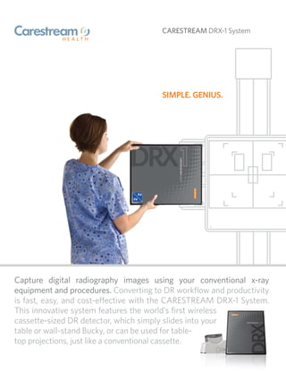
The Carestream DRX-1 system features a wireless cassette-sized digital radiography detector that slides into existing Bucky tables or wall stands. It allows healthcare providers to easily convert conventional x-ray rooms to digital radiography. The detector wirelessly transmits images to a compact console for viewing, editing, and sharing. The system requires only a one-day installation and minimal staff training, providing a simple and cost-effective transition to DR.
The document proposes new techniques called ERASE and ERASERS for more efficiently sanitizing computer media. ERASE calculates the entropy of disk blocks and overwrites those within a sensitive entropy range. ERASERS divides the area to be wiped into subpopulations, randomly samples blocks within each, and only overwrites subpopulations containing sensitive data. The techniques were implemented in a tool and showed improvements over current methods for wiping drives of increasing size.
The document summarizes Philips' SmartPath to dStream program, which upgrades existing MRI systems to digital broadband architecture without requiring a new magnet. It transforms systems into digital MRI with benefits like improved image quality, speed, and expandability. The program overhauls key system components, extends the lifetime by 10 years, and has lower costs than a new installation while achieving similar performance to the latest systems. It also has environmental benefits from reusing the existing magnet.
The document describes a multiprogramming system with a hierarchical structure implemented on a Dutch computer. The system divides all activities across sequential processes at different hierarchical levels, with independent abstractions at each level. This hierarchical structure proved vital for verifying the system's logical soundness and implementation correctness. The primary goal of the system is to smoothly process a continuous flow of user programs as a service to the university.
Recent observed environmental changes as well as projections in the fourth assessment report of the Intergovernmental Panel on Climate Change shed light on likely dramatic consequences of a changing mountain cryosphere following climate change. Some very destructive geological processes are triggered or intensified, influencing the stability of slopes and possibly inducing landslides. Unfortunately, the interaction between these complex processes is poorly understood. This project addresses the key issues in response to such changing conditons: monitoring and warning systems for the spatial and temporal detection of newly forming hazards, as well as extending the quantitative understanding of these changing natural systems and our predictive capabilities.
The Philips CT SyncRight option provides seamless integration between CT scanners and compatible injectors, allowing communication of scanning and injection parameters. SyncRight simplifies clinical workflow by streamlining operations and reducing decision points by over 60%, allowing clinicians to focus more on patients. Key benefits include automatically loading injection protocols, modifying protocols through the scanner interface, viewing injection progress in real-time, and producing reports.
The document proposes a trigger identification service to defend against reactive jammers in wireless sensor networks. The service aims to efficiently identify all sensor nodes whose transmissions trigger jamming attacks, known as trigger nodes. This is achieved through a group testing process using a randomized error-tolerant nonadaptive group testing scheme. The identification of trigger nodes can then be used to exclude them from routing paths and help locate reactive jammers. The paper presents models for the sensor network, jammer behaviors, and analyzes the performance of the proposed identification service.
The document summarizes Senso LAB, one of the most advanced wireless sensor network labs hosting hundreds of heterogeneous sensor nodes deployed around Middlesex University. It has over 20 members including professors and researchers studying topics such as energy-aware performance evaluation of wireless sensor networks, unequal clustering algorithms, intrusion detection systems, software modeling frameworks, and presents ongoing and future work. The lab utilizes the Castalia simulator to test distributed algorithms and protocols under realistic conditions.
NOISE REMOVAL TECHNIQUES FOR MICROWAVE REMOTE SENSING RADAR DATA AND ITS EVAL...cscpconf
Microwave Remote Sensing data acquired by a RADAR sensor such as SAR(Synthetic Aperture Radar) is affected by a peculiar kind of noise called speckle. This noise not only renders the
data ineffective for classification, texture analysis, segmentation etc. which are used for image analysis purposes, but also degrades the overall contrast and radiometric quality of the image. Here we discuss the various noise removal techniques which have been widely used by scientists all over the world. Different filtering methods have their pros and cons, and no single method can give the most satisfactory result. In order to circumvent those issues, better and better methods are being attempted. One of the recent methods is that based on Wavelet technique. This paper discusses the denoising techniques based on Wavelets and the results from some of those methods. The relative merits and demerits of the filters and their evaluation is also done.
Exposure factors such as kVp, mA, time, mAs, focal spot size, and distance influence the quality and quantity of the x-ray beam and the resulting radiographic image. KVp controls beam quality and penetration, mA controls quantity of x-rays, and mAs is the product of mA and time determining total exposure. Increasing kVp increases penetration but reduces contrast. Proper selection of these technical factors is needed to produce diagnostic radiographs with minimal radiation exposure.
The document summarizes a presentation given at an RPM working meeting at Bruce Power about using portable x-ray radiography equipment to conduct radiography tests. It discusses the goals of maintaining safety controls and reducing dose, costs, and exclusion zones while maintaining quality. It provides details on the portable x-ray tube and digital imaging equipment used, test setup, and results from a field application that showed significant savings in time, costs, and dose compared to traditional radiography.
Radiography clinical updates - session one menkantozz
The document discusses the radiographic critique of a cervical spine x-ray following a road traffic accident. It analyzes the request form, justifies the procedure, discusses optimization of exposure factors, and critiques the radiographic images. The lateral view shows all cervical vertebrae and the C7-T1 junction. The AP view demonstrates the cervical vertebrae and soft tissues of the neck. Both images have sufficient contrast and density to evaluate for fractures or injuries while minimizing radiation exposure to the patient.
This document provides an overview of dental radiography and x-ray physics. It defines key terms like atomic structure, ionization, and electromagnetic radiation. X-rays are produced when electrons collide with a tungsten target in the dental x-ray tube. The tubehead contains the x-ray tube, filters, and collimator to shape the beam. X-rays can interact with matter through absorption, Compton scattering, or coherent scattering. The document also outlines the parts of the x-ray machine and how alternating current is converted to produce x-rays when the exposure button is engaged.
This document provides information on periapical radiography. It discusses the history and types of dental x-rays, including periapical radiographs. Periapical radiographs are used to detect abnormalities of the root structure and surrounding bone. The document outlines techniques for proper positioning during periapical radiography, including the paralleling technique and bisected angle technique. It also discusses advantages and disadvantages of each technique as well as indications for periapical radiography.
Panoramic radiography, also called a panoramic x-ray, captures a single image of the entire mouth, including teeth, jaws, and surrounding structures. It is commonly used by dentists and oral surgeons to evaluate bones, teeth, and check for issues like tumors, cysts, or impacted teeth. The procedure involves a rotating x-ray tube that projects a beam through the patient's head and onto a rotating film or detector. It is painless, fast, and provides a wider view than intraoral x-rays. While it does not show the same detail as other imaging tests, panoramic x-rays are useful for initial evaluation of dental problems.
Computed radiography and direct/digital radiography are two digital imaging techniques. Computed radiography uses an imaging plate that captures x-ray data, which is then converted to a digital image. Direct digital radiography uses detectors like TFT or flat panel detectors to directly capture x-ray data digitally. Both techniques offer benefits over traditional film like faster imaging and easier sharing of images.
The document summarizes key aspects of radiographic film, including its composition, construction, types, handling, and the latent image formation process. Radiographic film consists of a base and emulsion layer containing light-sensitive silver halide crystals. X-rays interact with the crystals to form a latent image, which is developed into a visible image. Proper handling and storage of the film is required to avoid artifacts and ensure optimal image quality.
The document discusses different types of radiography technologies, including computed radiography (CR), direct digital radiography (DR), and the components and layers of imaging plates (IPs) used in CR. It also covers image processing techniques for CR/DR such as histogram generation, exposure compensation, and potential artifacts that can occur during acquisition, post-acquisition or display.
The document discusses various radiographic exposure factors and how they influence the quantity and quality of x-radiation exposure to patients. It describes how factors like kVp, mA, and exposure time determine the radiation dose and beam quality. It also discusses how the design of the x-ray machine like focal spot size, filtration, and high voltage generation impact technical settings. Film factors like sensitometry, contrast, and processing also influence radiographic image quality.
The document provides an overview of human anatomy and physiology. It begins with objectives for students to gain knowledge of body parts and systems. It then defines anatomy and physiology. The rest of the document describes the major functions of 7 body systems - integumentary, respiratory, cardiovascular, gastrointestinal, female and male genitourinary, musculoskeletal, and nervous systems. Each system section lists the main structures and their functions.
Radiographers are medical professionals who perform diagnostic imaging examinations and procedures to help physicians diagnose and treat diseases. They work under the supervision of radiologists to operate X-ray, CT, MRI, ultrasound and other medical imaging equipment and must have a strong understanding of human anatomy and pathology. Radiographers are responsible for correctly positioning patients, ensuring proper imaging techniques are used, and evaluating the quality of the resulting images.
The Kodak CR800 DirectView system is a single-cassette computed radiography system that provides fast, efficient, and productive x-ray imaging. It can process up to 62 cassettes per hour and make images available for review in under 33 seconds. The system features an easy-to-use interface, options for enhancing image quality, and capabilities for streamlining workflow and improving productivity.
DRX Plus Detectors: Going from Good to GreatCarestream
Carestream’s introduction in 2009 of the world’s first portable, wireless, cassette-sized detector – the CARESTREAM DRX-1 Detector – has fundamentally changed the digital radiography market with a versatile and cost-effective imaging solution. Now available, the third-generation design: the CARESTREAM DRX Plus Detector takes DR to a new level.
This paper describes many of the features and benefits of the DRX Plus at a detailed level to demonstrate that the DRX Plus takes digital radiography with portable wireless detectors from good to great.
For more information visit http://www.carestream.com/medical
The document describes the NETWORKSUPERVISIONTM DTX CableAnalyzer Series from Fluke Networks. It offers the following key benefits:
1) It significantly reduces the total time to certify fiber networks by improving every aspect of the testing process, including faster testing speeds, accurate results, long battery life, and easy setup and reporting.
2) It delivers fast certification of basic Tier 1 fiber through automated testing features and integrated fiber inspection tools.
3) It enables extended Tier 2 fiber certification by shooting traces with its optional Compact OTDR module to measure loss and reflectivity along fiber links.
This document provides an overview of EXAMION® Systems, a supplier of digital and conventional X-ray systems. It describes their portfolio of DR, CR, and portable X-ray generators. EXAMION® offers complete DR systems integrating an X-ray generator, detector, and acquisition software. They also provide CR digitizer systems that allow existing X-ray equipment to be used for digital radiography. A range of portable X-ray generators are presented for mobile and small animal use. The EXAMION® AQS software is highlighted as the heart of the digital systems, controlling image acquisition, processing, and archiving.
The document summarizes the key features and benefits of the Philips MobileDiagnost wDR mobile digital radiography system. It allows for wireless mobility, so digital radiography exams can be performed anywhere in the hospital. This is especially useful in acute and critical care areas where fast diagnoses are important. The system offers premium image quality, intuitive workflow, and wireless sharing of detectors between modalities to increase flexibility and efficiency. Lifecycle services are also available to help maximize system uptime and support.
Spread spectrum communication uses wideband noise-like signals that are hard to detect, intercept, or jam. It spreads data over multiple frequencies. There are two main techniques: direct sequence spread spectrum multiplies a data signal by a pseudorandom code, and frequency hopping spread spectrum modulates a narrowband carrier that hops between frequencies. Spread spectrum provides benefits like resistance to interference and jamming, better signal quality, and inherent security. It finds applications in wireless networks, Bluetooth, and CDMA cellular systems.
The document describes the Philips Juno DRF digital radiography and fluoroscopy system. It highlights that the 2-in-1 system allows for increased efficiency and workflow by performing both radiography and fluoroscopy examinations with a single detector. This enhances room utilization and maximizes return on investment. The versatile system also enables a wide range of applications like general radiography and fluoroscopy procedures.
The document describes the Philips Juno DRF digital radiography and fluoroscopy system. It highlights that the 2-in-1 system allows for increased efficiency and patient throughput by performing both radiography and fluoroscopy with one detector. This also maximizes room utilization. The system provides high quality images for a wide range of applications while simplifying the digital workflow.
This document discusses cognitive radio networks (CR). CR networks are defined as networks that are aware of their surroundings and can dynamically reconfigure their characteristics. The document outlines topics that will be discussed including CR antennas, mechanisms, accessibility, adaptivity, scalability, reliability and interconnectivity. It provides examples of how CR networks improve these qualities and can sense spectrum usage, analyze it, decide on parameters, and tune transmissions accordingly using reconfigurable antennas. The document compares CRs to conventional radios and outlines benefits of CRs such as functioning in challenging conditions and identifying unused spectrum. It mentions xG Technology developed CR network technology called xMax.
Cognitive radio is a form of wireless communication that detects available channels in the radio spectrum and moves into vacant channels to avoid occupied ones. It senses its operational environment to detect unused spectrum and adjusts transmission parameters accordingly. This allows for more efficient use of the spectrum by taking advantage of spectrum white spaces. The cognitive cycle involves spectrum sensing, decision, sharing, and mobility. Cognitive radio networks apply these principles across network resources as well to optimize performance. Techniques like software-defined radio, machine learning, game theory, and cross-layer design help enable cognitive capabilities. Cognitive radio could help address increasing mobile data usage and be applied in 5G networks and new applications.
The document discusses security risks associated with connecting smart meters and other devices to electricity networks. It notes that electricity networks are highly regulated due to safety concerns, but adding internet-connected devices introduces new cybersecurity risks. Specifically, the document outlines how wireless protocols used by smart meters could allow malware and denial of service attacks to spread and potentially interfere with network operations. It argues more testing is needed to ensure the security of smart meter networks before widespread deployment.
Electricity networks are highly regulated due to safety concerns. Previously, errors in control systems did not require immediate fixes unless they posed an operational threat. However, smart meters add wireless connections, potentially allowing cyber threats from the internet to impact infrastructure. Many smart meters and their wireless protocols have vulnerabilities. Extensive security testing is needed given that attacks have had serious consequences for other critical infrastructure networks.
The ZX10Se is an intelligent fire alarm control panel that supports five detection protocols and is suitable for new installations, expansions, and upgrades. It has a simple, modular design and intuitive user interface that makes it easy to use, maintain, expand, network, and install.
Reactive Systems with Data Distribution Service (DDS)Abdullah Ozturk
The principles of reactive systems and how OMG's Data Distribution Service (DDS) middleware supports them.
Based on the blog post: http://tech.aozturk.me/reactive-systems-with-dds/
Dr digital diagnost_with_wpd_at_dartmouth_hitchcock_medical_center-usa_custom...Jhon Arriaga Cordova
The document discusses a new wireless digital X-ray detector that provides more flexibility and freedom compared to traditional radiography systems. Technologists at Dartmouth Hitchcock Medical Center find the wireless detector allows exams to be done faster and more easily in any position without restrictions. It has enhanced workflow and improved image quality. After initial success in an outpatient room, the wireless detector will now be used in the emergency department to benefit trauma patients.
When you're building a solution to support 40,000 Department of Defense personnel from a central command center, one of the most demanding and security conscious customers in the world, you'd better know what's happening on your network. Join us for this webinar as we profile a recent use case where ensuring the validity of the data traveling on the network, and delivering time-sensitive information without delays was absolutely critical. Net Optics Tap technology provided the answer this Defense Contractor was looking for and proved mighty enough to conquer the needs of this demanding customer.
In this Webinar:
Understand the immediate impact and business value of deploying Network Taps
Learn how the ability to monitor and troubleshoot network issues remotely increases end-user satisfaction
Identify key points in your network where 100% visibility is critical to reducing mean-time-to-repair (MTTR) and improving network uptime
About Net Optics, Inc.
Net Optics is the leading provider of Intelligent Access and Monitoring Architecture solutions that deliver real-time IT visibility, monitoring and control. As a result, businesses achieve peak performance in network analytics and security. More than 7,500 enterprises, service providers and government organizations—including 85 percent of the Fortune 100—trust Net Optics’ comprehensive smart access hardware and software solutions to plan, scale and future-proof their networks through an easy-to-use interface. Net Optics maintains a global presence through leading OEM partner and reseller networks.
Web: http://www.netoptics.com
Phone: 408-737-7777
Twitter: @netoptics
Facebook: http://www.facebook.com/netoptics
LinkedIn: http://www.linkedin.com/company/net-optics-inc.
This document discusses wireless sensor networks. It describes how sensor networks are composed of densely deployed sensor nodes that can cooperatively monitor various environmental conditions. It outlines several applications of sensor networks including military surveillance, environmental monitoring, healthcare, home automation, and inventory management. It also examines factors that influence sensor network design such as fault tolerance, scalability, costs, and power consumption. Finally, it outlines the communication architecture of sensor networks including layers for applications, transport, network, data link, and physical.
LXS Scanning presentation at the International Security Expo 2018LXS Scanning
This presentation was delivered by our Chief Technology Officer Brendan Allman PhD at the International Security Expo in London in 2018. It discusses how our technology can upgrade existing security scanning machines by enhancing material discrimination capabilities to create safer and more secure checkpoints.
Digital radiography is replacing film-based radiography for inspecting munitions and explosives. It offers faster results, easier automation, and eliminates chemicals. While digital radiography has lower resolution than film, adjustments can improve image quality. Two cabinet x-ray systems at Indian Head have been qualified for digital radiography. This allows transitioning inspection of programs like the M21 cutter and CCU-22 impulse cartridge to digital. Computed radiography may also be used but requires further evaluation and qualification of systems. Indian Head is participating in working groups to advance digital radiography standards and qualifications.
1. CARESTREAM DRX-1 System
SIMPLE. GENIUS.
Capture digital radiography images using your conventional x-ray
equipment and procedures. Converting to DR workflow and productivity
is fast, easy, and cost-e ective with the CARESTREAM DRX-1 System.
This innovative system features the world’s first wireless
cassette-sized DR detector, which simply slides into your
table or wall-stand Bucky, or can be used for table-
top projections, just like a conventional cassette.
2. After exposure, this 35 x 43 cm detector can wirelessly Quick, easy transition
transmit digital images to the system’s capture console for • Minimize disruption and downtime with
one-day installation.
immediate viewing and manipulation. From the console, • Compared to DR room replacements, the
DRX-1 requires substantially less staff
images can be quickly forwarded to a printer, PACS, training time by building on user familiarity
archive, and other network destinations. The detector’s with existing equipment.
• The system’s compact modular capture
light weight (8.5 lbs/3.9 kg) and wireless operation provide console is used alongside your generator
console.
convenient handling – and unprecedented DR positioning
flexibility. Cost-e ective solution
• Minimize your investment and maximize
your efficiency cost-effectively.
Suitable for many general projection radiography imaging • Extend the life of your conventional x-ray
applications, the CARESTREAM DRX-1 System is designed equipment by using it for DR image
capture—the cassette-sized DRX-1 system
to be compatible with x-ray systems that use ISO detector makes it easy.
• Realize all the cost savings of this
4090-compliant 35 x 43 cm x-ray cassettes. There is no complete DR system.
need to modify your generator or Bucky, discard your wall
stand or table, replace grids, or radically alter procedures.
The DRX-1 system can be installed in your exam room in
just one day.
The same size as a conventional 35 x 43 cm x-ray cassette, the wireless DRX-1 system detector makes it easy to use your The DRX-1 system battery charger
conventional x-ray equipment for digital radiography imaging. simultaneously recharges up to
three DRX-1 system batteries.
Moving to DR is fast and easy – with the world’s first wireless, cassette-sized de
3. Better workflow and productivity • This system can also be installed as an Easy interfacing with your
• Images are immediately available at the
additional unit in a DR exam room. x-ray generator
• The capture console supports up to three
system’s capture console, and can be The DRX-1 system can synchronize with
detectors.
quickly forwarded to multiple network your x-ray generator for exposure control
• An optional power/communications
destinations. non-invasively:
tether for the detector enables wired
• Instant image availability can reduce • The generator’s hand-held exposure
communication with the console, and
exam time and recalls, improve patient control is connected to the DRX-1 system
trickle charging of the on-board battery
satisfaction, and provide quick answers in interface, which in turn is connected to the
when the detector is used in a Bucky.
the ER or trauma department. generator. The DRX-1 system interface
• This system is part of a broad family of
• The wireless battery-powered detector then assures that the DRX-1 system is
compatible DR and CR systems, laser
provides the cordless positioning freedom ready at the time of exposure.
imagers, PACS, archive options, worksta-
of a conventional cassette. • For generators with console mounted
tions, and other imaging-chain solutions
• A optional wall-mounted holder conve- exposure control, a special button is used
from Carestream Health.
niently stores the cassette when not in use. to manage the exposure, allowing the
DRX-1 system to synchronize with the
Broad versatility Proven image quality
exposure sequence.
• Count on Carestream Health’s proven
• The DRX-1 system is suitable for many
imaging technology found in KODAK
types of general radiography imaging
DirectView DR Systems and our broad
exams.
portfolio of medical imaging solutions.
• Conventional and CR cassettes can still
• Enhance image quality as never before
be exposed in your Bucky or used for
with optional KODAK DirectView EVP Plus
tabletop projections.
Software that provides both increased
latitude and high-contrast image detail.
The DRX-1 system capture console
simplifies user operation with the
same intuitive touch-screen user
interface found in KODAK DirectView
DR Systems.
SIMPLE. GENIUS.
etector. SIMPLE. GENIUS.