The document summarizes a study that used color Doppler ultrasonography to compare blood flow velocities in the central retinal artery between glaucoma patients and normal subjects. The study found:
1) Glaucoma patients had significantly lower end diastolic velocities and higher resistive indices compared to normal subjects.
2) There was no significant difference in peak systolic velocities between the two groups.
3) Glaucoma patients had significantly higher intraocular pressures compared to normal subjects.
A Comparative Study of Age Related Macular Degeneration In Relation To SD-OCT...iosrjce
IOSR Journal of Dental and Medical Sciences is one of the speciality Journal in Dental Science and Medical Science published by International Organization of Scientific Research (IOSR). The Journal publishes papers of the highest scientific merit and widest possible scope work in all areas related to medical and dental science. The Journal welcome review articles, leading medical and clinical research articles, technical notes, case reports and others.
In this case-based presentation, Dr. Lori Myers unscrambles the alphabet soup of Diabetic Retinopathy, providing clear explanations and outstanding images to describe the diagnosis, risk stratification, and treatment of diabetic retinopathy.
Electrophysiological assessment of optic neuritis: is there still a roleClare Fraser
Visual evoked potentials were once in the diagnostic criteria for Multiple Sclerosis, but have been left off the most recent criteria. However, there are newer techniques available which are still invaluable in the diagnosis of optic neuritis and its common mimics.
Purpose: To evaluate the corneal volume (CV) before and after Ferrara intrastromal corneal ring segments (ICRS) implantation and its influence in clinical outcomes in keratoconus patients.
Role of imaging in glaucoma management gunjan chadha
Glaucoma is chronic progressive optic neuropathy in which structural damage( Optic Nerve Head and Retinal Nerve Fiber Layer) proceeds the functional deterioration( Visual Field loss).
Hence structural imaging plays an important role in early diagnosis and follow up of a patient of glaucoma
Total corneal astigmatism in older adults taking into account posterior corne...Álvaro Rodríguez-Ratón
PURPOSE: To study the composition of corneal astigmatism in older adults, evaluating the difference made by the inclusion of posterior corneal astigmatism in a ray tracing calculation of total corneal astigmatism.
SETTING: Ophthalmology clinic.
METHODS: One hundred consecutive patients aged between 60 and 80 years were included in a prospective descriptive study. Their right eye was analysed by an integrated Placido disk and rotating Scheimpflug camera topographer (CSOTM Sirius). Several parameters were measured: anterior corneal astigmatism (ACA) and posterior corneal astigmatism (PCA), total astigmatism based on anterior topographic data (simK) and total corneal astigmatism (TCA) by merging anterior and posterior astigmatism using ray tracing.
RESULTS: Mean ACA was 1.51 diopters (D) and PCA was 0.38D. ACA was aligned 47% with-the-rule and PCA 87% against-the-rule. Cases with against-the-rule ACA showed low magnitude correlation between anterior and posterior surfaces. TCA had a mean deviation of 0.30D @ 3 over SimK in a vector calculation. Eighteen percent (18%) of cases differed by 0.50 D or more between SimK and TCA magnitude, and 53% had 10 or more degrees of axis discrepancy, the difference being higher at lower magnitudes of astigmatism.
CONCLUSIONS: Anterior WTR astigmatism tends to be compensated by posterior ATR astigmatism in older patients. Nevertheless, the high number of cases largely justifies the use of tomographic technology that takes into account the posterior corneal surface for managing individual total corneal astigmatism.
Discussion of clinical approach to typical (demyelnating) and atypical optic neuritis (immune/inflammatory/infectious) optic neuritis with evidence-based review.
Target: Ophthalmologists/Neurologists
Recomendações da OMS sobre cuidados maternos e neonatais para uma experiência pós-natal positiva.
Em consonância com os ODS – Objetivos do Desenvolvimento Sustentável e a Estratégia Global para a Saúde das Mulheres, Crianças e Adolescentes, e aplicando uma abordagem baseada nos direitos humanos, os esforços de cuidados pós-natais devem expandir-se para além da cobertura e da simples sobrevivência, de modo a incluir cuidados de qualidade.
Estas diretrizes visam melhorar a qualidade dos cuidados pós-natais essenciais e de rotina prestados às mulheres e aos recém-nascidos, com o objetivo final de melhorar a saúde e o bem-estar materno e neonatal.
Uma “experiência pós-natal positiva” é um resultado importante para todas as mulheres que dão à luz e para os seus recém-nascidos, estabelecendo as bases para a melhoria da saúde e do bem-estar a curto e longo prazo. Uma experiência pós-natal positiva é definida como aquela em que as mulheres, pessoas que gestam, os recém-nascidos, os casais, os pais, os cuidadores e as famílias recebem informação consistente, garantia e apoio de profissionais de saúde motivados; e onde um sistema de saúde flexível e com recursos reconheça as necessidades das mulheres e dos bebês e respeite o seu contexto cultural.
Estas diretrizes consolidadas apresentam algumas recomendações novas e já bem fundamentadas sobre cuidados pós-natais de rotina para mulheres e neonatos que recebem cuidados no pós-parto em unidades de saúde ou na comunidade, independentemente dos recursos disponíveis.
É fornecido um conjunto abrangente de recomendações para cuidados durante o período puerperal, com ênfase nos cuidados essenciais que todas as mulheres e recém-nascidos devem receber, e com a devida atenção à qualidade dos cuidados; isto é, a entrega e a experiência do cuidado recebido. Estas diretrizes atualizam e ampliam as recomendações da OMS de 2014 sobre cuidados pós-natais da mãe e do recém-nascido e complementam as atuais diretrizes da OMS sobre a gestão de complicações pós-natais.
O estabelecimento da amamentação e o manejo das principais intercorrências é contemplada.
Recomendamos muito.
Vamos discutir essas recomendações no nosso curso de pós-graduação em Aleitamento no Instituto Ciclos.
Esta publicação só está disponível em inglês até o momento.
Prof. Marcus Renato de Carvalho
www.agostodourado.com
- Video recording of this lecture in English language: https://youtu.be/kqbnxVAZs-0
- Video recording of this lecture in Arabic language: https://youtu.be/SINlygW1Mpc
- Link to download the book free: https://nephrotube.blogspot.com/p/nephrotube-nephrology-books.html
- Link to NephroTube website: www.NephroTube.com
- Link to NephroTube social media accounts: https://nephrotube.blogspot.com/p/join-nephrotube-on-social-media.html
263778731218 Abortion Clinic /Pills In Harare ,sisternakatoto
263778731218 Abortion Clinic /Pills In Harare ,ABORTION WOMEN’S CLINIC +27730423979 IN women clinic we believe that every woman should be able to make choices in her pregnancy. Our job is to provide compassionate care, safety,affordable and confidential services. That’s why we have won the trust from all generations of women all over the world. we use non surgical method(Abortion pills) to terminate…Dr.LISA +27730423979women Clinic is committed to providing the highest quality of obstetrical and gynecological care to women of all ages. Our dedicated staff aim to treat each patient and her health concerns with compassion and respect.Our dedicated group ABORTION WOMEN’S CLINIC +27730423979 IN women clinic we believe that every woman should be able to make choices in her pregnancy. Our job is to provide compassionate care, safety,affordable and confidential services. That’s why we have won the trust from all generations of women all over the world. we use non surgical method(Abortion pills) to terminate…Dr.LISA +27730423979women Clinic is committed to providing the highest quality of obstetrical and gynecological care to women of all ages. Our dedicated staff aim to treat each patient and her health concerns with compassion and respect.Our dedicated group of receptionists, nurses, and physicians have worked together as a teamof receptionists, nurses, and physicians have worked together as a team wwww.lisywomensclinic.co.za/
micro teaching on communication m.sc nursing.pdfAnurag Sharma
Microteaching is a unique model of practice teaching. It is a viable instrument for the. desired change in the teaching behavior or the behavior potential which, in specified types of real. classroom situations, tends to facilitate the achievement of specified types of objectives.
Knee anatomy and clinical tests 2024.pdfvimalpl1234
This includes all relevant anatomy and clinical tests compiled from standard textbooks, Campbell,netter etc..It is comprehensive and best suited for orthopaedicians and orthopaedic residents.
NVBDCP.pptx Nation vector borne disease control programSapna Thakur
NVBDCP was launched in 2003-2004 . Vector-Borne Disease: Disease that results from an infection transmitted to humans and other animals by blood-feeding arthropods, such as mosquitoes, ticks, and fleas. Examples of vector-borne diseases include Dengue fever, West Nile Virus, Lyme disease, and malaria.
Muktapishti is a traditional Ayurvedic preparation made from Shoditha Mukta (Purified Pearl), is believed to help regulate thyroid function and reduce symptoms of hyperthyroidism due to its cooling and balancing properties. Clinical evidence on its efficacy remains limited, necessitating further research to validate its therapeutic benefits.
Thyroid Gland- Gross Anatomy by Dr. Rabia Inam Gandapore.pptx
Retrobulbar hemodynamics in POAG
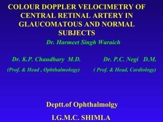
1. COLOUR DOPPLER VELOCIMETRY OF
CENTRAL RETINAL ARTERY IN
GLAUCOMATOUS AND NORMAL
SUBJECTS
Dr. Harmeet Singh Waraich
Dr. K.P. Chaudhary M.D. Dr. P.C. Negi D.M.
(Prof. & Head , Ophthalmology) ( Prof. & Head, Cardiology)
Deptt.of Ophthalmolgy
I.G.M.C. SHIMLA
2. Glaucoma can be defined as a multifactorial
optic neuropathy with a characteristic loss of
optic nerve fibres presenting as :-
Classical optic nerve head features.
Visual field changes.
May or may not be associated with elevated IOP and
angle anomalies in the presence or absence of any cause
for the disease.
Role of IOP in the current definition is only one of the
multiple factors responsible for the disease.
3. IOP and Glaucoma in the present context
Considering the intraocular pressure as a primary
variable, glaucoma can be classified as :-
1) High Tension Glaucoma-POAG (IOP> 21mm of Hg )
2) Normal Tension Glaucoma-NTG(IOP < 21mm of Hg )
3) Ocular Hypertension-OHT
4. Pathogenesis of Glaucomatous optic nerve damage
Two main mechanisms have been proposed
1) One suggests, mechanical damage directly to the
axons or small vessels by structural alterations at the
lamina cribrosa. – MECHANICAL THEORY
2) The other suggests, primary problem in the blood flow
of the optic nerve as a result of localised organic
change with or without a low perfusion pressure or
vasospasm. – VASCULAR THEORY
5. Evidence in support of Vascular
Theory.
1) In NTG patients, despite the IOP <21mm of Hg , there is
progressive damage of optic nerve head suggesting that
vascular factors have predominant role in NTG.
2) In OHT , IOP> 21 mm of Hg does not cause glaucomatous
damage , which supports that IOP alone is not sufficient to
cause optic nerve damage.
3) NTG is associated with various vascular disorders like
hypotension, migraine, Raynaud’s phenomenon which
suggests that same pathophysiological mechanism is
causing ONH damage.
6. VASCULAR COMPROMISE – NTG
Patients of NTG may suffer from ocular blood flow deficits.
Increased choroidal filling time.
Increased vascular downstream resistance in CRA & SPCA.
Increased areas of ICG hypofluorescence in peripapillary
region.
Diffuse cerebral ischemia.
7. Role of perfusion pressure & vascular
resistance of retrobulbar vessels.
Blood flow of optic nerve head = Perfusion pressure
Resistance
Blood flow has direct relation with perfusion pressure and inverse
relation with resistance of vessels.
Perfusion pressure = Mean arterial pressure – IOP
Fall in perfusion pressure due to decreased mean arterial pressure or
increased IOP results in decreased perfusion of ONH.
Increased resistance of retrobulbar vessels also decreases ONH
perfusion and thus accelerates glaucomatous damage.
8. Measurement of ONH Perfusion
Colour Doppler imaging is an ultrasound technique that
combines B scan gray scale imaging of tissue structure ,
coloured representation of blood flow based on Doppler
shifted frequencies and pulsed Doppler measurement of
blood flow velocities.
With the advent of colour Doppler it has become possible to
visualize retrobulbar vessels and to calculate the blood flow
velocities in these vessels.
12. COLOUR DOPPLER VELOCIMETRY OF CENTRAL RETINAL
ARTERY IN GLAUCOMATOUS AND NORMAL SUBJECTS.
STUDY CONDUCTED IN THE DEPTT. OF
OPHTHALMOLOGY I.G.M.C. SHIMLA
AIM OF STUDY
To assess the blood flow velocities ( Peak Systolic Velocity
and End Diastolic Velocity ) and Resistive Index in
Central Retinal Artery in patients with chronic open angle
glaucoma in comparison with non-glaucomatous control
population.
13. REVIEW OF LITERATURE
In 1989, Ericson – first to describe qualitative appearance of CDI
in normal orbits
Quantitative estimation – done by Lieb et al (1991),
Guthoff et al (1991) and Williamson et al( 1993).
In 1995, Rankin et al– found positive correlation of visual
field changes with retro bulbar hemodynamic parameters.
In 2000, Schumann et al suggested that eyes with progressive
visual field defects in pts with NTG had statistically significant
lower blood flow velocities and higher resistive indices in CRA
than with practically stable visual field defects.
14. Material & Methods
Group I.
30 patients of chronic open angle glaucoma fulfilling the selection
criteria.
Group II
30 normal age and sex matched healthy volunteers.
Criteria for selection of patients of Gr. I .Patients fulfilling 2 out
of 3 undermentioned selection criteria
IOP consistently above 21+1 mm of Hg.
Reproducible visual field defects.
Glaucomatous optic disc changes.
15. Exclusion criteria
Patients having history of
Intraocular surgery
Cardiovascular disease
Systemic hypertension or hypotension
Diabetes mellitus
Migraine
Active ocular infection or inflammation
16. Pre procedure evaluation
Written informed consent was obtained after explaining the
procedure.
Detailed history
Visual acuity
IOP recording – Goldmann applanation tonometer
Slit lamp Biomicroscopy – 78 D lens
Gonioscopy – Goldmann 3 Mirror Goniolens
PS Evaluation – Indirect ophthalmoscope
Visual field analysis- Humphrey visual field analyzer
17. Technique of Colour Doppler Imaging of Central
Retinal Artery
An ATL ( Advanced Technologies Limited) HDI 3000 Colour
Doppler with 7.5 MHz linear phased transducer was used for
colour Doppler imaging of Central Retinal Artery.
All the examinations were done with patients in supine position.
To examine the Central Retinal Artery the B scan image of the
optic nerve was used to localize the area of anterior optic nerve.
The sample volume was placed with the centre about 3mm
behind the surface of the disk.
20. From the spectral waveform of Central Retinal Artery, Peak
Systolic Velocity (PSV) and End Diastolic Velocity ( EDV)
were measured.
Resistive Index was calculated using the formula
PSV-EDV/ PSV
Resistive index is a ratio and is Doppler angle independent.
21. RESULTS
Std. Std. Error Significance
GROUP N Mean Deviation Mean (2-tailed)
AGE glaucoma 60 61.92 10.496 1.344 0.131
normal 60 59.60 5.428 .701
PSV glaucoma 60 9.48 3.348 .429 0.869
normal 60 9.57 2.554 .330
EDV glaucoma 60 2.89 1.066 .137 0.000
normal 60 3.93 1.219 .157
RI glaucoma 60 0.69 .062 .008 0.000
normal 60 0.59 .062 .008
22. RESULTS
Std. Std. Error Significance
GROUP N Mean Deviation of Mean (2-tailed)
IOP glaucoma 60 18.00 7.057 .904 0.039
normal 60 16.00 2.314 .299
SBP glaucoma 60 123.80 8.546 1.094 0.350
normal 60 122.67 3.909 .505
DBP glaucoma 60 80.82 6.136 .786 0.312
normal 60 79.93 2.875 .371
MEAN glaucoma 60 95.10 6.833 .875 0.374
normal 60 94.27 2.357 .304
PP glaucoma 60 77.16 6.778 .868 0.413
normal 60 77.93 2.603 .336
23. NORMAL 59.6
GLAUCOMA 61.92
0 20 40 60 80 100
GLAUCOMA NORMAL
Series1 61.92 59.6
Series1
Comparison of Age group of Normal & Glaucoma patients
( P value = 0.131)
24. NORMAL 16
IOP
GLAUCOMA
18
15 16 17 18 19
Series1
Comparison of IOP of Normal and Glaucoma patients ( P value=0.039)
25. NORMAL 77.93
PP
GLAUCOMA
77.16
76.6 76.8 77 77.2 77.4 77.6 77.8 78
Series1
Comparison of Perfusion pressure of Normal & Glaucoma patients
( P value = 0.413)
26. NORMAL 9.57
PSV
GLAUCOMA
9.48
9.4 9.45 9.5 9.55 9.6
Series1
Comparison of PSV of Normal & Glaucoma patients (P value = 0.869)
27. NORMAL 3.93
EDV
GLAUCOMA
2.89
0 0.5 1 1.5 2 2.5 3 3.5 4 4.5
Series1
Comparison of EDV in Normal & Glaucoma patients ( P value =0.000)
28. NORMAL 0.59
RI
GLAUCOMA
0.69
0.54 0.56 0.58 0.6 0.62 0.64 0.66 0.68 0.7
Series1
Comparison of RI of Normal & Glaucoma patients ( P value = 0.000)
29. DISCUSSION
Color Doppler Imaging and spectral analysis are attractive tools
for noninvasive vascular investigation of retrobulbar vessels.
Central retinal artery can be easily identified by its position within
the optic nerve head and its characteristic waveform.
Measurements from central retinal artery are most reproducible.
This study shows that there is significant decrease in mean End
Diastolic Velocity (EDV) and increase in Resistive Index (RI) in
glaucoma patients as compared to normal subjects substantiating
vascular theory of glaucoma.
Perfusion pressure was found to be lower in Glaucoma patients as
compared to normals but the difference was not significant.
30. DISCUSSION
A decrease in EDV is a sensitive indicator of downstream
resistance which leads to decreased perfusion.
Changes in resistance affect the diastolic flow velocity more
than systolic velocity.
Resistive Index represents a quantification of end organ
resistance, the formula is suited to low resistance vascular
beds typical of the cerebrovascular region.
Resistive index can be used to assess the effectiveness of various
pharmacological interventions directed at increasing the
31. SUMMARY AND
CONCLUSIONS
There is no doubt that vascular compromise plays an
important role in the pathogenesis of Glaucoma especially
NTG.
Colour Doppler can be used in investigating cases of POAG
& NTG on regular basis to assess the retrobulbar
haemodynamic variables . It can prove to be a promising
investigation to assess vascular compromise in these patients.
We can evaluate the severity of the disease, its progression
and efficacy of treatment in glaucoma patients with the help
of CDI which is a wonderful investigation to assess
retrobulbar haemodynamic profile.