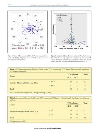
ARTICLE

Total corneal astigmatism in older adults taking into
account posterior corneal astigmatism by ray tracing
Alvaro Rodríguez Ratón, MD1; Javier Orbegozo Gárate, MD1; Iñaki Basterra Barrenetxea,OD1
PURPOSE: To study the composition of corneal astigmatism in older adults, evaluating the
difference made by the inclusion of posterior corneal astigmatism in a ray tracing calculation
of total corneal astigmatism.
SETTING: Ophthalmology clinic.
METHODS: One hundred consecutive patients aged between 60 and 80 years were included
in a prospective descriptive study. Their right eye was analysed by an integrated Placido
disk and rotating Scheimpflug camera topographer (CSO™ Sirius). Several parameters were
measured: anterior corneal astigmatism (ACA) and posterior corneal astigmatism (PCA),
total astigmatism based on anterior topographic data (simK) and total corneal astigmatism
(TCA) by merging anterior and posterior astigmatism using ray tracing.
RESULTS: Mean ACA was 1.51 diopters (D) and PCA was 0.38D. ACA was aligned 47%
with-the-rule and PCA 87% against-the-rule. Cases with against-the-rule ACA showed low
magnitude correlation between anterior and posterior surfaces. TCA had a mean deviation
of 0.30D @ 3 over SimK in a vector calculation. Eighteen percent (18%) of cases differed
by 0.50 D or more between SimK and TCA magnitude, and 53% had 10 or more degrees
of axis discrepancy, the difference being higher at lower magnitudes of astigmatism.
CONCLUSIONS: Anterior WTR astigmatism tends to be compensated by posterior ATR
astigmatism in older patients. Nevertheless, the high number of cases largely justifies the
use of tomographic technology that takes into account the posterior corneal surface for
managing individual total corneal astigmatism.
J Emmetropia 2013; 4: 179-184

Corneal astigmatism is gaining importance in cataract
surgery planning as new correction methods appear.
Although toric intraocular lenses (IOL) offer a more
predictive intervention than the incision techniques used
previously1, accurate calculation of corneal astigmatism
is essential for choosing the correct lens. While there are
many methods available for this purpose, Javal and Schiøtz
designed the first reliable astigmatic keratometer based on the
anterior corneal surface. This value was adapted to represent
Submitted: 05/17/2013
Revised: 08/14/2013
Accepted: 08/16/2013
Centro Oftalmológico Integral – COI Berri. Bilbao (Vizcaya), Spain.

1

Financial disclosure: None of the authors have any financial interest
in the devices mentioned in this article, nor have they received any
support for this research.
Corresponding Author: Alvaro Rodríguez Ratón
Address: C/ Rodríguez Arias nº 6, 2º. 48008 Bilbao, Spain.
Email: arraton@gmail.com
© 2013 SECOIR
Sociedad Española de Cirugía Ocular Implanto-Refractiva

the total corneal astigmatism using the keratometric index,
and from then on, all new topographers used that shortcut.
More recently, the posterior corneal surface has been
measured with slit-lamp, optical coherence tomography
(OCT) and Scheimpflug camera technology2-4. The
combination of anterior and posterior corneal astigmatism
has been calculated by vector analysis4,  5 but only the ray
tracing method, used here and in one previous study2,
obtains the total corneal astigmatism based on an exact
calculation and avoiding paraxial assumptions.
This paper aims to work in the same research line as
recent publications emphasizing the importance of the
posterior corneal surface in calculating the total astigmatism.
In our setting, astigmatism was scanned with an
integrated Placido disk and rotating Scheimpflug camera
topographer (CSO™ Sirius). We evaluated the correlation
of posterior astigmatism in magnitude and axis with the
anterior astigmatism. We accounted for the difference
found by including the posterior corneal astigmatism into a
ray tracing method to calculate the total corneal astigmatism
(TCA) versus simK.
ISSN: 2171-4703

179
180

CALCULATION OF TOTAL CORNEAL ASTIGMATISM BY RAY TRACING

PATIENTS AND METHODS
A prospective descriptive design was used to measure
astigmatism values from 100 consecutive patients at our
private practice who met the inclusion criteria. These were
patients aged between 60 and 80 years, with no previous
ocular surgery nor morbidity, and no contact lens usage.
Subjects were informed about the use of their personal
information, giving written consent. All research and
data collection followed the tenets of the Declaration of
Helsinki.
An integrated Placido disk and rotating Scheimpflug
camera topographer (CSO™ Sirius) was used. This device
was validated in a previous study6 and was calibrated by
the company. A green check mark in the results display was
required to guarantee the quality of each scan.
Only right eyes were analysed in a single measurement
for each patient; complete analysis was then performed by
the software. Four values were noted:
1. ACA: Anterior corneal astigmatism calculated by
a combination of Placido disk image and Scheimpflug
camera. Air (1.0) and true corneal refraction index (1.376)
are used for Gaussian optics calculation.
2. PCA: Posterior corneal astigmatism is evaluated by
the rotating Scheimpflug camera. True corneal (1.376) and
aqueous refraction index (1.336) are used for Gaussian
optics calculation.
3. SimK: Simulated keratometry uses the ACA
information but includes a lower refraction index
(keratometric index: 1.3375) to account for the posterior
divergent effect.
4. TCA: A ray tracing formula integrating pachymetry,
and anterior and posterior corneal information. It accurately
calculates how light rays impact the refractive surfaces and
obtains an exact corneal astigmatism value. We chose the
value for central 3 mm analysis.
A descriptive analysis was conducted to obtain the
mean values for each parameter and their standard
deviation (SD). Percentages showing the steep meridian
orientation were also calculated. A vertical anterior steep
meridian will generate a with-the-rule (WTR) astigmatism,

whereas a posterior one will add an against-the-rule (ATR)
astigmatism.
Vector analysis was performed to show the difference
between simK and TCA.
We also defined 1 D of astigmatism as the clinical
threshold above which a toric IOL may be necessary,
so this subgroup was analysed separately. A Chi-square
test was used to search for significant differences in
axis alignment of cylinder magnitude between simK
and TCA. Vector calculation and graph plotting
was performed using Eye Pro 2012©7. All statistics
were performed using IBM SPSS Statistics 21.00 for
Windows.

RESULTS
Our patient sample had a mean age of 70 years ± 4 (SD).
Mean astigmatism magnitude for anterior cylinder was
1.51 ± 1.28 D, posterior cylinder 0.38  0.20 D, simK
± 
1.16 ± 1.16 D and TCA 1.32 ± 1.11 D (Table 1). Results
are always stated in positive cylinder notation.
ACA was more frequently oriented WTR (47%)
whereas PCA was mostly ATR (87%). The most
prevalent combination was a corneal WTR anterior
astigmatism and posterior ATR astigmatism (45%;
Figure 1).
Anterior and posterior astigmatic magnitudes
were well correlated in the case of an anterior WTR
(R2 = 0.587) or oblique astigmatism (R2 = 0.462).
However, an anterior ATR astigmatism appeared to be
always related to a lower power posterior astigmatism,
thus showing poor correlation (R2 = 0.084; Figure 2).
The mean vector for the calculated simK was
0.25 @ 75 (Figure 3), and 0.20 @ 26 for the TCA
(Figure 4). Vector difference showed that simK tended
to underestimate TCA by 0.34 @ 3 (Figure 5).
We plotted the difference in the corneal astigmatism
axis calculated by simK and TCA. Below 1 D of
cylinder, the agreement was shown to be inferior to that
obtained for the higher astigmatism values. Therefore,

Table 1. Descriptive statistics (cylinder in diopters)
N

Min

Max

Mean

SD

Age

100

60

80

69.70

4.21

ACA cylinder

100

0.08

6.20

1.51

1.28

PCA cylinder

100

0.05

1.07

0.38

0.20

SimK cylinder

100

0.14

5.29

1.16

1.16

TCA cylinder

100

0.19

5.57

1.32

1.11

ACA, anterior corneal astigmatism; PCA, posterior corneal astigmatism; TCA, total corneal astigmatism

JOURNAL OF EMMETROPIA - VOL 4, OCTOBER-DECEMBER
CALCULATION OF TOTAL CORNEAL ASTIGMATISM BY RAY TRACING

181

due to the clinical interest, we analysed the differences
between simK and TCA by subgroups in patients with
more than 1 diopter of astigmatism measured by TCA
(Figure 6).
SimK and TCA had an astigmatism magnitude
disagreement of more than 0.5D in 25.5% of patients
with 1 D or higher astigmatism. Magnitude disagreement

was lower in the less than 1 D astigmatism group (11.3%),
but was not enough to indicate a significant difference
(p = 0.065; Table 2).
SimK and TCA had a  10 degree disagreement in
29.8% of the more than 1 D astigmatism group. This
disagreement reached 73.6% and was significantly higher
(p  0.05) in the lower astigmatism group (Table 3).

Figure 1. Distribution of anterior and posterior steep meridian in
our sample. Anterior and posterior steep meridians were most frequently oriented vertically. This resulted in an anterior WTR astigmatism and posterior ATR astigmatism in 45% of the cases.

Figure 3. SimK values plotted in a double-angle graph. Total astigmatism is taken from anterior scanning through the keratometric
index.

Figure 2. Correlation between anterior and posterior cylinder
magnitude, adjusting for anterior steep meridian orientation. Note
that in anterior ATR astigmatism, correlation between the anterior
and posterior cylinder is lower.

Figure 4. TCA values in a double-angle plot. This astigmatism is
calculated from a ray tracing formula merging anterior and posterior
corneal astigmatism.

JOURNAL OF EMMETROPIA - VOL 4, OCTOBER-DECEMBER
182

CALCULATION OF TOTAL CORNEAL ASTIGMATISM BY RAY TRACING

Figure 6. Steep axis difference between simK and TCA. The horizontal
line divides the cases with astigmatism over 1 D, which show better simK
and TCA steep axis correlation (p = 0.05). Even in those cases, 29.79%
show a more than 10 degree difference between simK and TCA.

Figure 5. Vector difference: simK-TCA. TCA shows a general tendency to be 0.34 @ 3 higher than simK, which would account for
posterior corneal astigmatism.

Table 2. Cylinder magnitude difference SimK versus TCA considering TCA above 1D as clinically significant
in a subgroup analysis.
TCA cylinder

Count

 1 D

Total

≥ 1 D

 0.5 D

47

35

82

≥ 0.5 D

6

12

18

53

Cylinder difference SimK versus TCA

47

100

Total
TCA, total cornea astigmatism. Chi-square test p = 0.065

Table 3. Steep axis difference SimK versus TCA considering TCA above 1D as clinically significant in a subgroup
analysis.
TCA cylinder

Count

 1 D

Total

≥ 1 D

 10°

14

33

47

≥ 10°

39

14

53

53

Steep axis difference SimK versus TCA

47

100

Total
Chi-square test p0.05

JOURNAL OF EMMETROPIA - VOL 4, OCTOBER-DECEMBER
CALCULATION OF TOTAL CORNEAL ASTIGMATISM BY RAY TRACING

DISCUSSION
Astigmatism is a low order aberration in an optical
system. In the eye, it is defined by the difference in
optical power between two orthogonal meridians of any
of the optical surfaces in the cornea and lens. These four
surfaces determine the astigmatism in the phakic eye,
which is correctable by a toric-shaped lens. However,
in pseudophakic eyes, the cornea alone determines the
refractive astigmatism, as some authors have described8.
In the quest for emmetropia, corneal astigmatism
must be neutralised during cataract surgery. Several
methods have been proposed in this respect, mainly
limbal and corneal relaxing incisions, as well as toric IOL.
Regardless of the method chosen for the correction,
precise pre-surgical corneal astigmatism measurement
is required. Since the introduction of the first Javal
manual keratometer, devices have been developed to
measure the total astigmatism from anterior surface
measurements through keratometric index conversion.
This thick-lens calculation of the ray passing the
cornea through convergent (anterior) and divergent
(posterior) corneal surfaces is replaced by a thin lens
calculation in which a keratometric index is employed
(1.3375). Automatic keratometers are based on a
projection of several points on the corneal surface;
analysis of their separation will determine the anterior
corneal curvature. While this is good for regular
surfaces, later Placido-based topography enabled
complete analysis of the anterior corneal surface,
building a reconstruction from thousands of measured
points. Different software was implemented to process
that information, and to give an approximation of the
keratometric corneal power, which was called simulated
K (simK). Srivannaboon et al. recently published an
article on the similarity and comparability between
simK and auto refractometer keratometry9.
In our study, we decided to take the simK
measurement from the same topographer as the TCA,
avoiding the use of any other keratometer in order not
to add any inter-device variability.
All previous devices have lacked the technology for
posterior corneal power measurement. Fortunately,
Scheimpflug camera devices have been designed to
measure the anterior and posterior curvature separately,
allowing independent analysis. This is a key input for
reproducing the ray behaviour through the cornea and,
logically, the way to obtain the total corneal astigmatism.
Some authors began this trend with a vector summation
of anterior and posterior astigmatism5, but later devices
incorporated ray tracing technology. This latter optical
analysis has two main advantages: it incorporates
pachymetry and the real angle of incidence of light
for each point on the cornea. It is also known to be
an exact calculation; opposing the paraxial optics that
keratometry had employed to simplify the formula.

183

With the technology available through the CSO
Sirius topographer, based on Placido disk and a
Scheimpflug camera, we decided to determine the
importance of PCA and its relationship to ACA.
We were particularly concerned about the impact
that obviating posterior astigmatism could have in
the overall astigmatism obviation if starting from
anterior information only.
Ho et al.10 confirmed that astigmatism varies
throughout life, so our patient selection was
restricted to older adults attending our clinic for
cataract surgery, obtaining a mean age of 69.7 ± 4.21
in our sample. This is the age at which most patients
will undergo cataract surgery, so we focused on that
astigmatism.
We were pleased to confirm that our mean
posterior astigmatism results (0.38 ± 0.20) were
consistent with previous studies using Pentacam4
and Galilei2 topographers regarding a 0.29 D and
0.30 D, respectively. Our results also agreed in axis
distribution, showing a vertical posterior steeper
meridian in 87% (Figure 1).
We concur with the paper by Koch et al.2 in
concluding that an anterior ATR astigmatism
seems to be poorly correlated with PCA magnitude,
whereas WTR and oblique astigmatism have a
direct correlation between anterior and posterior
magnitudes (Figure 2). This is important for refuting
some studies regarding a constant relationship
between anterior and posterior astigmatism in
normal eyes11.
Focusing now on the difference between the
astigmatism calculated for the anterior information
(simK) and that obtained through ray tracing from
the joint information from anterior and posterior
surfaces and pachymetry (TCA), there was a
tendency by simK towards underestimation of TCA
by 0.34 @ 3, as shown by a vector difference (Figure
5). Unfortunately, the SD was high and we were
unable to find a significant difference in the mean
due to the insufficient sample size.
The next step in the analysis was aimed at
detecting clinical differences in axis and magnitude
of the astigmatism measured by simK or TCA. This
would mean that a different toric IOL would have
been selected with a more than 0.5 D difference or a
different implantation axis would have been selected
with a more than 10-degree difference. Below 1 D
of TCA cylinder, agreement was lower in magnitude
and axis between simK and TCA, but it lacked
clinical interest, as toric IOL implantation is not
frequently indicated for such cases.
For patients with astigmatism more than 1 D,
TCA gave a significantly different axis in 25% and a
non-significantly different magnitude in 28% of the
sample, although it did show a trend.

JOURNAL OF EMMETROPIA - VOL 4, OCTOBER-DECEMBER
184

CALCULATION OF TOTAL CORNEAL ASTIGMATISM BY RAY TRACING

Conclusions should be interpreted with caution as
the sample size is small and most results show a tendency
rather than a significant difference.
Nevertheless, we believe that this finding should
encourage the surgeon to enter the TCA into the toric
IOL calculation, but further clinical studies should be
conducted to confirm this hypothesis.
We conclude that posterior corneal astigmatism
distribution in our population measured by the
Sirius topographer is comparable to that described in
previous studies with other topographers. A method
that includes pachymetry and anterior and posterior
corneal astigmatism in a ray tracing calculation results
in differences in axis and magnitude compared to the
traditional astigmatism calculation based on anterior
keratometry. It remains to be investigated in a clinical
setting whether treatment guided by TCA provides a
better refractive outcome for the patient.

2.	
3.	

5.	

6.	

7.	
8.	
9.	

10.	

REFERENCES
1.	

4.	

Visser N, Bauer NJC, Nuijts RMMA. Toric intraocular lenses:
Historical overview, patient selection, IOL calculation, surgical
techniques, clinical outcomes, and complications. J Cataract
Refract Surg. 2013; 39:624-37.
Koch DD, Ali SF, Weikert MP Shirayama M, Jenkins R, Wang
,
L. Contribution of posterior corneal astigmatism to total corneal
astigmatism. J Cataract Refract Surg. 2012; 38:2080-7.
Cheng L-S, Tsai C-Y, Tsai RJ-F, Liou S-W, Ho J-D. Estimation
accuracy of surgically induced astigmatism on the cornea when
neglecting the posterior corneal surface measurement. Acta
Ophthalmol. 2011; 89:417-22.

11.	

Ho J-D, Tsai C-Y, Liou S-W. Accuracy of corneal astigmatism
estimation by neglecting the posterior corneal surface measurement.
Am J Ophthalmol. 2009; 147:788-95, 795.e1-2.
Prisant O, Hoang-Xuan T, Proano C, Hernandez E, Awwad
ST, Azar DT, et al. Vector summation of anterior and posterior
corneal topographical astigmatism. J Cataract Refract Surg.
2002; 28:1636-43.
Savini G, Barboni P, Carbonelli M, Hoffer KJ. Repeatability
of automatic measurements by a new Scheimpflug camera
combined with Placido topography. J Cataract Refract Surg.
2011; 37:1809-16.
Eye Pro App 2012® for iPhone®, by EB Eye Limited.
Available
at:
https://itunes.apple.com/es/app/eye-pro/
id349481068?mt=8. Accessed: September 2013.
Tejedor J, Guirao A. Agreement between Refractive and
Corneal Astigmatism in Pseudophakic Eyes. Cornea.
2013;32:783-90.
Srivannaboon S, Soeharnila, Chirapapaisan C, Chonpimai
P. Comparison of corneal astigmatism and axis location in
cataract patients measured by total corneal power, automated
keratometry, and simulated keratometry. J Cataract Refract
Surg. 2012; 38:2088-93.
Ho J-D, Liou S-W, Tsai RJ-F, Tsai C-Y. Effects of aging on
anterior and posterior corneal astigmatism. Cornea. 2010;
29:632-7.
Montalbán R, Piñero DP, Javaloy J, Alio JL. Correlation of
the Corneal Toricity between Anterior and Posterior Corneal
Surfaces in the Normal Human Eye. Cornea. 2013; 32:791-8.

JOURNAL OF EMMETROPIA - VOL 4, OCTOBER-DECEMBER

First author:
Alvaro Rodríguez Ratón
Centro Oftalmológico Integral – COI Berri.
Bilbao, Spain

Total corneal astigmatism in older adults taking into account posterior corneal astigmatism by ray tracing

  • 1.
    ARTICLE Total corneal astigmatismin older adults taking into account posterior corneal astigmatism by ray tracing Alvaro Rodríguez Ratón, MD1; Javier Orbegozo Gárate, MD1; Iñaki Basterra Barrenetxea,OD1 PURPOSE: To study the composition of corneal astigmatism in older adults, evaluating the difference made by the inclusion of posterior corneal astigmatism in a ray tracing calculation of total corneal astigmatism. SETTING: Ophthalmology clinic. METHODS: One hundred consecutive patients aged between 60 and 80 years were included in a prospective descriptive study. Their right eye was analysed by an integrated Placido disk and rotating Scheimpflug camera topographer (CSO™ Sirius). Several parameters were measured: anterior corneal astigmatism (ACA) and posterior corneal astigmatism (PCA), total astigmatism based on anterior topographic data (simK) and total corneal astigmatism (TCA) by merging anterior and posterior astigmatism using ray tracing. RESULTS: Mean ACA was 1.51 diopters (D) and PCA was 0.38D. ACA was aligned 47% with-the-rule and PCA 87% against-the-rule. Cases with against-the-rule ACA showed low magnitude correlation between anterior and posterior surfaces. TCA had a mean deviation of 0.30D @ 3 over SimK in a vector calculation. Eighteen percent (18%) of cases differed by 0.50 D or more between SimK and TCA magnitude, and 53% had 10 or more degrees of axis discrepancy, the difference being higher at lower magnitudes of astigmatism. CONCLUSIONS: Anterior WTR astigmatism tends to be compensated by posterior ATR astigmatism in older patients. Nevertheless, the high number of cases largely justifies the use of tomographic technology that takes into account the posterior corneal surface for managing individual total corneal astigmatism. J Emmetropia 2013; 4: 179-184 Corneal astigmatism is gaining importance in cataract surgery planning as new correction methods appear. Although toric intraocular lenses (IOL) offer a more predictive intervention than the incision techniques used previously1, accurate calculation of corneal astigmatism is essential for choosing the correct lens. While there are many methods available for this purpose, Javal and Schiøtz designed the first reliable astigmatic keratometer based on the anterior corneal surface. This value was adapted to represent Submitted: 05/17/2013 Revised: 08/14/2013 Accepted: 08/16/2013 Centro Oftalmológico Integral – COI Berri. Bilbao (Vizcaya), Spain. 1 Financial disclosure: None of the authors have any financial interest in the devices mentioned in this article, nor have they received any support for this research. Corresponding Author: Alvaro Rodríguez Ratón Address: C/ Rodríguez Arias nº 6, 2º. 48008 Bilbao, Spain. Email: arraton@gmail.com © 2013 SECOIR Sociedad Española de Cirugía Ocular Implanto-Refractiva the total corneal astigmatism using the keratometric index, and from then on, all new topographers used that shortcut. More recently, the posterior corneal surface has been measured with slit-lamp, optical coherence tomography (OCT) and Scheimpflug camera technology2-4. The combination of anterior and posterior corneal astigmatism has been calculated by vector analysis4,  5 but only the ray tracing method, used here and in one previous study2, obtains the total corneal astigmatism based on an exact calculation and avoiding paraxial assumptions. This paper aims to work in the same research line as recent publications emphasizing the importance of the posterior corneal surface in calculating the total astigmatism. In our setting, astigmatism was scanned with an integrated Placido disk and rotating Scheimpflug camera topographer (CSO™ Sirius). We evaluated the correlation of posterior astigmatism in magnitude and axis with the anterior astigmatism. We accounted for the difference found by including the posterior corneal astigmatism into a ray tracing method to calculate the total corneal astigmatism (TCA) versus simK. ISSN: 2171-4703 179
  • 2.
    180 CALCULATION OF TOTALCORNEAL ASTIGMATISM BY RAY TRACING PATIENTS AND METHODS A prospective descriptive design was used to measure astigmatism values from 100 consecutive patients at our private practice who met the inclusion criteria. These were patients aged between 60 and 80 years, with no previous ocular surgery nor morbidity, and no contact lens usage. Subjects were informed about the use of their personal information, giving written consent. All research and data collection followed the tenets of the Declaration of Helsinki. An integrated Placido disk and rotating Scheimpflug camera topographer (CSO™ Sirius) was used. This device was validated in a previous study6 and was calibrated by the company. A green check mark in the results display was required to guarantee the quality of each scan. Only right eyes were analysed in a single measurement for each patient; complete analysis was then performed by the software. Four values were noted: 1. ACA: Anterior corneal astigmatism calculated by a combination of Placido disk image and Scheimpflug camera. Air (1.0) and true corneal refraction index (1.376) are used for Gaussian optics calculation. 2. PCA: Posterior corneal astigmatism is evaluated by the rotating Scheimpflug camera. True corneal (1.376) and aqueous refraction index (1.336) are used for Gaussian optics calculation. 3. SimK: Simulated keratometry uses the ACA information but includes a lower refraction index (keratometric index: 1.3375) to account for the posterior divergent effect. 4. TCA: A ray tracing formula integrating pachymetry, and anterior and posterior corneal information. It accurately calculates how light rays impact the refractive surfaces and obtains an exact corneal astigmatism value. We chose the value for central 3 mm analysis. A descriptive analysis was conducted to obtain the mean values for each parameter and their standard deviation (SD). Percentages showing the steep meridian orientation were also calculated. A vertical anterior steep meridian will generate a with-the-rule (WTR) astigmatism, whereas a posterior one will add an against-the-rule (ATR) astigmatism. Vector analysis was performed to show the difference between simK and TCA. We also defined 1 D of astigmatism as the clinical threshold above which a toric IOL may be necessary, so this subgroup was analysed separately. A Chi-square test was used to search for significant differences in axis alignment of cylinder magnitude between simK and TCA. Vector calculation and graph plotting was performed using Eye Pro 2012©7. All statistics were performed using IBM SPSS Statistics 21.00 for Windows. RESULTS Our patient sample had a mean age of 70 years ± 4 (SD). Mean astigmatism magnitude for anterior cylinder was 1.51 ± 1.28 D, posterior cylinder 0.38  0.20 D, simK ±  1.16 ± 1.16 D and TCA 1.32 ± 1.11 D (Table 1). Results are always stated in positive cylinder notation. ACA was more frequently oriented WTR (47%) whereas PCA was mostly ATR (87%). The most prevalent combination was a corneal WTR anterior astigmatism and posterior ATR astigmatism (45%; Figure 1). Anterior and posterior astigmatic magnitudes were well correlated in the case of an anterior WTR (R2 = 0.587) or oblique astigmatism (R2 = 0.462). However, an anterior ATR astigmatism appeared to be always related to a lower power posterior astigmatism, thus showing poor correlation (R2 = 0.084; Figure 2). The mean vector for the calculated simK was 0.25 @ 75 (Figure 3), and 0.20 @ 26 for the TCA (Figure 4). Vector difference showed that simK tended to underestimate TCA by 0.34 @ 3 (Figure 5). We plotted the difference in the corneal astigmatism axis calculated by simK and TCA. Below 1 D of cylinder, the agreement was shown to be inferior to that obtained for the higher astigmatism values. Therefore, Table 1. Descriptive statistics (cylinder in diopters) N Min Max Mean SD Age 100 60 80 69.70 4.21 ACA cylinder 100 0.08 6.20 1.51 1.28 PCA cylinder 100 0.05 1.07 0.38 0.20 SimK cylinder 100 0.14 5.29 1.16 1.16 TCA cylinder 100 0.19 5.57 1.32 1.11 ACA, anterior corneal astigmatism; PCA, posterior corneal astigmatism; TCA, total corneal astigmatism JOURNAL OF EMMETROPIA - VOL 4, OCTOBER-DECEMBER
  • 3.
    CALCULATION OF TOTALCORNEAL ASTIGMATISM BY RAY TRACING 181 due to the clinical interest, we analysed the differences between simK and TCA by subgroups in patients with more than 1 diopter of astigmatism measured by TCA (Figure 6). SimK and TCA had an astigmatism magnitude disagreement of more than 0.5D in 25.5% of patients with 1 D or higher astigmatism. Magnitude disagreement was lower in the less than 1 D astigmatism group (11.3%), but was not enough to indicate a significant difference (p = 0.065; Table 2). SimK and TCA had a 10 degree disagreement in 29.8% of the more than 1 D astigmatism group. This disagreement reached 73.6% and was significantly higher (p 0.05) in the lower astigmatism group (Table 3). Figure 1. Distribution of anterior and posterior steep meridian in our sample. Anterior and posterior steep meridians were most frequently oriented vertically. This resulted in an anterior WTR astigmatism and posterior ATR astigmatism in 45% of the cases. Figure 3. SimK values plotted in a double-angle graph. Total astigmatism is taken from anterior scanning through the keratometric index. Figure 2. Correlation between anterior and posterior cylinder magnitude, adjusting for anterior steep meridian orientation. Note that in anterior ATR astigmatism, correlation between the anterior and posterior cylinder is lower. Figure 4. TCA values in a double-angle plot. This astigmatism is calculated from a ray tracing formula merging anterior and posterior corneal astigmatism. JOURNAL OF EMMETROPIA - VOL 4, OCTOBER-DECEMBER
  • 4.
    182 CALCULATION OF TOTALCORNEAL ASTIGMATISM BY RAY TRACING Figure 6. Steep axis difference between simK and TCA. The horizontal line divides the cases with astigmatism over 1 D, which show better simK and TCA steep axis correlation (p = 0.05). Even in those cases, 29.79% show a more than 10 degree difference between simK and TCA. Figure 5. Vector difference: simK-TCA. TCA shows a general tendency to be 0.34 @ 3 higher than simK, which would account for posterior corneal astigmatism. Table 2. Cylinder magnitude difference SimK versus TCA considering TCA above 1D as clinically significant in a subgroup analysis. TCA cylinder Count  1 D Total ≥ 1 D 0.5 D 47 35 82 ≥ 0.5 D 6 12 18 53 Cylinder difference SimK versus TCA 47 100 Total TCA, total cornea astigmatism. Chi-square test p = 0.065 Table 3. Steep axis difference SimK versus TCA considering TCA above 1D as clinically significant in a subgroup analysis. TCA cylinder Count  1 D Total ≥ 1 D 10° 14 33 47 ≥ 10° 39 14 53 53 Steep axis difference SimK versus TCA 47 100 Total Chi-square test p0.05 JOURNAL OF EMMETROPIA - VOL 4, OCTOBER-DECEMBER
  • 5.
    CALCULATION OF TOTALCORNEAL ASTIGMATISM BY RAY TRACING DISCUSSION Astigmatism is a low order aberration in an optical system. In the eye, it is defined by the difference in optical power between two orthogonal meridians of any of the optical surfaces in the cornea and lens. These four surfaces determine the astigmatism in the phakic eye, which is correctable by a toric-shaped lens. However, in pseudophakic eyes, the cornea alone determines the refractive astigmatism, as some authors have described8. In the quest for emmetropia, corneal astigmatism must be neutralised during cataract surgery. Several methods have been proposed in this respect, mainly limbal and corneal relaxing incisions, as well as toric IOL. Regardless of the method chosen for the correction, precise pre-surgical corneal astigmatism measurement is required. Since the introduction of the first Javal manual keratometer, devices have been developed to measure the total astigmatism from anterior surface measurements through keratometric index conversion. This thick-lens calculation of the ray passing the cornea through convergent (anterior) and divergent (posterior) corneal surfaces is replaced by a thin lens calculation in which a keratometric index is employed (1.3375). Automatic keratometers are based on a projection of several points on the corneal surface; analysis of their separation will determine the anterior corneal curvature. While this is good for regular surfaces, later Placido-based topography enabled complete analysis of the anterior corneal surface, building a reconstruction from thousands of measured points. Different software was implemented to process that information, and to give an approximation of the keratometric corneal power, which was called simulated K (simK). Srivannaboon et al. recently published an article on the similarity and comparability between simK and auto refractometer keratometry9. In our study, we decided to take the simK measurement from the same topographer as the TCA, avoiding the use of any other keratometer in order not to add any inter-device variability. All previous devices have lacked the technology for posterior corneal power measurement. Fortunately, Scheimpflug camera devices have been designed to measure the anterior and posterior curvature separately, allowing independent analysis. This is a key input for reproducing the ray behaviour through the cornea and, logically, the way to obtain the total corneal astigmatism. Some authors began this trend with a vector summation of anterior and posterior astigmatism5, but later devices incorporated ray tracing technology. This latter optical analysis has two main advantages: it incorporates pachymetry and the real angle of incidence of light for each point on the cornea. It is also known to be an exact calculation; opposing the paraxial optics that keratometry had employed to simplify the formula. 183 With the technology available through the CSO Sirius topographer, based on Placido disk and a Scheimpflug camera, we decided to determine the importance of PCA and its relationship to ACA. We were particularly concerned about the impact that obviating posterior astigmatism could have in the overall astigmatism obviation if starting from anterior information only. Ho et al.10 confirmed that astigmatism varies throughout life, so our patient selection was restricted to older adults attending our clinic for cataract surgery, obtaining a mean age of 69.7 ± 4.21 in our sample. This is the age at which most patients will undergo cataract surgery, so we focused on that astigmatism. We were pleased to confirm that our mean posterior astigmatism results (0.38 ± 0.20) were consistent with previous studies using Pentacam4 and Galilei2 topographers regarding a 0.29 D and 0.30 D, respectively. Our results also agreed in axis distribution, showing a vertical posterior steeper meridian in 87% (Figure 1). We concur with the paper by Koch et al.2 in concluding that an anterior ATR astigmatism seems to be poorly correlated with PCA magnitude, whereas WTR and oblique astigmatism have a direct correlation between anterior and posterior magnitudes (Figure 2). This is important for refuting some studies regarding a constant relationship between anterior and posterior astigmatism in normal eyes11. Focusing now on the difference between the astigmatism calculated for the anterior information (simK) and that obtained through ray tracing from the joint information from anterior and posterior surfaces and pachymetry (TCA), there was a tendency by simK towards underestimation of TCA by 0.34 @ 3, as shown by a vector difference (Figure 5). Unfortunately, the SD was high and we were unable to find a significant difference in the mean due to the insufficient sample size. The next step in the analysis was aimed at detecting clinical differences in axis and magnitude of the astigmatism measured by simK or TCA. This would mean that a different toric IOL would have been selected with a more than 0.5 D difference or a different implantation axis would have been selected with a more than 10-degree difference. Below 1 D of TCA cylinder, agreement was lower in magnitude and axis between simK and TCA, but it lacked clinical interest, as toric IOL implantation is not frequently indicated for such cases. For patients with astigmatism more than 1 D, TCA gave a significantly different axis in 25% and a non-significantly different magnitude in 28% of the sample, although it did show a trend. JOURNAL OF EMMETROPIA - VOL 4, OCTOBER-DECEMBER
  • 6.
    184 CALCULATION OF TOTALCORNEAL ASTIGMATISM BY RAY TRACING Conclusions should be interpreted with caution as the sample size is small and most results show a tendency rather than a significant difference. Nevertheless, we believe that this finding should encourage the surgeon to enter the TCA into the toric IOL calculation, but further clinical studies should be conducted to confirm this hypothesis. We conclude that posterior corneal astigmatism distribution in our population measured by the Sirius topographer is comparable to that described in previous studies with other topographers. A method that includes pachymetry and anterior and posterior corneal astigmatism in a ray tracing calculation results in differences in axis and magnitude compared to the traditional astigmatism calculation based on anterior keratometry. It remains to be investigated in a clinical setting whether treatment guided by TCA provides a better refractive outcome for the patient. 2. 3. 5. 6. 7. 8. 9. 10. REFERENCES 1. 4. Visser N, Bauer NJC, Nuijts RMMA. Toric intraocular lenses: Historical overview, patient selection, IOL calculation, surgical techniques, clinical outcomes, and complications. J Cataract Refract Surg. 2013; 39:624-37. Koch DD, Ali SF, Weikert MP Shirayama M, Jenkins R, Wang , L. Contribution of posterior corneal astigmatism to total corneal astigmatism. J Cataract Refract Surg. 2012; 38:2080-7. Cheng L-S, Tsai C-Y, Tsai RJ-F, Liou S-W, Ho J-D. Estimation accuracy of surgically induced astigmatism on the cornea when neglecting the posterior corneal surface measurement. Acta Ophthalmol. 2011; 89:417-22. 11. Ho J-D, Tsai C-Y, Liou S-W. Accuracy of corneal astigmatism estimation by neglecting the posterior corneal surface measurement. Am J Ophthalmol. 2009; 147:788-95, 795.e1-2. Prisant O, Hoang-Xuan T, Proano C, Hernandez E, Awwad ST, Azar DT, et al. Vector summation of anterior and posterior corneal topographical astigmatism. J Cataract Refract Surg. 2002; 28:1636-43. Savini G, Barboni P, Carbonelli M, Hoffer KJ. Repeatability of automatic measurements by a new Scheimpflug camera combined with Placido topography. J Cataract Refract Surg. 2011; 37:1809-16. Eye Pro App 2012® for iPhone®, by EB Eye Limited. Available at: https://itunes.apple.com/es/app/eye-pro/ id349481068?mt=8. Accessed: September 2013. Tejedor J, Guirao A. Agreement between Refractive and Corneal Astigmatism in Pseudophakic Eyes. Cornea. 2013;32:783-90. Srivannaboon S, Soeharnila, Chirapapaisan C, Chonpimai P. Comparison of corneal astigmatism and axis location in cataract patients measured by total corneal power, automated keratometry, and simulated keratometry. J Cataract Refract Surg. 2012; 38:2088-93. Ho J-D, Liou S-W, Tsai RJ-F, Tsai C-Y. Effects of aging on anterior and posterior corneal astigmatism. Cornea. 2010; 29:632-7. Montalbán R, Piñero DP, Javaloy J, Alio JL. Correlation of the Corneal Toricity between Anterior and Posterior Corneal Surfaces in the Normal Human Eye. Cornea. 2013; 32:791-8. JOURNAL OF EMMETROPIA - VOL 4, OCTOBER-DECEMBER First author: Alvaro Rodríguez Ratón Centro Oftalmológico Integral – COI Berri. Bilbao, Spain