8/23/2017 4:11 PM 1
UNIVERSITY OF KANDAHAR
MAR/01/2014
DES BY: H.S
In thisPresentation
• Introduction to Retina
• Microscopic Anatomy
• Functional Anatomy
• Bloodsupply to Retina
• Introduction to Vision
• VisionMechanism
• VisualPathway
• Conclusion
• References
Presenters:
4
1. Mohd Shafiq Amizai 2. Muhammad Haroon
Stanikzai
Muhammad Fahim Zafar
• Sense of vision is a complex function of the two eyes andtheir central
connections.
The physiologicalactivities involvedin the normal functioning of the eyes are:
• Maintenanceof clear ocular media,
• Maintenanceof normal intraocularpressure,
• The imageforming mechanism,
• Physiology of vision,
• Physiology of binocularvision,
• Physiology of pupil,and
• Physiology of ocular motility. 5
Retina & The Vision
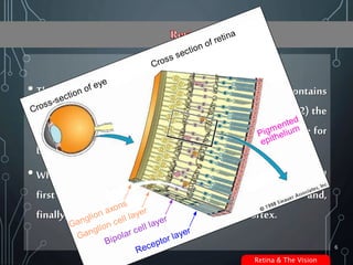
• The retina is the light-sensitive portion of the eye that contains
(1) the cones, which are responsible for color vision, and (2) the
rods, which can detect dim light and are mainly responsible for
black and white vision and vision in the dark.
• When either rods or cones are excited, signals are transmitted
first through successive layers of neurons in the retina and,
finally, into optic nervefibers and the cerebralcortex.
6
Cross-section of eye
Ganglion cell layer
Bipolar cell layer
Receptor layer
Pigmented
epithelium
Ganglion axons
Cross section of retina
Retina & The Vision
•The totalarea of the retinaisapproximately1,100 mm2 .
•The circumferential diameter at the equator of the adult eye
averages 69mm.
•The average healthy retina is 250-µm thick immediately
adjacent tothe temporal marginof theoptic nerve.
7
Retina & The Vision
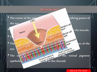
• The center of the retina provides the greatest resolving power of
the eye.
• This area, responsible for central vision, is known as themacula.
• The centerof themacula is called the fovea.
• The inner surface of the retina is adjacent to the vitreous of the
eye.
• The outermost layer of the retina, the retinal pigment
epithelium, is tightly attached to thechoroid.
8
Retina & The Vision
•The retinais the sensor or filmof your eye.
•Its layers do three things
• Provide blood and nutrients (choroid)
• Absorb light and convert to an electrical signal
(photoreceptors)
• Transfer the signal tothe brain (nervecells)
9
Retina & The Vision
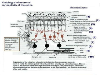
• pigmentedlayer
• layerof rodsandconesprojectingtothepigment
• outernuclearlayercontainingthecellbodiesoftherods andcones
• outerplexiformlayer
• innernuclearlayer
• innerplexiformlayer
• ganglioniclayer
• layerof opticnervefibers
• innerlimitingmembrane.
10
Retina & The Vision
• The inner limiting membrane is the boundary between the
retina and the vitreousbody.
• It is formed by astrocytes and the footplates of Muller cells
together with a basal lamina.
• The nerve fiber layer is the layer of optic nerve fibers consisting
of ganglion cell axon fibers, which course towards the optic
nervehead.
11
Retina & The Vision
• The ganglion cells layer contains the nuclei of ganglion cells, the
axons of which becometheoptic nervefibers for messages.
• Thereare also somedisplaced amacrine cells within this layer.
• The inner plexiform layer contains the synapses between
dendrites of ganglion cells and amacrine cells and the axons of
bipolar cells.
12
Retina & The Vision
• The inner nuclear layer contains the nuclei of horizontal, bipolar
and amacrine cells.
• The inner nuclear layer is thicker in the central area of the retina
compared with peripheral retina because of a greater density of
cone-connecting second-order neurons (cone bipolar cells) and
smaller and more closely-spaced horizontal cells and amacrine
cells concernedwith the conepathways.
• Thereare also nuclei of the supporting Muller cells. 13
Retina & The Vision
• The outer plexiform layer contains the rod and cone axons
horizontal cell dendrites, and bipolar cells dendrites.
• Synapses among thesestructures occur within this layer.
• The outer plexiform layer is also known as the outer synaptic
layer.
14
Retina & The Vision
• The outer nuclear layer consists of the cell bodies of the retinal
rodsand cones.
• In the peripheral retina, the rod cell bodies outnumber the cone
cell bodies (will be discussed Latter) , whereas the reverse is true for the
central retina.
• The outer limiting membrane (external limiting membrane) is
the layer that separates the inner segment portions of the
photoreceptorsfrom their cell nuclei. 15
Retina & The Vision
• Thepigment epithelium is the mostexternal layer of the retina.
• It abuts onthe choroidal layer of the eye.
• It contains a single layer of cuboidal-supporting cells for the
neural portion of the retina.
• These cells contain melanin, which absorbs light and decreases
light scatter within theeye.
16
Retina & The Vision
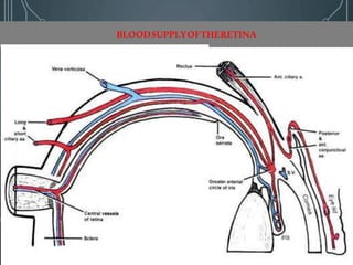
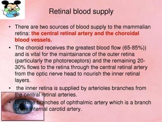
BLOODSUPPLYOFTHERETINA
•‫د‬choriod‫د‬choriocapillariesā€«Ł¾ŁˆŲ§Ų³Ų·Ł‡ā€¬ā€«ŲÆā€¬pigment epithelium،rod
and coneā€«Ų§Łˆā€¬Outernuclear layerā€«ŪŒā€¬ā€«Ų§Ś©ŪŒŚ–ā€¬ā€«Łˆā€¬ā€«Ų§Ų±ā€¬.
•‫د‬retinaā€«ŪŒā€¬ā€«Ų±ā€¬ā€«Ł†Łˆā€¬ā€«Ų·ŲØŁ‚ŪŒā€¬ā€«ŲÆā€¬central retinal arteryā€«Ł¾ŁˆŲ§Ų³Ų·Ł‡ā€¬ā€«Ų³Ų±Ł‡ā€¬ā€«ŪŒā€¬ā€«Ų§Ś©ŪŒŚ–ā€¬ā€«Łˆā€¬ā€«Ų§Ų±ā€¬ā€«Ś©ŁˆŁ…ā€¬
‫چه‬‫د‬ophthalmic arteryā€«ŪŒā€¬ā€«Ų§Ų®Ų±ā€¬ā€«Ų“Ų¹ŲØŁ‡ā€¬ā€«ŲÆŁ‡ā€¬.
•‫د‬retinaā€«ŲÆŲ§Ų®Ł„ŪŒā€¬ā€«ŪŒŁ†Ł‡ā€¬ā€«Łˆā€¬ā€«Ų·ŲØŁ‚Łˆā€¬ā€«ŲÆā€¬central veinā€«Ł¾ŁˆŲ§Ų³Ų·Ł‡ā€¬ā€«ŲŖŲ®Ł„ŪŒŁ‡ā€¬ā€«ŪŒā€¬ā€«Ś©ŪŒŚ–ā€¬ā€«Ų¬ŪŒā€¬ā€«Ų±ā€¬ā€«Ų§ŁˆŲÆŲ®Ų§ā€¬
ā€«ŪŒŁ†Ł‡ā€¬ā€«Łˆā€¬ā€«Ų·ŲØŁ‚Łˆā€¬ā€«ŪŒŪŒā€¬ā€«ŲÆā€¬Vortex veinsā€«Ł¾ŁˆŲ§Ų³Ų·Ł‡ā€¬ā€«Ų³Ų±Ł‡ā€¬ā€«ŲŖŲ®Ł„ŪŒŁ‡ā€¬ā€«ŪŒā€¬ā€«Ś©ŪŒŚ–ā€¬.
17
Retina & The Vision
• Physiology of vision is a complex phenomenon which is still
poorly understood. The main mechanisms involved in
physiology of vision are :
•Initiationof vision(Photo transduction)
•Processing and transmission of visualsensation
•Visualperception
20
Retina & The Vision
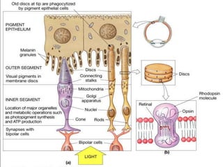
• The rods and cones serve as sensory nerve endings for visual
sensation.
• Light falling upon the retina causes photochemical changes
which in turn trigger a cascade of biochemical reactions that
result in generation of electrical changes.
• Photochemical changes occurring in the rods and cones are
essentially similar but the changes in rod pigment (rhodopsin or
visual purple) havebeenstudied in moredetail.
21
Retina & The Vision
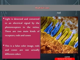
• Light is detected and converted
to an electrical signal by the
photoreceptors in the retina.
There are two main kinds of
receptors,rodsand cones
• This is a false color image, rods
and cones are not actually
different colors
rod
cone
Retina & The Vision
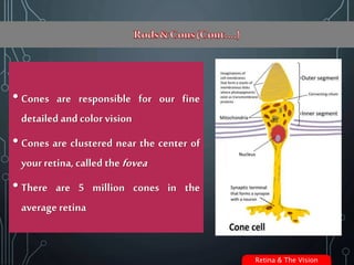
• Cones are responsible for our fine
detailed and color vision
• Cones are clustered near the center of
your retina, called thefovea
• There are 5 million cones in the
average retina
Retina & The Vision
• Rods are responsible for low light and
peripheral vision
• They are present everywhere in the
retina except the fovea
• There are 125 million rods in the average
retina
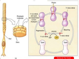
PHOTOCHEMICAL CHANGES
• Rhodopsinbleaching
• Rhodopsinregeneration
• Visual cycle : In the retina of living animals,
under constant light stimulation, a steady
state must exist under which the rate at
which the photochemicals are bleached is
equal to the rate at which they are
regenerated. This equilibrium between the
photodecomposition and regeneration of
visual pigments is referred to as visual
25
Retina & The Vision
PROCESSINGANDTRANSMISSIONOFVISUAL
IMPULSE
• The receptor potential generated in the photoreceptors is
transmitted by electrotonic conduction (i.e., direct flow of
electric current, and not as action potential) to other cells of the
retina viz. horizontal cells, amacrine cells, and ganglion cells.
• However, the ganglion cells transmit the visual signals by means
of action potential to the neurons of lateral geniculate body and
the later to the primary visual cortex.
26
Retina & The Vision
• It is a complex integration of light sense, form sense, sense of
contrast and colour sense.
• The receptive field organization of the retina and cortex are
usedto encodethis information about a visual image.
•The lightsense
• It is awareness of the light. The minimum brightness required to
evokea sensation of light is called thelight minimum.
27
Retina & The Vision
• It should be measured when the eye is dark adapted for at least
20-30 minutes.
• The human eye in its ordinary use throughout the day is capable
of functioning normally over an exceedingly wide range of
illumination by a highly complex phenomenon termed as the
visual adaptation.
28
Retina & The Vision
• The process of visual adaptation primarily involves :
• Dark adaptation (adjustment in dim illumination),
• Light adaptation (adjustment to bright illumination)
29
Retina & The Vision
•The form sense
• It is the ability to discriminate between the shapes of the
objects.
• Cones play a major role in this faculty.
• Therefore, form sense is most acute at the fovea, where there are
maximum number of cones and decreases very rapidly towards
the periphery
30
Retina & The Vision
•Sense of contrast
• It is the ability of the eye to perceive slight changes in the
luminance between regions which are not separated by definite
borders. Loss of contrast sensitivity results in mild fogginess of
the vision. Contrast sensitivity is affected by various factors like
age, refractive errors, glaucoma, amblyopia, diabetes, optic
nerve diseases and lenticular changes. the presence of normal
visual acuity.
31
Retina & The Vision
• Color sense
• It is the ability of the eye to discriminate betweendifferent
colours excited by light of different wavelengths.
• Colour vision is a function of the conesand thus better
appreciated in photopic vision. In dim light (scotopic vision), all
colours are seen grey and this phenomenonis called Purkinje
shift.
32
Retina & The Vision
• Theories of color vision
• The process of colour analysis begins in the retina and is not
entirely a function of brain.
• Many theories have been put forward to explain the colour
perception, but two have beenparticularly influential:
33
Retina & The Vision
1. Trichromatictheory/Young-Helmholtztheory.
2. Opponent-process theory.
34
Retina & The Vision
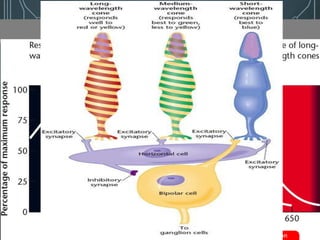
TRICHROMATICTHEORY
• Color perception occurs through the relative rates of response
by three kinds of cones.
• Short wavelength, medium-wavelength, long-wavelength.
• Each coneis maximally sensitive to a different set of
wavelengths.
• The ratio of activity across the three types of conesdetermines
thecolor. Moreintense light increases the brightness of the
color but does not change the ratio and thusdoesnot change
the perception of the color itself.
35
Retina & The Vision
OPPONENT-PROCESSTHEORY
• Theopponent-processtheorysuggests that weperceivecolor in
terms of paired opposites.
• The brain has a mechanism that perceivescolor on a continuum
from red togreen and another from yellow to blue.
• A possible mechanism for the theory is that bipolar cells are
excited by one set of wavelengths and inhibited by another.
36
Retina & The Vision
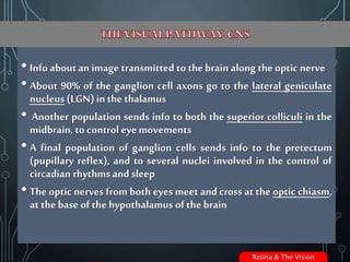
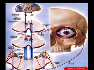
• Info about an image transmitted to thebrain along the optic nerve
• About 90% of the ganglion cell axons go to the lateral geniculate
nucleus (LGN)in the thalamus
• Another population sends info to both the superior colliculi in the
midbrain, to control eyemovements
• A final population of ganglion cells sends info to the pretectum
(pupillary reflex), and to several nuclei involved in the control of
circadian rhythms and sleep
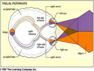
• The optic nerves from both eyes meet and cross at the optic chiasm,
at thebase of thehypothalamus of the brain
Retina & The Vision
• Info from both eyes is combined and splits according to the
visual field
• the right side of the visual cortex deals with the left half of
the field of view from both eyes
• the left side of the visual cortex deals with the right half of
the field of view from both eyes
• small region in the center processed by both halves of the
brain
• The neurons of the LGN relay the visual image to the visual
cortex which is located at the rear of the brain in the occipital
lobe abovethecerebellum
• The visual cortex is the most massive system in the human brain
and is responsible for higher-level processing of the visual image 38
Retina & The Vision
• Sense of vision is a complex function of the two eyes andtheir central
connections.
The physiologicalactivities involvedin the normal functioning of the eyes are:
• Maintenanceof clear ocular media,
• Maintenanceof normal intraocularpressure,
• The imageforming mechanism,
• Physiology of vision,
• Physiology of binocularvision,
• Physiology of pupil,and
• Physiology of ocular motility. 40
Retina & The Vision
• pigmentedlayer
• layerof rodsandconesprojectingtothepigment
• outernuclearlayercontainingthecellbodiesoftherods andcones
• outerplexiformlayer
• innernuclearlayer
• innerplexiformlayer
• ganglioniclayer
• layerof opticnervefibers
• innerlimitingmembrane.
41
Retina & The Vision
• Physiology of vision is a complex phenomenon which is still
poorly understood. The main mechanisms involved in
physiology of vision are :
•Initiationof vision(Photo transduction)
•Processing and transmission of visualsensation
•Visualperception
42
Retina & The Vision
•Guyton's Physiology
•Ganong'sPhysiology
•Comprehensive Ophthalmology
•www.Medscape.com
•The Images WereRetrieved Fromwww.google.com
For Comments:
Haroon_kand@yahoo.com
43
Retina & The Vision
Retina & the vision
Retina & the vision