Structure of Spermand Ovum
Dr. M. Mukilan,
Assistant Professor,
Sri Ramakrishna College of Arts & Science (Autonomous),
Coimbatore – 641 006
17/09/2024
2.
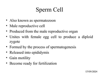
Sperm Cell
• Alsoknown as spermatozoon
• Male reproductive cell
• Produced from the male reproductive organ
• Unites with female egg cell to produce a diploid
zygote
• Formed by the process of spermatogenesis
• Released into epididymis
• Gain motility
• Become ready for fertilization
17/09/2024
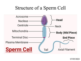
Structure of aSperm Cell
• Three different structure
• Head
• Body
• Tail
Head
• Flat pear-shaped structure
• Pointed tip
• Broad base
• Contains genetic material within the cell nucleus
17/09/2024
5.
Structure of aSperm Cell
• Covered by acrosome
• Contains hydrolytic enzymes
• Penerate the egg layers
• Acrosome reaction
• Contains haploid chromosomes
• Human sperm – 23 number of chromosomes
• After fertilization – Zygote – 46 number of
chromosomes
17/09/2024
6.
Structure of aSperm Cell
Body
• Also called as mid piece
• Contains mitochondria
• Provides energy needed for the movement
Tail
• 80 % of its entire length
• Needed for sperm movement
• Contains axoneme
• Axoneme – bundle of microtubules surrounded by
mitochondria
17/09/2024
7.
Structure of aSperm Cell
• Axoneme – two central singlet microtubule
surrounded by nine microtubule doublets
• Dyenin proteins
• Needed for movement
• Uses ATP hydrolysis for energy creation
17/09/2024
8.
Types of Sperm
•Two types
• X sperm
• Y sperm
X sperm
• Combines with the X chromosome of the female
• Forms a zygote with XX chromosomes
• Produces a female offspring
17/09/2024
9.
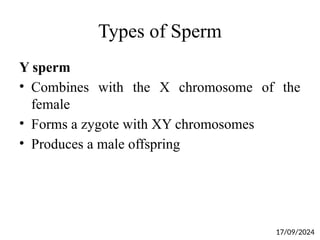
Types of Sperm
Ysperm
• Combines with the X chromosome of the
female
• Forms a zygote with XY chromosomes
• Produces a male offspring
17/09/2024
10.
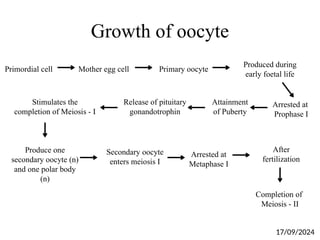
Growth of oocyte
Primordialcell Mother egg cell Primary oocyte
Produced during
early foetal life
Arrested at
Prophase I
Attainment
of Puberty
Release of pituitary
gonandotrophin
Stimulates the
completion of Meiosis - I
Produce one
secondary oocyte (n)
and one polar body
(n)
Secondary oocyte
enters meiosis I
Arrested at
Metaphase I
After
fertilization
Completion of
Meiosis - II
17/09/2024
11.
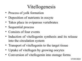
Vitellogenesis
• Process ofyolk formation
• Deposition of nutrients in oocyte
• Takes place in oviparous vertebrates
• Sequential process
• Consists of four events
• Induction of vitellogenin synthesis and its release
into the circulation system
• Transport of vitellogenin to the target tissue
• Uptake of vitellogen by growing oocytes
• Conversion of vitellogenin into storage forms
17/09/2024
12.
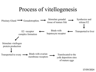
Process of vitellogenesis
PituitaryGland Gonadotrophins
Stimulate gonadal
tissue of mature fish
Synthesize and
release E2
Transported to liver
Binds with
hepatocyte receptor
E2 –receptor
complex formation
Stimulate vitellogen
protein production
Transported to ovary Binds with ovarian
membrane receptors
Translocated to the
yolk deposition sites
of mature eggs
17/09/2024
13.
Ovum
• Female reproductivecell
• Produced from pair of ovaries
• fuses with sperm during the process of fertilisation
• Develops into its mature form via a process
called oogenesis
• In viviparous animals, this ovum is fertilised inside
the body of the females.
• The embryo development takes place in the uterus
17/09/2024
14.
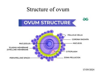
Structure of ovum
•Largest cells
• Spherical and non-motile
• Diameter of 0.15 mm
• 10-15 cm length
• Has large, centrally located nucleus
• Covered by cytoplasm
• Oocyte nucleus – Germinal vesicle
• Oocyte nucleolus – Germinal Disc
• Oocyte yolk – ooplasm
• Human – less ooplasm
• Alecithal
17/09/2024
15.
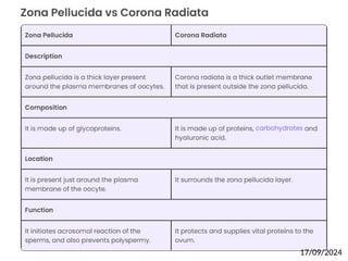
Structure of ovum
•Covered by cortex
• Cortex contains many microvilli
• Microvilli needed for transport of substance in
and out of cytoplasm
• Ovum has three layers
• Inner thin vitelline membrane
• Middle zona pellucida
• Outer corona radiata
17/09/2024
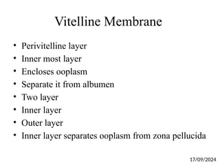
Vitelline Membrane
• Perivitellinelayer
• Inner most layer
• Encloses ooplasm
• Separate it from albumen
• Two layer
• Inner layer
• Outer layer
• Inner layer separates ooplasm from zona pellucida
17/09/2024
19.
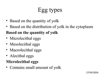
Egg types
• Basedon the quantity of yolk
• Based on the distribution of yolk in the cytoplasm
Based on the quantity of yolk
• Microlecithal eggs
• Mesolecithal eggs
• Macrolecithal eggs
• Alecithal eggs
Microlecithal eggs
• Contains small amount of yolk
17/09/2024
20.
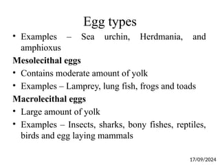
Egg types
• Examples– Sea urchin, Herdmania, and
amphioxus
Mesolecithal eggs
• Contains moderate amount of yolk
• Examples – Lamprey, lung fish, frogs and toads
Macrolecithal eggs
• Large amount of yolk
• Examples – Insects, sharks, bony fishes, reptiles,
birds and egg laying mammals
17/09/2024
21.
Egg types
Alecithal Eggs
•Absence/little amount of yolk
• Example - Human
Based on the distribution of yolk in the cytoplasm
Homolecithal Eggs
• Yolk is uniformly distributed
• Example – annelids, molluscs, echinoderms and
protochordates
Telolecithal eggs
• Concentrated in the vegetal half
• Example - Amphibians
17/09/2024
22.
Egg types
Meiolecithal eggs
•Yolk is very large
• Occupies entire cytoplasm
• Leave small disc like area for nucleus
• Example – Birds, reptiles, and egg laying mammals
Centerolecithal eggs
• Yolk is localized at the centre
• Example - Insects
17/09/2024
23.
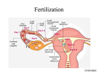
Fertilization
• Fusion ofmale and female gametes
• Results in the formation of gamete
• Takes place in fallopian tube
• Starts with the entry of sperm into female
reproductive system
• After entry, sperm moves towards uterus and
reach fallopian tube
• 24 hours – Fertilization – In the presence of
secondary oocyte
17/09/2024
24.
Fertilization
• Secondary oocyte– fuses with one sperm
• Polarization of ovum membrane
• Once sperm fuses with layers of ovum membrane –
Depolarization
• Avoid polyspermy
• Sperms induces secondary oocyte to complete
meiosis – II
• Secondary oocyte – Egg
• Active for 24 hours
• Sperm – 48-72 hours
17/09/2024
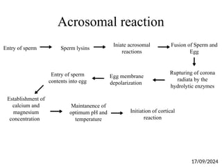
Acrosomal reaction
Entry ofsperm Sperm lysins
Iniate acrosomal
reactions
Fusion of Sperm and
Egg
Entry of sperm
contents into egg
Rupturing of corona
radiata by the
hydrolytic enzymes
Egg membrane
depolarization
Establishment of
calcium and
magnesium
concentration
Maintanence of
optimum pH and
temperature
Initiation of cortical
reaction
17/09/2024
28.
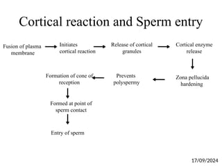
Cortical reaction andSperm entry
Fusion of plasma
membrane
Initiates
cortical reaction
Release of cortical
granules
Cortical enzyme
release
Entry of sperm
Zona pellucida
hardening
Prevents
polyspermy
Formation of cone of
reception
Formed at point of
sperm contact
17/09/2024
29.
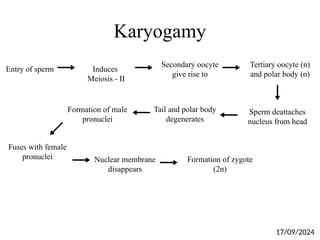
Karyogamy
Entry of spermInduces
Meiosis - II
Secondary oocyte
give rise to
Tertiary oocyte (n)
and polar body (n)
Fuses with female
pronuclei
Sperm deattaches
nucleus from head
Formation of male
pronuclei
Nuclear membrane
disappears
Tail and polar body
degenerates
Formation of zygote
(2n)
17/09/2024
30.
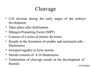
Cleavage
• Cell divisionduring the early stages of the embryo
development
• Takes place after fertilization
• Mitogen-Promoting Factor (MPF)
• Consists of a series of mitotic divisions
• Results in the formation of smaller and nucleated cells –
blastomeres
• Grouped together to form morula
• Morula consists of 8-16 blastomeres
• Termination of cleavage results in the development of
blastula
17/09/2024
Cleavage of Zygotein Humans
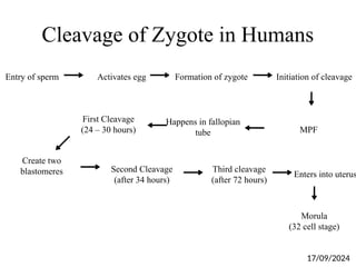
Entry of sperm Activates egg Formation of zygote Initiation of cleavage
First Cleavage
(24 – 30 hours) MPF
Happens in fallopian
tube
Create two
blastomeres Second Cleavage
(after 34 hours)
Third cleavage
(after 72 hours)
Enters into uterus
Morula
(32 cell stage)
17/09/2024
33.
Types of Cleavage
•Based on reorganization and cytoplasmic contents
• Determinate cleavage
• Indeterminate cleavage
• Holoblastic cleavage
• Discodial cleavage
• Superficial cleavage
• Transitional cleavage
17/09/2024
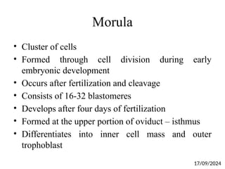
34.
Morula
• Cluster ofcells
• Formed through cell division during early
embryonic development
• Occurs after fertilization and cleavage
• Consists of 16-32 blastomeres
• Develops after four days of fertilization
• Formed at the upper portion of oviduct – isthmus
• Differentiates into inner cell mass and outer
trophoblast
17/09/2024
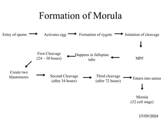
35.
Formation of Morula
Entryof sperm Activates egg Formation of zygote Initiation of cleavage
First Cleavage
(24 – 30 hours) MPF
Happens in fallopian
tube
Create two
blastomeres Second Cleavage
(after 34 hours)
Third cleavage
(after 72 hours)
Enters into uterus
Morula
(32 cell stage)
17/09/2024
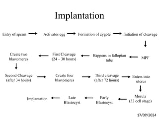
36.
Implantation
Entry of spermActivates egg Formation of zygote Initiation of cleavage
First Cleavage
(24 – 30 hours) MPF
Happens in fallopian
tube
Create two
blastomeres
Second Cleavage
(after 34 hours)
Third cleavage
(after 72 hours)
Enters into
uterus
Morula
(32 cell stage)
Create four
blastomeres
Early
Blastocyst
Late
Blastocyst
Implantation
17/09/2024
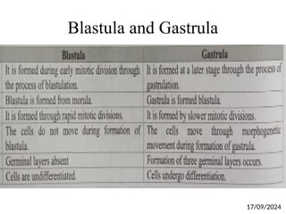
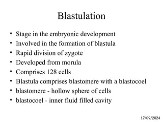
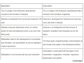
Blastulation
• Stage inthe embryonic development
• Involved in the formation of blastula
• Rapid division of zygote
• Developed from morula
• Comprises 128 cells
• Blastula comprises blastomere with a blastocoel
• blastomere - hollow sphere of cells
• blastocoel - inner fluid filled cavity
17/09/2024
39.
Blastulation
• Absence ofcell layers
• Pre embryo
• Less cells are present
• Needed for
1. Cell polarity
2. Axis formation
3. Cell specification
4. Regulation of gene expression
• Not involved with any cell movement
17/09/2024
40.
Gastrulation
• Stage inthe embryonic development
• Involved in the formation of Gastrula
• Slow division of zygote
• Developed from blastula
• Comprises more cells than in blastula
• Multilayered cell
• Consists of three layers
• Ectoderm, endoderm and mesoderm
17/09/2024
41.
Gastrulation
• Needed forthe formation of new tissues and organs
in the developing embryo
• Shows cell movement
• Five types of cell movements
• Invagination
• Involution
• Ingression
• Delamination
• Epiboly
17/09/2024
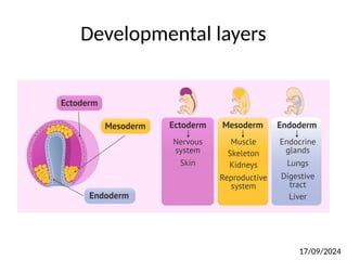
Germ Layers
• Primarylayers formed during embryonic
development
• Three germ layers
• Endoderm, mesoderm, and ectoderm
• Coordinate and function to develop new organs
• Formed during the gastrulation stage
17/09/2024
44.
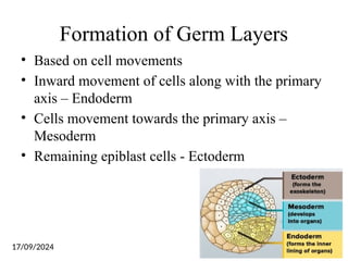
Formation of GermLayers
• Based on cell movements
• Inward movement of cells along with the primary
axis – Endoderm
• Cells movement towards the primary axis –
Mesoderm
• Remaining epiblast cells - Ectoderm
17/09/2024
Axis formation
• Followedby gastrulation
• Controlled by specific set of genes
• Fusion of egg and sperm results in the formation
of zygote
• Formed zygote undergoes sequential cleavage and
differentiation process to form blastula
• Gastrula formed from blastula
• After gastrulation, axis formation takes place
17/09/2024
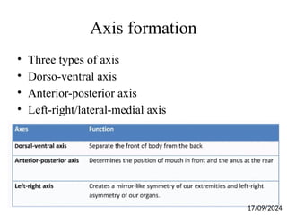
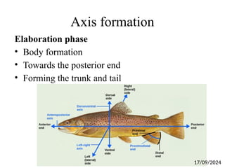
Axis formation
• Firstembryonic axis
• Needed for unidirectional movement
• Head-tail axis
• Consists of two phase
• Initiation phase
• Elaboration phase
Initiation phase
• Embryo is divided into head and tail
17/09/2024
Axis formation
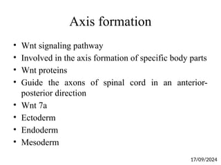
• Wntsignaling pathway
• Involved in the axis formation of specific body parts
• Wnt proteins
• Guide the axons of spinal cord in an anterior-
posterior direction
• Wnt 7a
• Ectoderm
• Endoderm
• Mesoderm
17/09/2024