Premium Call Girls Cottonpet Whatsapp 7001035870 Independent Escort Service
Strain cardíaco na avaliação da função cardíaca fetal
1. C
2008, the Authors
Journal compilation C
2008, Wiley Periodicals, Inc.
DOI: 10.1111/j.1540-8175.2008.00761.x
Global Longitudinal Cardiac Strain and Strain
Rate for Assessment of Fetal Cardiac Function:
Novel Experience with Velocity Vector Imaging
Piers C.A. Barker, M.D.,∗
Helene Houle, B.A.,† Jennifer S. Li, M.D.,∗
Stephen Miller, M.D.,∗
James Rene Herlong, M.D.,∗
and Michael G.W. Camitta, M.D.∗
∗
Duke Children’s Heart Program, Duke University Medical Center, Durham, North Carolina;
and †Siemens Medical Solutions, Mountain View, California
Background: Cardiac strain and strain rate are new methods to quantitate fetal cardiac function.
Doppler-based techniques are regional measurements limited by angle of insonation. Newer feature-
tracking algorithms permit angle independent measurements from two-dimensional datasets. This
report describes the novel measurement of global strain, strain rate, and velocity using Velocity Vector
Imaging (VVI) in a group of fetuses with and without heart disease. Methods: Global and segmental
longitudinal measurements were performed on the right and left ventricles in 33 normal fetuses and 15
fetuses with heart disease. Segmental measurements were compared to global measurements. Clinical
outcome data were recorded for fetuses with heart disease. Results: Forty-eight fetuses were evaluated
with VVI. Cardiac strain and strain rate in normal fetuses were similar to normal adult values, but
lower than pediatric values (LV strain = −17.7%, strain rate −2.4/sec; RV strain = −18.0%, strain
rate −1.9/sec). No difference was present between segmental and global measurements of cardiac
strain and strain rate, although basal and apical velocities were significantly different from global
velocities for both right and left ventricles. In fetuses with heart disease, lower global cardiac strain
appeared to correlate with clinical status, although there was no correlation with visual estimates
of cardiac function or outcome. Conclusion: Measurement of global longitudinal cardiac strain and
strain rate is possible in fetuses using VVI. Segmental measurements are not significantly different
from global measurements; global measurements may be a useful tool to quantitate fetal cardiac
function. (ECHOCARDIOGRAPHY, Volume 26, January 2009)
fetal echocardiography, cardiac strain, velocity vector imaging
Quantification of fetal cardiac function has
long been an elusive goal in the evaluation
of fetal cardiac physiology and adaptation to
disease. The fetal circulation is unique in its
source of oxygenated blood, degree of intracar-
diac and extracardiac mixing, and output of the
right and left ventricles.1
Measurements of car-
diac function validated in adults, such as the
shortening fraction or ejection fraction, often
fail to provide accurate results in fetuses due
to intrinsic differences in fetal wall motion and
small ventricular volumes that magnify mea-
surement error. More recently, measurement of
fetal cardiac strain and strain rate has been
Address for correspondence and reprint requests: Piers C.
A. Barker, M.D., Room 7502D, Duke Hospital North, Box
3090, Durham, NC 27710. Fax: +1-919-681-7892; E-mail:
piers.barker@duke.edu
attempted to overcome the limitations of two-
dimensional and M-mode imaging.2–4
Myocardial strain is defined as the change
in length of an object relative to its baseline
length caused by an applied stress, with5
strain
rate being derived from the velocity of the de-
formation over time.6
In the practice of cardiac
ultrasound, the strain rate is typically mea-
sured using tissue Doppler imaging to calculate
the velocities of two points set a small, fixed
distance apart, with cardiac strain then calcu-
lated as the integral of the strain rate measure-
ment.6
By analyzing segments of myocardium
directly rather than changes in ventricular di-
mensions or volumes, cardiac strain, and strain
rate may be better measurements of ventricu-
lar contractility.7
However, assessment of only
certain small segments of myocardium limits
the extrapolation of these segmental results to
global cardiac function.
28 ECHOCARDIOGRAPHY: A Jrnl. of CV Ultrasound Allied Tech. Vol. 26, No. 1, 2009
2. FETAL GLOBAL LONGITUDINAL CARDIAC STRAIN AND STRAIN RATE
Both regional cardiac strain and strain rate
have been reported and validated as measures
of ventricular function in adults and children.6
However, the majority of these studies have
been based upon tissue or color Doppler mea-
surements, including the first fetal studies.4,8,9
Tissue Doppler measurements have the advan-
tage of less reliance on image quality and bor-
der detection, and permit the acquisition of
data at much higher frame rates than those
available by traditional two-dimensional ultra-
sound or cardiac magnetic resonance imaging.6
However, tissue Doppler is inherently limited
by its dependence on the angle of insonation,
which permits analysis of only those limited
segments of myocardium that are parallel to
the ultrasound beam, and can be affected by re-
gional cardiac translation.10
Both of these lim-
itations pose significant problems in fetal pa-
tients, given the variation in fetal position, and
prevent measurement of global indices for the
left or right ventricle.
Speckle or feature tracking is a novel way
of assessing myocardial motion from the two-
dimensional B-mode image. As opposed to
tissue Doppler, “speckles” derived from the sta-
ble interference and backscatter of the ultra-
sound signal in the myocardium are tracked
from frame to frame with reference to their pre-
vious position and distance of movement.7,11,12
From these data, both the velocity and the di-
rection of myocardial motion (the velocity vec-
tor) can be calculated for any region of the
myocardium, regardless of angle to the ultra-
sound beam, with strain rate and strain cal-
culated by comparing adjacent velocity vectors.
Further refinements of this tracking technique
allow for the incorporation of manually traced
borders, annuli position, and speckle periodic-
ity to create the potentially more accurate “fea-
ture” tracking software used in this study.7,11
This method has been validated in adult pa-
tients for the calculation of cardiac strain and
strain rate,13
but the application to fetal pa-
tients has only recently been reported, and
only in normal fetuses.2,3,14
Recently, feature-
tracking techniques have been applied to assess
global cardiac strain and strain rate in animal
infarct models and humans after myocardial in-
farction, in whom regional measurements may
not accurately reflect cardiac function due to
injured segments,15
as well as in adults with
systemic right ventricles to overcome the lim-
itations of right ventricular (RV) geometry.16
However, this method has not yet been fully
studied in fetal patients, whose small cardiac
size and different physiology limit the useful-
ness of regional measurements.
We therefore report our experience in the
novel use of velocity vector imaging (VVI) to
calculate global cardiac strain, strain rate, and
velocity in a series of fetuses with and without
heart disease.
Methods
Longitudinal cardiac strain, strain rate, and
velocity analysis was performed on the fe-
tal right ventricle and fetal left ventricle (if
present) obtained during a clinically indicated
fetal echocardiogram. The study was approved
by the Duke University Medical Center Institu-
tional Review Board for Human Research and
all subjects consented to participate. A research
version of the commercially available VVI soft-
ware (Siemens Medical Solutions, Mountain
View, CA, USA) was used for all measurements.
For each fetus, a high-resolution, zoomed
loop of the apical four-chamber view incorpo-
rating at least one complete cardiac cycle was
recorded, with machine settings adjusted to
maximize frame rate. This image was stored
digitally and transferred to the offline worksta-
tion (Syngo USWP, Siemens Medical Solutions)
for later analysis. Syngo VVI was launched
from review of each DICOM digital clip. R-wave
gating was performed using a superimposed
M-mode tracing of left or RV wall motion to
define the onset of ventricular systole (initial in-
ward motion of the ventricular wall) as a corol-
lary of the electrical QRS and therefore the be-
ginning and end of a cardiac cycle. This method
of R-wave gating was also used for fetuses eval-
uated during an arrhythmia, with the cardiac
cycle selected as representative of baseline si-
nus rhythm (i.e., not during or at the onset or
termination of the abnormal rhythm).
After definition of the cardiac cycle, the en-
docardium of the right and left ventricles was
traced manually from a single frame of the
digital loop that provided the clearest still-
frame endocardial border definition (typically
mid-systole). The same cardiac cycle was used
for both the left ventricular (LV) and RV trac-
ing, except in three normal fetuses and two
abnormal fetuses in which separate apical
four-chamber views were required. Endocar-
dial tracing began at the edge of the atrioven-
tricular valve annulus, extended to the apex
of the ventricle without incorporation of the
papillary muscle complex, and returned basally
to the other edge of the atrioventricular valve
Vol. 26, No. 1, 2009 ECHOCARDIOGRAPHY: A Jrnl. of CV Ultrasound Allied Tech. 29
3. BARKER, ET AL.
annulus. This therefore provided both the bor-
der and annuli position information necessary
for the “feature-tracking” component of the
VVI algorithm. Twenty-two individual, equally
spaced velocity vectors were then automatically
calculated for each frame of the cardiac cycle
by the VVI algorithm and displayed for the
complete loop. Accuracy of border tracking was
visually confirmed by viewing the cardiac cy-
cle with only border information displayed (i.e.,
with velocity vectors removed). If necessary, in-
dividual regions of the border were adjusted
until the border was correctly tracked for each
frame.
Cardiac strain, strain rate, and velocity data
were automatically calculated from the veloc-
ity vector information, and displayed in a six-
segment model for both fetal ventricles. In addi-
tion, the global peak systolic strain, global peak
systolic strain rate, and global peak systolic ve-
locities were calculated from the entire velocity
vector dataset as an average of all segments of
ventricular motion, and displayed as a separate
curve.
Statistical Testing
Global longitudinal cardiac strain, strain
rate, and velocities were compared to regional
measurements using Student’s t-test for both
normal fetuses and fetuses with heart disease.
A P-value of 0.05 was used to define a signif-
icant difference. Interobserver variability was
tested between two observers (PB and HH) on
ten randomly selected datasets and intraob-
server variability was tested for two observers
(PB and HH) on five randomly selected datasets
using coefficient of variation analysis.
For fetuses with heart disease, global longitu-
dinal cardiac strain and strain rate were com-
pared to visually estimated function (hypercon-
tractile, normal, mildly decreased, moderately
decreased, and severely decreased, as recorded
by a skilled independent observer (MC) blinded
to the results of the strain analysis) and ulti-
mate fetal outcome. No comparisons were made
between abnormal fetuses as a group and nor-
mal fetuses due to the heterogeneity of fetal
cardiac diagnoses.
Results
Forty-eight fetal patients were enrolled in
the study, consisting of 33 fetuses with normal
cardiac anatomy and function, and 15 fetuses
with congenital or functional heart disease. The
median gestational age was 24 weeks (range
17–38 weeks). Four fetuses with congenital or
functional heart disease underwent multiple
echocardiograms, permitting serial analysis of
fetal strain. Accurate endocardial border track-
ing and calculation of velocity vectors were ac-
complished on all right and left ventricles in
all fetuses despite limitations in image qual-
ity secondary to fetal position or maternal body
habitus, with the exception of one left ventricle
in a single abnormal fetal patient due to exces-
sive fetal motion. Longitudinal cardiac strain
measurements were possible in all tracked fe-
tuses, while strain rate and velocity measure-
ments were limited to 22 normal fetuses and 12
abnormal fetuses due to compression of frame
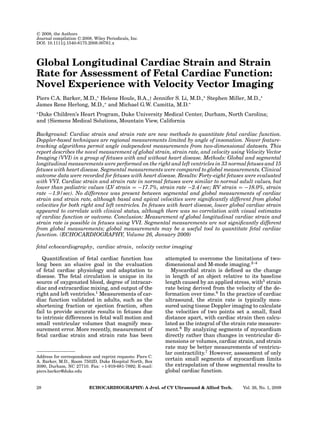
rate/time data in the other fetuses. Figure 1
demonstrates typical LV velocity vectors and
the resultant strain calculations for a normal
24-week fetus.
Table I demonstrates the results of global
and segmental longitudinal strain analysis for
both left and right ventricles in normal fe-
tuses. The mean LV global peak systolic strain
was −17.7% (standard deviation 6.4) with a
median of −16.6% (range −9.2% to −32.9%).
The mean RV global peak systolic strain was
−18.0% (standard deviation 6.4) with a median
of −17.4% (range −6.7% to −33.4%). There was
no statistical difference between global strain
and segmental strain measurements for either
ventricle.
Table II demonstrates the results of global
and segmental longitudinal strain rate analy-
sis for both left and right ventricles in normal
fetuses. The mean LV global peak systolic strain
rate was −2.4/sec (standard deviation 1.2/sec)
with a median of −1.9/sec (range −5.9/sec
to −0.7/sec). The mean RV global peak sys-
tolic strain rate was −1.9/sec (standard devi-
ation 0.8/sec) with a median of −1.7/sec (range
−3.8/sec to −0.5/sec). There was no statisti-
cal difference between global strain rate and
segmental strain rate measurements for either
ventricle.
Table III demonstrates the results of global
and segmental longitudinal velocity analysis
for both left and right ventricles in normal
fetuses. The mean LV global peak systolic
velocity was 1.6 cm/sec (standard deviation
0.6 cm/sec) with a median of 1.5 cm/sec (range
0.5–3.0 cm/sec). The mean RV global peak sys-
tolic velocity was 1.6 cm/s (standard devia-
tion 0.5 cm/sec) with a median of −1.6 cm/sec
(range 0.8–2.3 cm/sec). In contrast to strain and
strain rate measurements, the basal segmental
30 ECHOCARDIOGRAPHY: A Jrnl. of CV Ultrasound Allied Tech. Vol. 26, No. 1, 2009
4. FETAL GLOBAL LONGITUDINAL CARDIAC STRAIN AND STRAIN RATE
Figure 1. Velocity vector tracing
of the left ventricular endocardium
(endo) in a normal fetus at 24
weeks of gestation with correspond-
ing global and segmental strain
curves. Global (average) peak sys-
tolic strain curve is shown in black.
Base left = septal base; mid-left =
mid-septal; apex left = apical septal;
apex right = apical free wall; mid-
right = mid free wall; base right =
basal free wall.
velocities for both the left and right ventricles
were significantly higher than the global ve-
locity measurement, while the apical segmen-
tal velocities were significantly lower than the
global velocity measurement.
Fetuses with congenital or functional heart
disease demonstrated similar results, with no
significant difference detected between global
strain and global strain rate measurements
compared to regional measurements. Segmen-
tal velocities did differ, however, with the LV
apical septal and apical free wall velocities sig-
nificantly lower than the global velocity, and
the basal free wall significantly higher. For the
right ventricle, the mid-septal and apical sep-
tal velocities were significantly lower, and the
basal free wall significantly higher compared to
the global RV peak velocity.
TABLE I
Ventricular Peak Global and Regional Strain Measurements in Normal Fetuses (n=33)
LV Mean LV Median LV Range LV SD RV Mean RV Median RV Range RV SD
Global strain −17.7 −16.6 −32.9 to −9.2 6.4 −18.0 −17.4 −33.4 to −6.7 6.4
Septal base −15.9 −15.4 −44.8 to −2 8.7 −17.3 −15.2 −34.3 to −5.6 7.9
Mid-septal −14.9 −13.4 −41.1 to −1.5 7.7 −17.4 −16.8 −31.5 to −6 6.7
Apical septal −18.5 −19.4 −37.9 to −4.7 8.5 −16.1 −15.0 −39.2 to −2.5 9.3
Apical free wall −19.3 −19.1 −41.9 to −2.6 9.5 −16.7 −15.5 39.2 to −1.5 10.1
Mid free wall −19.1 −19.0 −39 to −6.3 8.3 −19.4 −19.5 −33.1 to −8.2 7.0
Base free wall −17.8 −15.1 −37 to −5.7 9.4 −20.2 −18.2 −40 to −1.5 9.1
All values expressed as percent change in length.
P 0.05 for all regional strain measurements compared to global strain.
LV = left ventricular; RV = right ventricular.
Table IV demonstrates global peak longitu-
dinal strain and strain rate measurements in
fetuses with structural or functional heart dis-
ease, compared with visually estimated func-
tion and clinical outcome. There was an over-
all trend toward lower global strain and strain
rate compared to normal fetuses, but this was
not uniform and varied depending upon disease
state, with one fetus with aortic valve stenosis
demonstrating global peak cardiac strain more
than 1 standard deviation above the global peak
systolic strain in normal fetuses. There was no
correlation between calculated cardiac strain
and strain rate and visually estimated ventric-
ular function, although in one patient followed
serially (chaotic atrial tachycardia [CAT 1]),
the improvement in ventricular function
matched an improvement from a low cardiac
Vol. 26, No. 1, 2009 ECHOCARDIOGRAPHY: A Jrnl. of CV Ultrasound Allied Tech. 31
5. BARKER, ET AL.
TABLE II
Ventricular Peak Global and Regional Strain Rate Measurements in Normal Fetuses (n=22)
LV Mean LV Median LV Range LV SD RV Mean RV Median RV Range RV SD
Global strain rate −2.4 −1.9 −5.9 to −0.7 1.2 −1.9 −1.7 −3.8 to −0.5 0.8
Septal base −1.9 −1.7 −4.9 to −0.6 1.0 −1.9 −1.9 −4.1 to −0.9 0.8
Mid-septal −2.1 −1.9 −7.5 to −0.6 1.5 −1.9 −2.0 −3.8 to −0.9 0.8
Apical septal −2.7 −2.4 −8.2 to −0.6 1.7 −2.1 −1.8 −5.5 to −0.2 1.3
Apical free wall −2.8 −2.7 −5.7 to −0.2 1.5 −2.5 −1.9 −5.9 to −0.4 1.6
Mid free wall −2.4 −1.9 −5.7 to −0.7 1.4 −2.2 −2.3 −3.8 to −1 0.8
Base free wall −2.5 −1.9 −7.8 to −0.9 1.7 −2.3 −2.2 −4.6 to −0.9 1.1
All values expressed as rate of change in length (per second).
P 0.05 for all regional strain rate measurements compared to global strain rate.
LV = left ventricular; RV = right ventricular.
strain to closer to the normal value. Similarly,
there was no correlation between calculated
strain and strain rate and ultimate fetal out-
come.
Intraobserver variability ranged between 5–
12% for the left ventricle and 5–6% for the right
ventricle. Interobserver variability ranged be-
tween 10% for the left ventricle and 13% for the
right ventricle.
Discussion
Myocardial strain and strain rate have been
proposed as useful tools in the evaluation of car-
diac mechanics. Myocardial strain and strain
rate, being regional measurements, are rela-
tively free of confounding factors such as car-
diac translation, which may occur with respi-
ration or motion of structures adjacent to the
heart.10
The presence of multiple confounding
variables such as fetal motion, high heart rates,
and limited maternal transabdominal imaging
TABLE III
Ventricular Peak Global and Regional Velocity Measurements in Normal Fetuses (n=22)
LV Mean LV Median LV Range LV SD RV Mean RV Median RV Range RV SD
Global velocity 1.6 1.5 0.5–3.0 0.6 1.6 1.6 0.8–2.3 0.5
Septal base 2.1∗ 1.9 1.0–4.6 1.0 2.0∗ 2.1 0.8–3.1 0.6
Mid-septal 1.4 1.2 0.2–3.4 0.9 1.4 1.3 0.6–2.6 0.5
Apical septal 0.6∗ 0.6 0.0–1.5 0.4 0.8∗ 0.6 0.2–3.1 0.7
Apical free wall 1.0∗ 0.8 0.2–2.4 0.6 1.1∗ 1.1 0.1–2.4 0.7
Mid free wall 1.9 1.7 0.3–3.6 0.9 1.9 1.8 0.7–3.8 0.8
Base free wall 2.5∗ 2.5 0.8–4.9 1.0 2.6∗ 2.5 1.2–4.7 0.9
All values reported as cm/sec.
∗P 0.05 for regional velocity measurement compared to global velocity measurement.
LV = left ventricular; RV = right ventricular.
windows therefore makes these new measure-
ments appealing for assessment of fetal cardiac
function.
The majority of published studies have
measured cardiac strain and strain rate us-
ing tissue or color Doppler-derived velocities,
although more recent speckle-tracking algo-
rithms have permitted these measurements to
be performed on two-dimensional data at ac-
ceptably high frame rates.11
These measure-
ments have been validated in vivo and in vitro
for both tissue Doppler and two-dimensionally
derived data, and have compared favorably to
MRI-tagging techniques.7,10,11,13
While tissue
Doppler has the advantage of less reliance on
image quality and visual border detection, it
has the inherent disadvantage of all Doppler
technologies by being dependent on angle of in-
sonation.5
This therefore limits the number of
cardiac segments available for analysis to those
parallel to the transducer beam, resulting in ex-
clusion of the cardiac apex.
32 ECHOCARDIOGRAPHY: A Jrnl. of CV Ultrasound Allied Tech. Vol. 26, No. 1, 2009
6. FETAL GLOBAL LONGITUDINAL CARDIAC STRAIN AND STRAIN RATE
TABLE
IV
Summary
of
Fetuses
with
Congenital
or
Functional
Heart
Disease
Gest.
Age
Visual
Visual
Diagnosis
(weeks)
LV
Strain
LV
SR
Function
RV
Strain
RV
SR
Function
Clinical
Outcome
SVT
1,
intermittent
20
−13.8
−1.3
Normal
−9.3
−1.1
Normal
Term
delivery,
stable
postnatally
HLHS
1
(mitral
atresia,
aortic
atresia)
25
N/A
N/A
Normal
−13.5
−1.3
Normal
Term
delivery,
stable
s/p
palliation
HLHS
2
(mitral
stenosis,
aortic
stenosis)
23
N/A
N/A
Normal
−13.3
−1.1
Normal
Deceased
in
utero,
unclear
etiology
SVT
2,
intermittent
26
−27.1
−2.7
Normal
−27.6
−3.2
Normal
Term
delivery,
stable
postnatally
TTTS
1,
donor
(A),
oligohydramnios
25
−11.2
−0.9
Normal
−16.8
−1.4
Normal
Preterm
delivery,
stable
postnatally
TTTS
1,
recipient
(B),
polyhydramnios
25
−13.3
−1
Normal
−13.3
−1.3
Normal
Preterm
delivery,
stable
postnatally
Ebstein’s
anomaly
of
tricuspid
valve
27
−16.3
−1.5
Normal
−18.5
−1.9
Normal
Hydrops
at
29
weeks,
deceased
D-TGA/IVS
33
Inc.
view
Inc.
view
Normal
−11.3
−0.9
Normal
Term
delivery
TTTS
2,
recipient
(A),
pulm
stenosis
29
−13.6
−1.1
Normal
−12.8
−1.2
Normal
Preterm
delivery,
deceased
day
2
TTTS
2,
donor
(B),
normal
29
−18.8
−2.3
Normal
−25.6
−3.5
Normal
Preterm
delivery,
survived
VSD/AS/Coa
31
−20.5
−2.3
Normal
−18.5
−1.8
Normal
Term
delivery,
stable
s/p
repair
CCTGA
1
20
−14.5
−1.4
Normal
−10.6
−0.8
Normal
Term
delivery,
no
intervention
needed
CCTGA
1
38
−13.9
−0.9
Normal
−7.8
−0.5
Normal
Aortic
stenosis
1
24
−25.7
−3.4
Normal
−21.1
−2.2
Normal
Term
delivery
Aortic
stenosis
1
28
−28.1
−3.6
Normal
−23.4
−4.5
Normal
s/p
BAV
day
2,
repeat
BAV
6
weeks
Aortic
stenosis
1
32
−27.7
−2.8
Hypercontractile
−27.3
−3.9
Normal
s/p
Ross
procedure
at
2
months
SVT
3,
early
return
of
sinus
rhythm
25
−13.2
−1.3
Normal
−15.5
−1.4
Normal
Hydrops
SVT
3,
hydrops
resolved
33
−16.6
−1.5
Normal
−14.1
−1.2
Normal
Hydrops
resolved,
term
delivery
CAT
1,
predominantly
in
arrhythmia
33
−10
−1.3
Mildly
decreased
−11.7
−1.2
Mildly
decreased
Preterm
delivery,
stable
postnatally
CAT
1,
predominantly
in
sinus
rhythm
35
−27.9
−4
Normal
−16.5
−1.9
Normal
All
strain
values
expressed
as
percent
change
in
length.
All
strain
rate
values
expressed
as
rate
of
change
in
length
(per
second).
Gest.
age
=
gestational
age;
Inc.
view
=
incomplete
view;
BAV
=
balloon
aortic
valvuloplasty;
CAT
=
chaotic
atrial
tachycardia;
CCTGA
=
{S,L,L}
congenitally
corrected
transposition
of
the
great
arteries;
D-TGA/IVS
=
{S,D,D}
transposition
of
the
great
arteries
with
intact
ventricular
septum;
HLHS
=
hypoplastic
left
heart
syndrome;
SVT
=
supraventricular
tachycardia;
TTTS
=
twin-twin
transfusion
syndrome;
VSD/AS/Coa
=
ventricular
septal
defect
with
aortic
stenosis
and
coarctation
of
the
aorta;
LV
=
left
ventricular;
RV
=
right
ventricular;
SR
=
strain
rate.
Vol. 26, No. 1, 2009 ECHOCARDIOGRAPHY: A Jrnl. of CV Ultrasound Allied Tech. 33
7. BARKER, ET AL.
Dependency on angle of insonation is particu-
larly problematic for fetal cardiology, given the
extremely variable position of the fetus rela-
tive to a transducer placed on the maternal ab-
domen. In the first fetal study published using
tissue Doppler to calculate fetal cardiac strain,
this angle dependence limited analysis to only
75 of 120 fetuses (63%),8
although this im-
proved in subsequent tissue/color Doppler stud-
ies.4,9
A similar study reporting measurement
of fetal tissue Doppler velocities, rather than
myocardial strain, excluded 16% of potential
subjects for similar reasons.17
In contrast, the two-dimensional feature-
tracking program used in this study permit-
ted the analysis of all visible ventricular seg-
ments, independent of fetal position or angle
of insonation. This resulted in only 1 ventricle
out of a total of 104 ventricles being excluded
for analysis due to limited views (1% of at-
tempted measurements). Additionally, the in-
clusion of all six segments permitted the calcu-
lation of global peak longitudinal systolic strain
and strain rate as novel measurements of fetal
ventricular function.
This study demonstrates that the feature-
based VVI software can be successfully ap-
plied to fetal 2-dimensional echocardiographic
datasets. This finding is similar to recently
published fetal studies examining normal fe-
tuses.2,3
Calculated global and regional peak
systolic strain measurements for normal fe-
tuses were similar for both the fetal left and
right ventricles at approximately −18%, and
−2.4 s−1
and −1.9 s−1
, respectively. These mea-
surements are similar to those published from
in vitro, adult, and fetal studies.3,6,8,10,13,18
However, calculated cardiac strain and strain
rate were lower than two recently reported fe-
tal and pediatric studies using tissue or color
Doppler methods,4,6,8,10,13,19
with the exception
of LV peak strain rate, which was similar to the
reported pediatric values. While overall there
has been a good reported correlation between
tissue Doppler and the two commonly used
feature-tracking algorithms, discrepancies be-
tween these methods have also been recently
reported that prevent the final definition of a
normal range for these values in fetuses.20–23
Calculated myocardial velocities were lower
than previously reported studies,2–4,17
although
this study did not specifically analyze the veloc-
ity at the atrioventricular annulus. It is not sur-
prising that there was more variability between
regional segments and global measurements
of velocity, based upon fiber orientation vari-
ation for both the left and right ventricles from
base to apex.12
Previous studies have shown fe-
tal myocardial velocity to vary with gestational
age,2,4
consistent with fetal somatic growth, al-
though the effect of gestational age was not as-
sessed in this study.
The finding that global measurements of
peak longitudinal strain and strain rate are
not significantly different from multiple seg-
mental measurements suggests that global
measurements may be a more useful tool to
quantitate fetal cardiac function, and may be
superior to tissue Doppler measurements.
Specifically, global measurements based on
two-dimensional datasets permit angle inde-
pendent analysis and avoid any variation in
the placement of the sample volumes or re-
gions of interest in such a small structure as
the fetal heart. In adult patients with systemic
right ventricles, global measurements have
been proposed as a method to avoid confound-
ing wall motion abnormalities and local noise
which may more greatly impact regional mea-
surements.16
To this end, a lower global mea-
surement may also provide a clue to look more
closely at the individual segments for regional
hypokinesis.
For fetuses with congenital or functional
heart disease, the global peak longitudinal
strain and strain rate demonstrated a tendency
toward lower values, although this was not uni-
form as demonstrated by the fetus with aortic
valve stenosis, the fetus with ventricular sep-
tal defect/aortic stenosis and coarctation of the
aorta, the fetus recovered from CAT 1, and the
fetal right ventricle in the donor in one case
of twin-twin transfusion syndrome (TTTS 2).
It is possible to speculate that the increased
strain and strain rate in these fetuses repre-
sent myocardial compensation for the struc-
tural heart disease (increased afterload in the
case of aortic stenosis and ventricular septal de-
fect/coarctation) and functional heart disease
(increased cardiac output of the right ventri-
cle in the donor twin). However, this theory
does not fully explain the increase in strain
and strain rate in the recovering fetus with ar-
rhythmia, or the lower strain and strain rate
throughout gestation of the fetus with con-
genitally corrected transposition of the great
arteries (CCTGA 1). Instead, these differences
more likely underscore the limitations of our
understanding of fetal cardiac adaptation to
disease.
The lack of significant correlation between
calculated strain and strain rate, and visually
34 ECHOCARDIOGRAPHY: A Jrnl. of CV Ultrasound Allied Tech. Vol. 26, No. 1, 2009
8. FETAL GLOBAL LONGITUDINAL CARDIAC STRAIN AND STRAIN RATE
estimated cardiac function and outcomes in fe-
tuses with heart disease further highlights our
limitations in assessing fetal cardiac function
and estimating prognosis. In the case of the
two fetuses who died in utero, it is possible that
they were well compensated at the time of the
fetal study, and decompensated before the next
visit. While the small number of abnormal fe-
tuses and the variability in pathology limited
our ability to analyze this group in more detail,
the application of VVI to much larger groups
of abnormal fetuses opens the field for further
investigation.
Limitations
The small size of the current study prevents
definition of normal values for fetuses at dif-
ferent gestational ages, as well as prevents
more detailed assessment of the relationship
between calculated measurements and postna-
tal outcome. RV strain and strain rate were cal-
culated using a LV-derived six-segment model,
which may not accurately reflect the more com-
plex geometry of the right ventricle, but is
similar in approach to previous studies using
tissue Doppler from an apical view as the mea-
surement tool. Circumferential and radial mea-
surements were not analyzed in this study,
and could provide useful comparisons to LV
measurements. Unfortunately, compression of
frame rate/time data limited the calculation of
strain rate and velocity in a few fetuses, but this
did not affect the strain measurement as strain
is calculated directly from speckle motion by the
VVI algorithm. Finally, the very nature of fetal
imaging, due to the effect of fetal movement,
size, position, and maternal factors complicate
efforts to obtain two-dimensional datasets for
analysis, although it is reassuring that ade-
quate images with accurate border tracking
could be obtained for all patients but one in
this study.
Conclusion
Fetal global peak longitudinal strain, strain
rate, and velocity can be successfully calculated
independent of angle of insonation using VVI.
Global peak longitudinal strain and strain rate
do not differ from regional measurements. Pre-
liminary experience suggests that normal fetal
left and right ventricular global peak longitu-
dinal strain and strain rate measurements are
similar to those of the normal adult heart. This
novel use of VVI is a promising tool for further
investigation into fetal cardiac physiology.
Acknowledgments: The authors are particularly in-
debted to the sonographers and staff of the Duke University
Pediatric Echo Laboratory for their assistance with image
acquisition for this project.
References
1. Kiserud T: Physiology of the fetal circulation. Semin
Fetal Neonatal Med 2005;10:493–503.
2. Younoszai AK, Saudek DE, Emery SP, Thomas JD:
Evaluation of myocardial mechanics in the fetus
by velocity vector imaging. J Am Soc Echocardiogr
2008;21:470–474.
3. Ta-Shma A, Perles Z, Bavri S, et al: Analysis of seg-
mental and global function of the fetal heart us-
ing novel automatic functional imaging. J Am Soc
Echocardiogr 2008;21:146–150.
4. Perles Z, Nir A, Gavri S, Rein AJ: Assessment of fetal
myocardial performance using myocardial deforma-
tion analysis. Am J Cardiol 2007;99:993–996.
5. D’hooge JHA, Jamal F, Kukulski T, et al: Regional
strain and strain rate measurements by cardiac ul-
trasound: Principles, implementation and limitations.
Eur J Echocardiogr 2000;1:154–170.
6. Voight JUFF. Strain and strain rate: New and clini-
cally relevant echo parameters of regional myocardial
function. Z Kardiol 2004;93:249–258.
7. Perk G, Tunick PA, Kronzon I: Non-Doppler two-
dimensional strain imaging by echocardiography—
from technical considerations to clinical applications.
J Am Soc Echocardiogr 2007;20:234–243.
8. Di Salvo GRM, Paladini D, Pacileo G, et al: Quantifica-
tion of regional left and right ventricular longitudinal
function in 75 normal fetuses using ultrasound-based
strain rate and strain imaging. Ultrasound Med Biol
2005;31: 1159–1162.
9. Larsen LU, Petersen OB, Norrild K, et al: Strain rate
derived from color Doppler myocardial imaging for as-
sessment of fetal cardiac function. Ultrasound Obstet
Gynecol 2006;27:210–213.
10. Urheim SET, Torp H, Angelsen B, et al: Myocardial
strain by Doppler echocardiography: Validation of a
new method to quantify regional myocardial function.
Circulation 2000;102:1158–1164.
11. Stefani L, Toncelli L, Gianassi M, et al: Two-
dimensional tracking and TDI are consistent methods
for evaluating myocardial longitudinal peak strain in
left and right ventricle basal segments in athletes.
Cardiovasc Ultrasound 2007;5:7.
12. Sengupta PP, Krishnamoorthy VK, Korinek J, et
al: Left ventricular form and function revisited: Ap-
plied translational science to cardiovascular ultra-
sound imaging. J Am Soc Echocardiogr 2007;20:539–
551.
13. Korinek J, Wang J, Sengupta PP, Miyazaki C, et al:
Two-dimensional strain—a Doppler-independent ul-
trasound method of quantitation of regional deforma-
tion: Validation in vitro and in vivo. J Am Soc Echocar-
diogr 2005;18:1247–1253.
14. Lorch SMSA, Johnson MC, Singh GK, et al: Does
strain and strain rate predict myocardial hypertro-
phy in fetuses of insulin-dependent diabetic mothers?
J Am Soc Echocardiogr 2006;19:599.
Vol. 26, No. 1, 2009 ECHOCARDIOGRAPHY: A Jrnl. of CV Ultrasound Allied Tech. 35
9. BARKER, ET AL.
15. Pirat B, Khoury DS, Hartley CJ, et al: A novel feature-
tracking echocardiographic method for the quantita-
tion of regional myocardial function: Validation in an
animal model of ischemia-reperfusion. J Am Coll Car-
diol 2008;51:651–659.
16. Chow PC, Liang XC, Cheung EW, et al: Novel two-
dimensional global longitudinal strain and strain rate
imaging for assessment of systemic right ventricular
function. Heart 2008;94:855–859.
17. Paladini DLA, Teodoro A, Arienzo M, et al: Tissue
Doppler imaging of the fetal heart. Ultrasound Obstet
Gynecol 2000;16:530–535.
18. Weidemann E, Kowalski M, D’hooge J, et al: Doppler
myocardial imaging. A new tool to assess regional in-
homogeneity in cardiac function. Basic Res Cardiol
2001;96:595–605.
19. Weidemann F, Eyskens B, Jamal F, et al: Quantifi-
cation of regional left and right ventricular radial
and longitudinal function in healthy children using
ultrasound-based strain rate and strain imaging. J
Am Soc Echocardiogr 2002;15:20–28.
20. Korinek J, Kjaergaard J, Sengupta PP, et al: High
spatial resolution speckle tracking improves accuracy
of 2-dimensional strain measurements: An update on
a new method in functional echocardiography. J Am
Soc Echocardiogr 2007;20:165–170.
21. Leitman M, Lysyansky P, Sidenko S, et al:
Two-dimensional strain-a novel software for real-
time quantitative echocardiographic assessment
of myocardial function. J Am Soc Echocardiogr
2004;17:1021–1029.
22. Modesto KM, Cauduro S, Dispenzieri A, et al: Two-
dimensional acoustic pattern derived strain param-
eters closely correlate with one-dimensional tissue
Doppler derived strain measurements. Eur J Echocar-
diogr 2006;7:315–321.
23. Teske AJ, De Boeck BW, Olimulder M, et al:
Echocardiographic assessment of regional right ven-
tricular function: A head-to-head comparison be-
tween 2-dimensional and tissue Doppler-derived
strain analysis. J Am Soc Echocardiogr 2008;21:275–
283.
36 ECHOCARDIOGRAPHY: A Jrnl. of CV Ultrasound Allied Tech. Vol. 26, No. 1, 2009