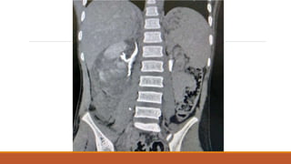
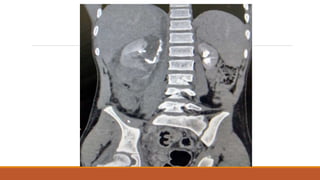
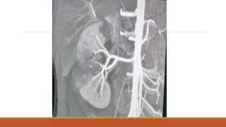
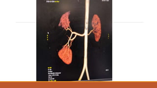
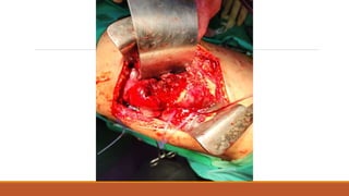
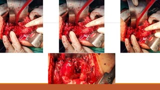
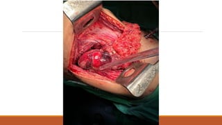
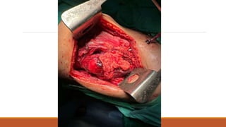
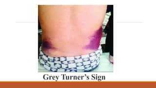
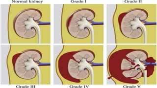
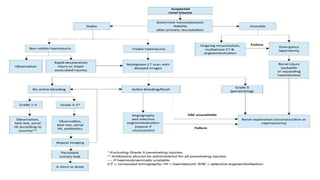
Renal Trauma
Case Scenario
14 year old boy, presented to ER department with history of RTA on 16/2/23 ( hit by bike while
crossing the road), his abdomen was distended and tender, he was having vomiting and
hematuria, after resuscitation USG done which showed sub-capsular hematoma of about 150-200
ml, Renal Tri-phasic CT scan showed.
Questions:
Which mechanism of injury is it?
Which type & grade of renal trauma is it?
How to evaluate?
How to manage?
What are the options to manage the patient?
Epidemiology:
Road Traffic accidents are major cause of long-term morbidity and mortality in developing countries.
Genitourinary Injuries accounts for 10% of all abdominal trauma.
Renal trauma accounts 5% of all trauma cases and 84% of genitourinary injuries.
Early Diagnosis is essential to prevent serious complications.
Signs of Renal Injury
Large Retroperitoneal Hematoma (Grey Turner’s Sign).
A palpable mass may represent a large Retroperitoneal Hematoma or urinary extravasation but if retro-
peritoneum has been torn, free blood may be noted in peritoneal cavity and no palpable mass will be
appreciated.
Kidney:
Microscopic / Gross Hematuria
Tenderness or ecchymosis over flanks or upper quadrant of abdomen
Diffuse abdominal tenderness on palpation (acute abdomen)
Distended abdomen with absent bowel sounds
Grey Turner’s Sign
Imaging:
Indications for imaging are
 Visible hematuria.
 Non-visible hematuria and one episode of hypotension.
 A history of rapid deceleration injury and/or significant associated injuries.
 Penetrating trauma.
 Clinical signs suggesting renal trauma e.g. flank pain, abrasions, fractured ribs, abdominal
distension and/or a mass and tenderness.
 Multiphase CT Scan is the imaging modality of choice in renal trauma.
 Ultrasound is inferior to CT scan but can be used for follow up.
 Diagnostic accuracy of MRI is similar to CT scan in renal trauma .
 IVP should only be performed when CT isn’t available .
 CT urography is the examination of choice when ureteral injuries are suspected.
Renal Trauma Classification:
Blunt trauma (80-90%)
Direct blow to the kidney (fall from ladder, direct fall onto flank)
Rapid acceleration or rapid deceleration ( RTA etc.)
A combination of above
Penetrating injury (10-20%)
Gunshot Injuries (more common)
Stab Injuries
Penetrating Injury is more severe and less predictable.
Bullets are mostly associated with multiple organ injuries (77%).
Penetrating Trauma anterior to anterior axillary line disrupts renal pedicle & hilum, hence more
damaging .
Penetrating Trauma posterior to anterior axillary line damages renal parenchyma .
AAST Renal Injury Grading Scale
American Association of Surgery for Trauma has developed this validated system which has
clinical relevance.
It helps to predict the need for any intervention.
Can predicts morbidity and mortality.
Evaluation
Assess hemodynamic stability upon admission.
Record past renal surgery, and known pre-existing renal abnormalities (ureter-o-pelvic junction
obstruction, solitary kidney, uro-lithiasis).
Test for hematuria in a patient with suspected renal injury.
Management
 Manage stable patients with blunt renal trauma non-operatively with close monitoring and re-imaging
as required.
Manage isolated Grade 1-4 stab and low-velocity gunshot wounds in stable patients non-operatively.
 Use selective angio-embolisation for active renal bleeding if there are no other indications for immediate
surgical exploration.
Proceed with renal exploration in the presence of:
Persistent hemodynamic instability
Grade 5 vascular or penetrating injury
Expanding or pulsatile peri-renal hematoma.
 Repeat imaging in high-grade and penetrating injuries and in cases of fever, worsening flank pain,
or falling hematocrit.
 Measure blood pressure annually to diagnose Reno-vascular hypertension.
The overall exploration rate for blunt trauma is low.
The goals of exploration following renal trauma are control of hemorrhage and renal salvage.
Most series recommend the trans-peritoneal approach for surgery.
Entering the retro peritoneum and leaving the confined hematoma undisturbed within the perinephric
fascia is recommended; temporarily packing the fossa tightly with laparotomy pads can salvage the
kidney in instances of intra-operative hemorrhage.
Access to the pedicle is obtained either through the posterior parietal peritoneum, which is incised over the
aorta, just medial to the inferior mesenteric vein or by bluntly dissecting along the plane of the psoas muscle
fascia, adjacent to the great vessels, and directly placing a vascular clamp on the hilum.
Feasibility of renal reconstruction should be judged during the operation.
The overall rate of patients who undergo a nephrectomy during exploration is approximately 30%.
Other intra-abdominal injuries also increase the likelihood of nephrectomy.
Mortality is associated with overall severity of the injury and not often a consequence of the renal
injury itself.
High velocity gunshot injuries make reconstruction difficult and nephrectomy is usually required.
Since nephrectomy is independently associated with increased risk of mortality in injured patients it
should be avoided when possible.
Renorrhaphy is the most common reconstructive technique.
Partial nephrectomy is required when non-viable tissue is detected.
Watertight closure of the collecting system is desirable, although closing the parenchyma over the injured
collecting system is acceptable.
The use of hemostatic agents and sealants in reconstruction is helpful. In all cases, drainage of the ipsilateral
retro-peritoneum is recommended.
The repair of vascular injuries is seldom, if ever, effective.
Repair should be attempted in patients with a solitary kidney or bilateral injuries.
Nephrectomy for main artery injury has outcomes similar to those of vascular repair and does not worsen
post-treatment renal function in the short-term. Bleeding or dissection of the main renal artery may also be
managed with a stent.
Renal trauma.pptx
Renal trauma.pptx

Renal trauma.pptx

  • 1.
  • 2.
    Case Scenario 14 yearold boy, presented to ER department with history of RTA on 16/2/23 ( hit by bike while crossing the road), his abdomen was distended and tender, he was having vomiting and hematuria, after resuscitation USG done which showed sub-capsular hematoma of about 150-200 ml, Renal Tri-phasic CT scan showed.
  • 7.
    Questions: Which mechanism ofinjury is it? Which type & grade of renal trauma is it? How to evaluate? How to manage? What are the options to manage the patient?
  • 12.
    Epidemiology: Road Traffic accidentsare major cause of long-term morbidity and mortality in developing countries. Genitourinary Injuries accounts for 10% of all abdominal trauma. Renal trauma accounts 5% of all trauma cases and 84% of genitourinary injuries. Early Diagnosis is essential to prevent serious complications.
  • 13.
    Signs of RenalInjury Large Retroperitoneal Hematoma (Grey Turner’s Sign). A palpable mass may represent a large Retroperitoneal Hematoma or urinary extravasation but if retro- peritoneum has been torn, free blood may be noted in peritoneal cavity and no palpable mass will be appreciated.
  • 14.
    Kidney: Microscopic / GrossHematuria Tenderness or ecchymosis over flanks or upper quadrant of abdomen Diffuse abdominal tenderness on palpation (acute abdomen) Distended abdomen with absent bowel sounds
  • 15.
  • 16.
    Imaging: Indications for imagingare  Visible hematuria.  Non-visible hematuria and one episode of hypotension.  A history of rapid deceleration injury and/or significant associated injuries.  Penetrating trauma.  Clinical signs suggesting renal trauma e.g. flank pain, abrasions, fractured ribs, abdominal distension and/or a mass and tenderness.
  • 17.
     Multiphase CTScan is the imaging modality of choice in renal trauma.  Ultrasound is inferior to CT scan but can be used for follow up.  Diagnostic accuracy of MRI is similar to CT scan in renal trauma .  IVP should only be performed when CT isn’t available .  CT urography is the examination of choice when ureteral injuries are suspected.
  • 18.
    Renal Trauma Classification: Blunttrauma (80-90%) Direct blow to the kidney (fall from ladder, direct fall onto flank) Rapid acceleration or rapid deceleration ( RTA etc.) A combination of above Penetrating injury (10-20%) Gunshot Injuries (more common) Stab Injuries
  • 19.
    Penetrating Injury ismore severe and less predictable. Bullets are mostly associated with multiple organ injuries (77%). Penetrating Trauma anterior to anterior axillary line disrupts renal pedicle & hilum, hence more damaging . Penetrating Trauma posterior to anterior axillary line damages renal parenchyma .
  • 20.
    AAST Renal InjuryGrading Scale American Association of Surgery for Trauma has developed this validated system which has clinical relevance. It helps to predict the need for any intervention. Can predicts morbidity and mortality.
  • 23.
    Evaluation Assess hemodynamic stabilityupon admission. Record past renal surgery, and known pre-existing renal abnormalities (ureter-o-pelvic junction obstruction, solitary kidney, uro-lithiasis). Test for hematuria in a patient with suspected renal injury.
  • 24.
    Management  Manage stablepatients with blunt renal trauma non-operatively with close monitoring and re-imaging as required. Manage isolated Grade 1-4 stab and low-velocity gunshot wounds in stable patients non-operatively.
  • 25.
     Use selectiveangio-embolisation for active renal bleeding if there are no other indications for immediate surgical exploration. Proceed with renal exploration in the presence of: Persistent hemodynamic instability Grade 5 vascular or penetrating injury Expanding or pulsatile peri-renal hematoma.
  • 26.
     Repeat imagingin high-grade and penetrating injuries and in cases of fever, worsening flank pain, or falling hematocrit.  Measure blood pressure annually to diagnose Reno-vascular hypertension.
  • 27.
    The overall explorationrate for blunt trauma is low. The goals of exploration following renal trauma are control of hemorrhage and renal salvage. Most series recommend the trans-peritoneal approach for surgery. Entering the retro peritoneum and leaving the confined hematoma undisturbed within the perinephric fascia is recommended; temporarily packing the fossa tightly with laparotomy pads can salvage the kidney in instances of intra-operative hemorrhage.
  • 28.
    Access to thepedicle is obtained either through the posterior parietal peritoneum, which is incised over the aorta, just medial to the inferior mesenteric vein or by bluntly dissecting along the plane of the psoas muscle fascia, adjacent to the great vessels, and directly placing a vascular clamp on the hilum. Feasibility of renal reconstruction should be judged during the operation. The overall rate of patients who undergo a nephrectomy during exploration is approximately 30%. Other intra-abdominal injuries also increase the likelihood of nephrectomy.
  • 29.
    Mortality is associatedwith overall severity of the injury and not often a consequence of the renal injury itself. High velocity gunshot injuries make reconstruction difficult and nephrectomy is usually required. Since nephrectomy is independently associated with increased risk of mortality in injured patients it should be avoided when possible.
  • 30.
    Renorrhaphy is themost common reconstructive technique. Partial nephrectomy is required when non-viable tissue is detected. Watertight closure of the collecting system is desirable, although closing the parenchyma over the injured collecting system is acceptable.
  • 31.
    The use ofhemostatic agents and sealants in reconstruction is helpful. In all cases, drainage of the ipsilateral retro-peritoneum is recommended. The repair of vascular injuries is seldom, if ever, effective. Repair should be attempted in patients with a solitary kidney or bilateral injuries. Nephrectomy for main artery injury has outcomes similar to those of vascular repair and does not worsen post-treatment renal function in the short-term. Bleeding or dissection of the main renal artery may also be managed with a stent.