Downloaded 698 times


![Two Component Model
single
lethal
hits
n
1.0
D0 =
1
reciprocal
initial slope
S.F.
0.1
• Two Component Model
(or single target, single hit +
multi-target (n), single hit)
• S.F.=e-D/1D0[1-(1-e-D/nD0)n]Extrapolation
Number
Single hit
0.01
Accumulation
of sublethal
damage
0.001
Accumulate
d
damage
D0 =
n
reciprocal
final slope
DOSE Gy
WMcB2009
www.radbiol.ucla.edu](https://image.slidesharecdn.com/radiobiologybehinddosefractionation-131220073848-phpapp01/75/Radiobiology-behind-dose-fractionation-12-2048.jpg)


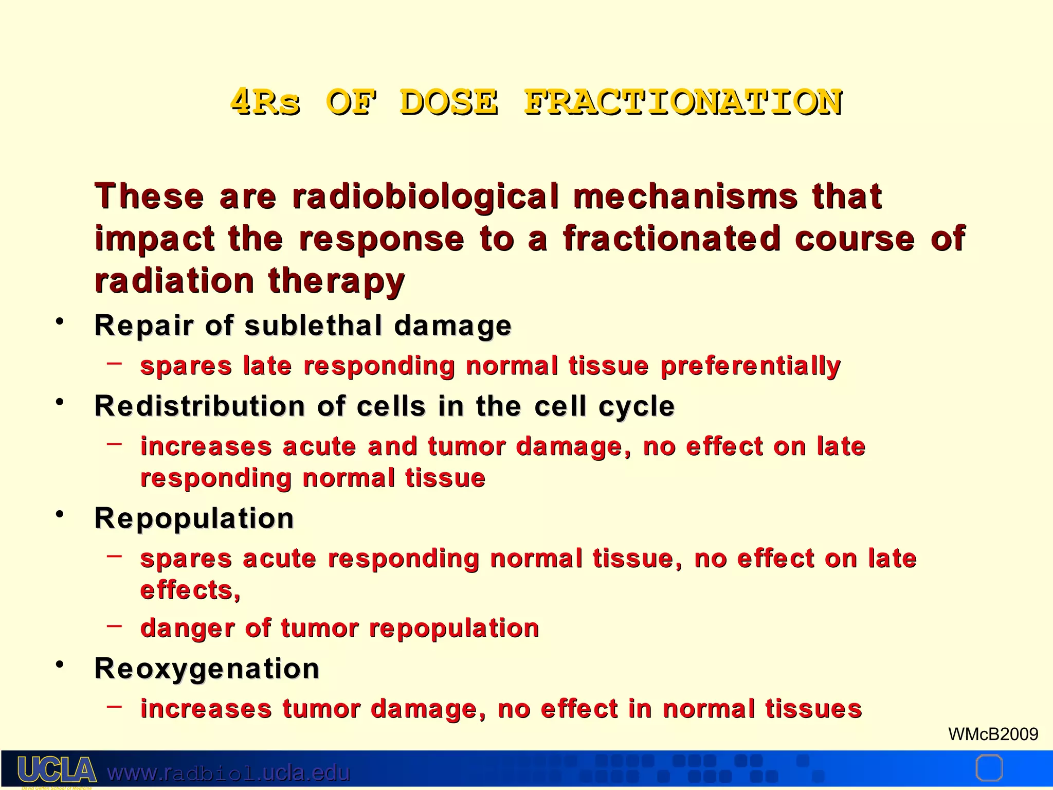
![What are α/β ratios for human
cancers?
In fact, for some tumors e.g. prostate, breast, melanoma, soft tissue
sarcoma, and liposarcoma α/β ratios may be moderately low
Prostate
– Brenner and Hall IJROBP 43:1095, 1999
• comparing implants with EBRT
∀ α/β ratio is 1.5 Gy [0.8, 2.2]
– Lukka JCO 23: 6132, 2005
• Phase III NCIC 66Gy 33F in 45days vs 52.5Gy 20F in 28 days
• Compatible with α/β ratio of 1.12Gy (-3.3-5.6)
Breast
– Owen, J.R., et al. Lancet Oncol, 7: 467-471, 2006 and Dewar et al JCO,
ASCO Proceedings Part I. Vol 25, No. 18S: LBA518, 2007.
• UK START Trial
– 50Gy in 25Fx c.w. 39Gy in 13Fx; or 41.6Gy in 13Fx [or 40Gy in 15Fx (3
wks)]
• Breast Cancer α/β = 4.0Gy (1.0-7.8)
• Breast appearance α/β = 3.6Gy; induration α/β
If fractionation sensitivity of a cancer is similar to = 3.1Gy
dose-limiting
healthy
tissues, it may be possible to give fewer, larger fractions without
compromising effectiveness or safety
WMcB2009
www.radbiol.ucla.edu](https://image.slidesharecdn.com/radiobiologybehinddosefractionation-131220073848-phpapp01/75/Radiobiology-behind-dose-fractionation-19-2048.jpg)


The document discusses the radiobiology behind dose fractionation in radiation therapy. It provides an overview of the linear quadratic model which describes how cell survival changes with dose and is used to determine biologically equivalent doses for different fractionation schedules. The model assumes equal effect per fraction but may not be accurate at high or low doses. Fractionation takes advantage of the four R's - repair, repopulation, redistribution, and reoxygenation - to better kill tumors while sparing normal tissues. The alpha/beta ratio indicates a tissue's sensitivity to fractionation and is used to estimate equivalent total doses for different fraction sizes.


