
Recommended
More Related Content
Similar to Normal abd xray rjj.pptx
Similar to Normal abd xray rjj.pptx (20)
More from rohanjohnjacob
More from rohanjohnjacob (20)
Recently uploaded
Recently uploaded (20)
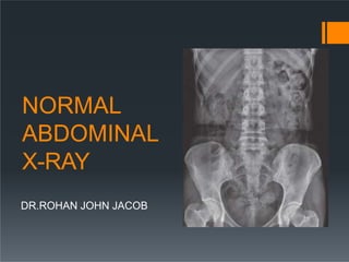
Normal abd xray rjj.pptx
- 2. PLANES AND REGIONS EXTEND: Inferior surface of diaphragm (superior) to the pelvic inlet (inferior) and contained by muscles of abdominal walls. PLANES: Divided into nine regions by two transverse and two parasagittal planes I. Transpyloric plane: midway between the suprasternal notch and the symphysis pubis (level of L1 vertebra and tips of Rt and Lt 9th CC) II. Transtubercular plane: level of tubercles of iliac crest and upper border of L5 III. 2 X Parasagittal planes: run at Right angles to the transverse planes vertically passing through a point midway between ASIS and symphysis pubis on each side in the mid clavicular line.
- 3. REGIONS:
- 4. x- Five basic densities on rays Gas: Black Fat: Dark grey Soft tissue: Light grey Bone / calcification: White Metal: Intense white
- 5. Abdominal Organs Liver right upper quadrant extends to the hemidiaphragm and past the midline Spleen left upper quadrant extends to the hemidiaphragm Its lower pole may be outlined by fat Measurement of its length from the dome of the diaphragm to the tip. This is usually less than 14 cm Relationship of the spleen to the ninth, tenth and eleventh ribs
- 6. Normal gallbladder or biliary system are not visible. Gas may be seen in the extrahepatic ducts in the elderly where the ampullary tone is low, after sphincterotomy, or after surgical anastomosis of bile ducts to small bowel Pancreas is not visible unless calcified. If calcification is distributed throughout the gland it is seen as a transverse structure at L 1 level, with a larger head on the right side and a body and tail extending to the left and upwards. Psoas muscle symmetrical triangles either side of the lumbar spine Arise from the transverse processes of lumbar vertebrae and combine with iliacus muscles to insert to lesser trochanter of femur narrowest near the diaphragm, widest at the pelvis
- 7. Stomach left of midline, beneath hemidiaphragm Gastric fundus fixed in location: within 2.5cm of left hemidiaphragm. sometimes just a small volume of gas in the fundus do not mistake a rim of gas for pneumoperitoneum Kidneys sit on the psoas muscles at level of T12 to L3 often just see the rounded lower pole Perirenal fat often makes part or all of the renal outlines visible Renal size is variable, with a normal range of 10 – 15 cm on a radiograph or approximately three-and-a-half vertebral bodies in height The left kidney is usually larger, but a difference in size of more than 2 cm is abnormal The kidneys are relatively larger in the child (approximately four vertebral bodies in height) Adrenal glands visible only if calcified.
- 8. Small bowel less than 3 cm wide tends to be central only seen if it contains gas 3 or more air fluid levels - abnormal mucosal folds (valvulae conniventes) traverse the bowel lumen Large bowel less than 6 cm wide, caecum and sigmoid up to 9 cm peripheral ascending and descending colon in fixed positions laterally transverse colon and sigmoid variable position on a mesentery Haustral folds do not go all the way across the lumen Any air fluid levels – abnormal (?) Numerous gas – fluid levels may be normal and 18% of normal films have fluid levels in the caecum contains faeces - mottled appearance THE 3/6/9 RULE
- 9. VALVULAE CONNIVENTES HAUSTRAL FOLDS Faecoliths or fluid levels of the appendix may be visible on plain films of the abdomen in the right iliac fossa in approximately 10% of individuals.
- 10. Haustra: I. The sacculation of the colon by the taeniae coli gives rise to septa called haustra II. The haustra are fixed anatomical structures in the proximal colon, but in the distal colon require active con- traction for their formation III. Haustra may be absent distal to the midtransverse colon.
- 11. Normal portal veins are not visible • Gas in the portal vein and its radicles may occur in cases of ischaemic bowel • Portal vein gas may also be seen in well patients after insertion of feeding tubes into the jejunum because of physical mucosal damage caused by tunnelling of the tube. Lung bases pulmonary vessels in the bases projected over upper abdomen Also look for free intra abdominal air below the diaphragm, costophrenic angles, or for a raised or flattened diaphragm.
- 12. Bladder: has variable appearance depending on how full it is. It has the same density as other soft tissue structures, due to its water content.
- 14. Bones and Joints Spine lower thoracic and lumbar spine should be of similar height intervertebral disc spaces should be similar spinous processes should be visible Lower ribs Sacrum and pelvis Sacroiliac Joints And Hip Joints are often visualised on abdominal radiographs. Make sure that you look at the bones to check for other causes of abdominal pain. Evidence of discitis, bony metastases etc. Bones can be used as landmarks for invisible soft tissue structures. E.g. the transverse processes of the lumbar vertebrae(L2 to L5) act as landmarks for the course of the ureters. The vesico-ureteric junctions are located at the level of the ischial spines.
- 15. Vessels Aorta is visible only if calcified It is then seen as linear calcification vertically in the midline and to the left The shadow of the inferior vena cava can be identified as it pierces the right hemidiaphragm and enters the heart. On a lateral chest radiograph it identifies a hemidiaphragm as being the right-sided one
- 16. Factors affecting position and surface marking of organs: a) Body build b) Phase of respiration c) Posture d) Age: loss of tone of abdominal musculature e) Pathology of organs f) Contents of hollow viscera g) Presence of abnormal mass h) Normal variants within the population
- 17. Normal Variant Riedel’s Lobe I. is a tongue-like, inferior projection of the right lobe of the liver beyond the level of the most inferior costal cartilage on cross-sectional images. II. It is not considered a true accessory lobe of the liver but an anatomical variant of the right lobe of the liver.
- 18. Referral criteria A preliminary evaluation of bowel gas in an emergent setting: 50% sensitivity for acute bowel obstruction Evaluation of radiopaque tubes and lines Evaluation for radiopaque foreign bodies Evaluation for post procedural intraperitoneal/retroperitoneal free gas Monitoring the amount of bowel gas in postoperative ileus Monitoring the passage of contrast through the bowel Monitoring renal calculi: 80 – 90% sens if radiolucent stone
- 19. Procedure The patient should be gowned with minimum clothing. Radiopaque materials (zippers, belts, etc.) should be removed. If relevant, enteric tube suction should be avoided before the study. Ideally, the patient's bladder should be emptied as well. Abdominal radiographs may be obtained in the radiology department or may be performed portably. Portable abdominal radiographs may be necessary due to patient immobility but are of much poorer quality. Gonadal shielding may be provided for men Views should generally include either the diaphragm or inferior pubic ramus
- 20. projections Basic: Antero-posterior - supine Alternative: Postero-anterior - prone Supplementary: * Antero-posterior – erect * Antero-posterior or Postero-anterior - left lateral decubitus * Lateral – dorsal decubitus * Anterior/Posterior obliques
- 21. AP Supine POSITION of patient: I. Supine with pelvis adjusted so that ASIS are equidistant from the tabletop. Arms placed alongside the trunk or above the head. III. II. CR casette positioned so that region below symphysis pubis included. Centre of image receptor located 1 cm below line joining iliac crests. IV. Ideally respiration arrested on full expiration.
- 22. Picture Criteria: I. Bowel pattern should be demonstrable with minimal unsharpness II. Diaphragm to symphysis pubis III. Lateral abdominal wall and peritoneal fat layer IV. Sharply demonstrated outline of psoas muscles, lower border of liver, kidney. V. Ribs and spinous processes of lumbar vertebrae VI. Whole of urinary tract VII. The abdomen should be free from rotation with symmetry of the: ribs (superior), iliac crests (middle), obturator foramen (inferior)
- 24. Free intraperitoneal gas may outline the umbilical ligaments and falciform ligament making them visible, thus making a diagnosis of pneumoperitoneum possible on a supine radiograph.
- 25. PA PRONE When kidneys are not of primary interest Reduces gonad dose POSITION of the patient: I. Prone with median sagittal plane at right angles to table II. Arms up beside head and both legs extended. III. CR, equipment setting and picture criteria same as supine projection.
- 26. PA ERECT Valuable projection in assessing air fluid levels, and free air in the abdominal cavity. Perforation of a hollow abdominal viscus: most sensitive to detect the presence of free gas in the abdomen IS ERECT CHEST X-RAY AND NOTABDOMEN ERECT .
- 27. POSITION of the patient: I. Patient stands with back against the receptor or vertical Bucky II. Legs separated well apart to maintain comfortable position III. Pelvis is adjusted so that theASIS are equidistant IV. Horizontal central ray directed perpendicular to midpoint at the level of iliac crests.
- 28. Picture criteria same as that of supine with both domes of diaphragm visible to visualize any free air in the peritoneal cavity. Air fluid levels
- 29. Lateral For identification and localization of foreign bodies. POSITION of the patient: I. Patient turned onto the side of examination with hands resting near the head II. Hips and knees flexed for stability III. Median sagittal plane parallel to table IV. Vertebral column positioned over midline of the table V. Immobilization band applied across pelvis VI. Cassette centered at the level of iliac crest VII. Vertical central ray directed to the center of the cassette. Picture criteria: The prevertebral space along with the abdominal aorta.
- 31. Lateral Decubitus Performed as an alternative to the PA erect view to assess for free gas in the abdominal cavity if the patient is unable to sit or stand. POSITION of the patient: I. Patient in lateral recumbent position II. Elbows and arms flexed and hands resting near head III. Cassette positioned in vertical bucky against the posterior aspect of the trunk. IV. Central ray is directed perpendicular to the midpoint at the level of iliac crest with x-ray tube horizontally
- 32. Picture Criteria: elevated lateral abdominal wall included on the image to detect any free intraperitoneal gas.
- 33. Dorsal Decubitus Used when it is unsafe to perform both a PAerect or a lateral decubitus view This projection requires no patient movement. Xray beam: 5 cm above the iliac crests at the midcoronal plane of the patient Picture Criteria: III. I. The anterior abdominal wall and the diaphragms are included on the image to detect any free intraperitoneal gas. II. There should be no blurring of the bowel gas due to respiratory motion. Due to the high exposure of this examination and the need to demonstrate soft tissue, the use of an aluminium filter over the anterior portion of the patient is advantageous to even out density and filter out higher energy x-rays
- 35. Pediatric Abdominal X-ray Pockets of gas scattere in several areas such as Small bowel Colon Rectum No excessive dilated bowel No air fluid levels
- 36. Contraindications Pregnancy is a relative contraindication I. Ten day rule : Whenever possible, one should confine the radiological examination of the lower abdomen and pelvis to the 10-day interval following the onset of menstruation. Now this is applied only to examinations falling under high dose. II. 28 day rule: In case if the women confirms she is certain she is not pregnant and the LMP is within 28 days, it is regarded as safe.
- 37. Things to look for Name, Date Position of film and view Adequate area covered or not Bowel preparation Pre- Peritoneal fat lines Visualized organs are normal in size Visualized bones and joints are normal Visualized shadows Any Radio opacity Any artifacts Any calcification