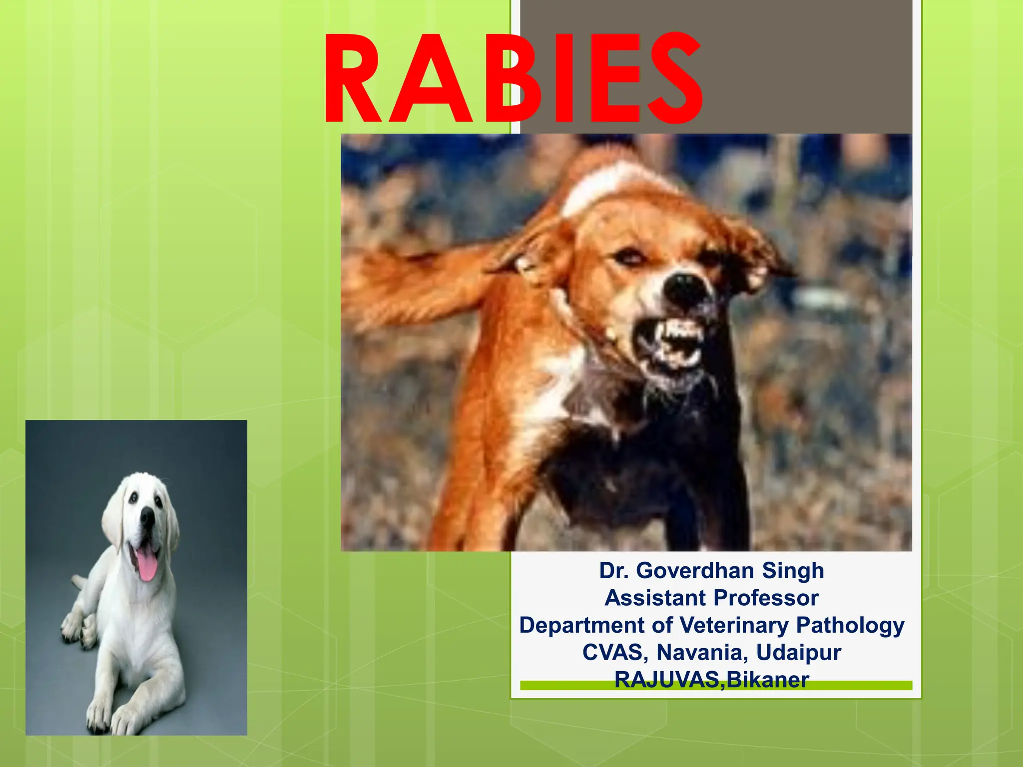
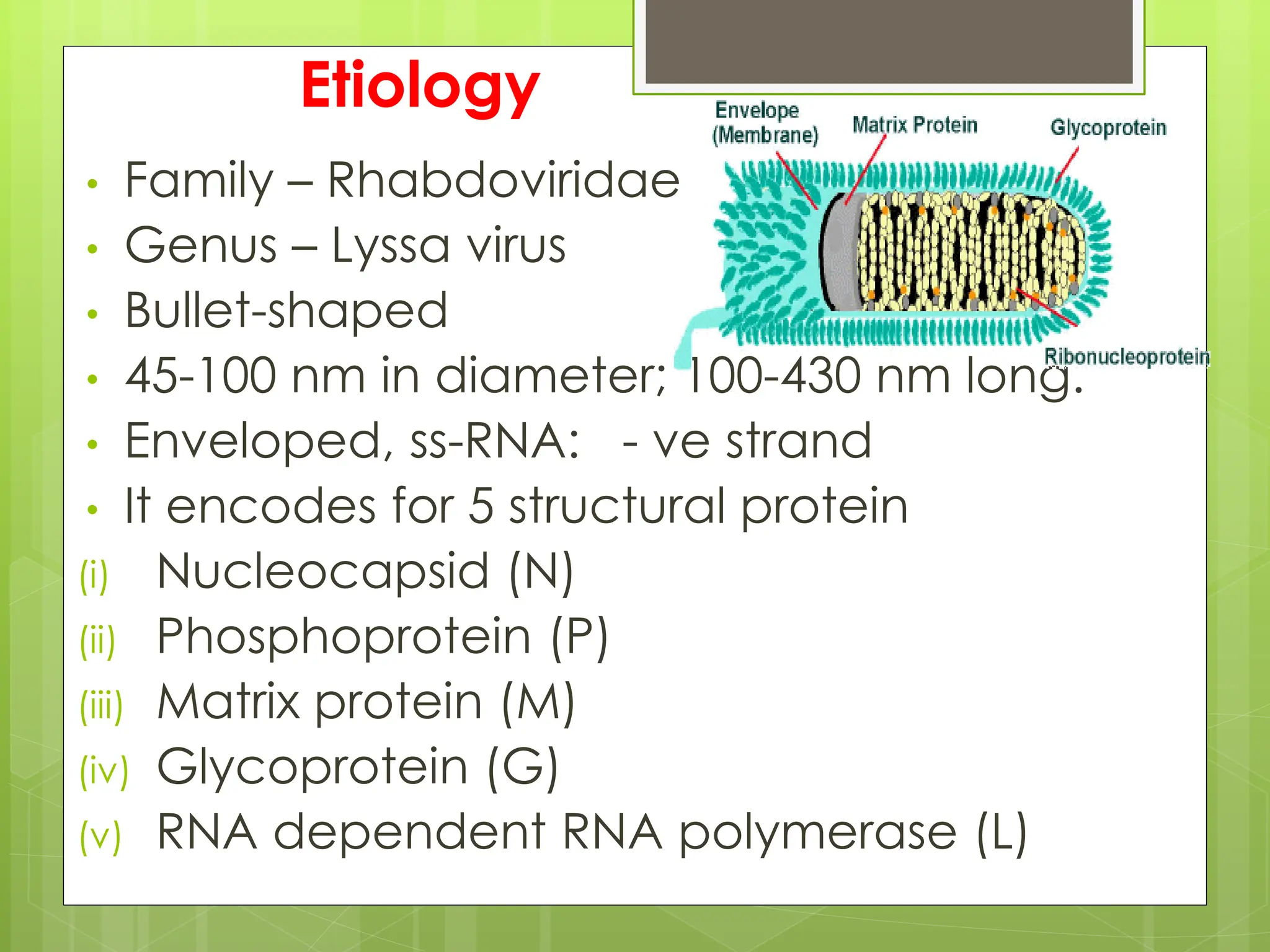
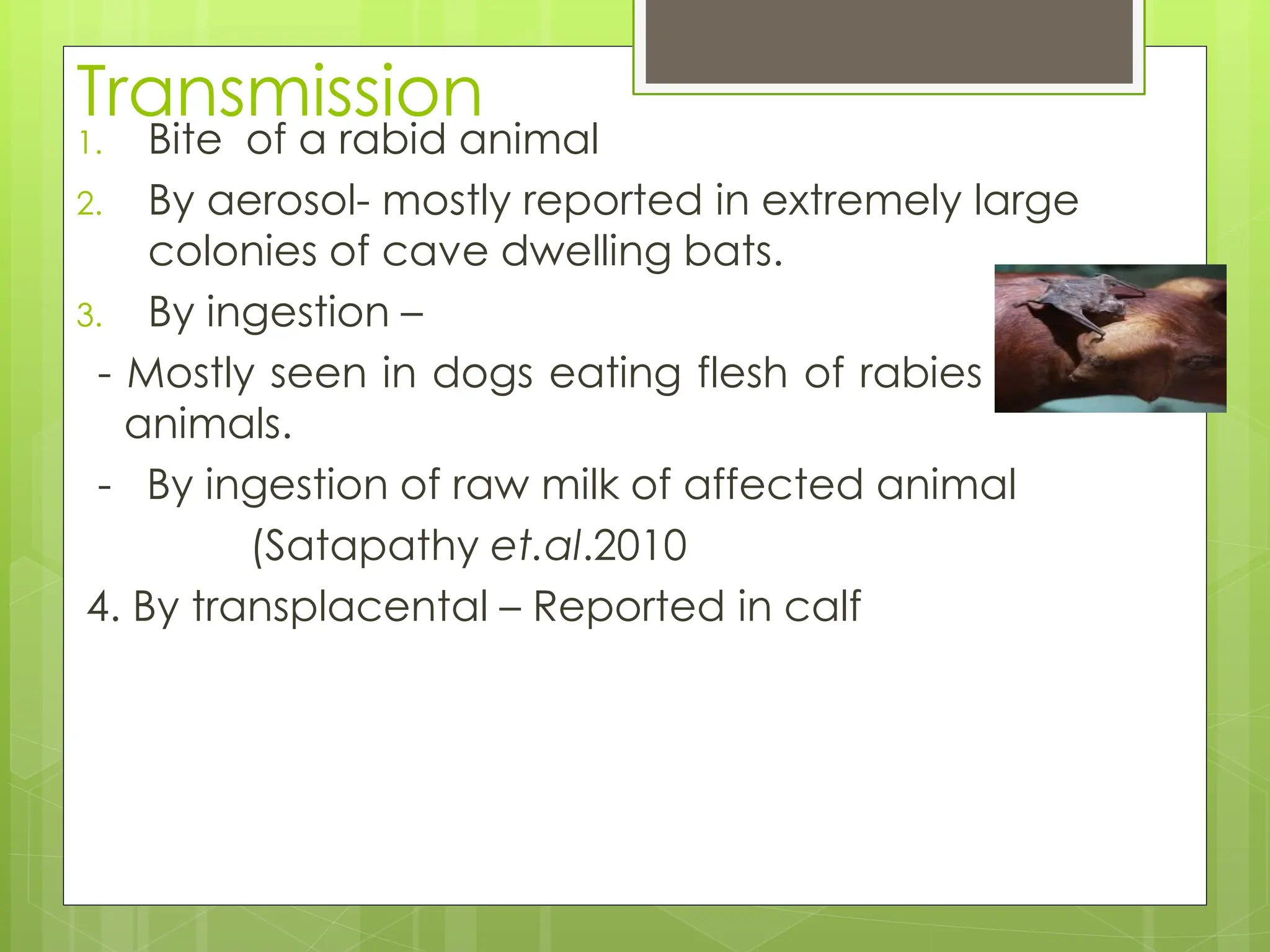
2. Synonyms- HYDROPHOBIA, LYSSA,
JALATANKA
Rabies is an acute,viral encephalomyelitis
(inflammation of both brain and
spinal cord), which affects all warm-
blooded animals, including humans.
3. Highly fatal disease.
Mortality rate being close to 100%.
Rabies occurs throughout the world.
A few countries are free of the disease by virtue of
their island status, or due to successful eradication
programmes, or because of strict quarantine
regulations.
4. New Zealand and Australia never had the
disease.
Britain, Hawaii, Japan and Scandinavia
are currently free of the disease.
In United States, Canada, and Western
Europe, rabies in dogs has been
effectively controlled through
vaccination.
5.
6. Etiology
• Family – Rhabdoviridae
• Genus – Lyssa virus
• Bullet-shaped
• 45-100 nm in diameter; 100-430 nm long.
• Enveloped, ss-RNA: - ve strand
• It encodes for 5 structural protein
(i) Nucleocapsid (N)
(ii) Phosphoprotein (P)
(iii) Matrix protein (M)
(iv) Glycoprotein (G)
(v) RNA dependent RNA polymerase (L)
7. 7 different genotype of rabies virus
reported.
UV light and heat destroy virus quickly.
Rabies virus can remain viable in carcass
for several days at 20 °C.
Storage at ultra low temperature (-30°c
to - 80 °c ) prolongs viral activity for
years in untreated frozen samples.
8. Transmission
1. Bite of a rabid animal
2. By aerosol- mostly reported in extremely large
colonies of cave dwelling bats.
3. By ingestion –
- Mostly seen in dogs eating flesh of rabies affected
animals.
- By ingestion of raw milk of affected animal
(Satapathy et.al.2010
4. By transplacental – Reported in calf
9. Host and range
Dog, and cat, act as a principal source of
infection.
Foxes, wolves, skunks, blood-sucking
vampire bats, insectivorous and
frugivorous (fruit-eating) bats, racoons,
mongoose, and squirrels may provide the
major source of infection in countries
where domestic carnivore are well
controlled.
11. • The rabies virus is maintained by interrelated cycle-
• Urban cycle rabies: dogs,cats
• Sylvatic cycle rabies: foxes, wolves, bats etc.
• Rabies in humans results from the bite of a rabid
dog, cat, fox, or wolf.
• These animals also transmit the disease to cattle,
horses, and sheep, which seldom spread it further.
• Bats are the important species in which
symptomless carriers are known to occur.
Multiplication of the virus without invasion of the
nervous system is known to occur in fatty tissues in
bats, and may be the basis of the 'reservoir'
mechanism known to occur in this species.
17. Symptoms
• Incubation period- vary from 10 days to 3 months in dogs
• There are two types – (1) Furious form (2) Dumb form
Furious form – can be divided into two stages
a) Stage of melancholy- Change in behavior, unusual violence
and frenzy behavior, bite inanimate objects, alert condition,
eat non-edible object e.g. stone, bone, grass pupils get
dilated, altered facial expression.
b) Stage of excitement- Becomes aggressive due to excitability
& irritability, hide in dark place due to photophobia,
hydrophobia, champing of jaw & dribbling of saliva, lick
their Genital & show sign of heat.
• At the end, the lower jaw will hang, head will drop down,
ascending paralysis, coma & death.
18. Dumb form:–
It is also known as PARALYTIC FORM .
In this form, paralysis of lower jaw, tongue, larynx & hind
quarters.
Dog cannot bite, saliva remain infective.
Peculiar voice is produced known as HOWL. (deep cry)
Dog used to seek solitude and appear sluggish &
morose.
Lasts for 1-7 days Coma and finally death..
22. Sign in horse -
Weakness and lameness is initial sign.
may show furious form & become virulent &
uncontrollable fall & roll on the ground
Shows signs of colic
chew foreign object drooling of saliva &
convulsion, sexual excitement
23. Cattle
Rabies in cattle follows the same general pattern.
Those with the furious form are dangerous,
attacking and pursuing man and other animals.
Lactation stops abruptly in dairy cattle.
Dribbling of urine (frequent urination)
A most typical clinical sign in cattle is a
characteristic bellowing (making a deep loud cry).
This may continue intermittently until shortly before
death.
24. Rabies in wild animals
Symptoms
The incubation period is usually two to three weeks.
Different species show different signs of the disease.
Expect variations even within the same species, because
few animals show all of the signs of rabies. Some signs are
subtle and easily missed.
unprovoked aggression ("furious" rabies). Some animals
may attack anything that moves, or even inanimate objects.
unusual friendliness ("dumb" rabies).
animal may stumble, fall, appear disoriented or
uncoordinated, or wander aimlessly.
25. Paralysis, often beginning in the hind legs or throat. Paralysis
of the throat muscles can cause the animal to bark, whine,
drool, choke, or froth at the mouth.
Vocalizations ranging from chattering to shrill screams.
Nocturnal animals may become unusually active during the day
Raccoons walk as if they're on very hot pavement.
Skunks, raccoons, foxes, and dogs usually display furious
rabies.
Bats often display dumb rabies, and may be found on the
ground, unable to fly.
28. Gross examination
No gross changes are detected in the CNS with Rabies
In few cases post-mortem revealed severe congestion of meninges,
cerebellum and cerebral hemisphere with hemorrhage.
Edema of the cerebellum and cerebral hemisphere, in addition to,
widening of gyri and narrowing of sulci of cerebral hemisphere were seen.
Congestion of meningeal blood vessels with flattening gyri and narrowing
sulci (arrow);
29. Congestion and edema in the wall of the lateral ventricle (arrow)
with bulging into the lumen.
30. Gross picture of rabbid cow showing congestion in cerebral
hemisphere
31. Microscopical examination
The lesions of rabies are microscopic, limited to the central
nervous system, and extremely variable in extent.
Necrosis of neurons with specific cytoplasmic inclusion
bodies in the affected nerve cells.
Spherical cytoplasmic inclusion bodies with specific
tinctorial (staining) characteristics Negri bodies present in
neurons of dogs, cats.
Negri bodies are not always present in rabies. Certain strains
of rabies virus do not produce inclusion bodies, indicating that
Negri bodies are not necessary for viral replication.
32. Negri bodies are formed in only 20%(Appx.) of cases.
In the dog, they are found mostly in the hippocampus.
In cattle more numerous in the Purkinje cells of the cerebellum.
In impression smears, Seller's stain is effective. The inclusion
body is bright red or magenta.
34. In some cases, there is diffuse non suppurative encephalitis
characterized by perivascular cuffing, neuronophagic nodules
and other changes of destruction of neurons throughout the
brain. These changes are particularly prominent in the brain
stem, hippocampus and the gasserian ganglia. (Gasserian
ganglion lies on the sensory root of the trigeminal nerve, and
from it originate the three branches - ophthalmic, maxillary,
mandibular).
Lesions in the gasserian ganglia consists of the collections of
proliferating glial cells encroaching on the neurons and
replacing them. These collections of proliferating glial cells are
known as "Babes' nodules".
36. Collection of samples:
Usually the brain is collected following the opening of the skull in a
necropsy room, and the appropriate samples are collected.
This step may be hazardous if laboratory technicians are not fully trained,
or under field conditions.
In such cases, there are two possible methods of collecting some brain
samples without opening the skull:
Occipital foramen route for brain sampling:
A 5 mm drinking straw or a 2 ml disposable plastic pipette is introduced into
the occipital foramen in the direction of an eye. Samples can be collected
from the rachidian bulb, the base of the cerebellum, hippocampus, cortex,
and medulla oblongata.
Retro-orbital route for brain sampling:
In this technique, a trocar is used to make a hole in the posterior wall of the
eye socket, and a plastic pipette is then introduced through this hole. The
sampled parts of the brain are the same as in the former technique, but
they are taken in the opposite direction.
38. 1. Direct Microscopy: Histological Identification of
Characteristic Cell Lesions
2. Demonstration of Viral Antigen
2.1. Fluorescent Antibody Technique (FAT)
2.2. Rapid Rabies Enzyme Immunodiagnosis (RREID)
3. Virus Isolation
3.1. Mouse inoculation test
3.2.Rapid tissue culture infection test(RTCT)
4. Demonstration of Antibodies
4.1. The Mouse Neutralization Test (MNT),
4.2.Rapid Fluorescent Focus Inhibition Test (RFFIT)
4.3.Fluorescence Antibody Virus Neutralization Test (FAVN)
39. 1. Direct Microscopy: Histological Identification of Characteristic
Cell Lesions
Infected neuronal cells reveal aggregates of viral particles
“Negri bodies” intracytoplasmic inclusion bodies .
Demonstrated by histological tests (Seller’s Technique) on
smears taken from various areas of the brain.
40. simple, rapid test,
Seller’s method on unfixed tissue smears has a very
low sensitivity and is only suitable for fresh specimens.
Limitation
Techniques that stain sections of paraffin embedded
brain tissues are time consuming, less sensitive, and
more expensive.
Histological techniques are much less sensitive than
immunological methods, especially in the case of
autolysed specimens, and are no longer
recommended for primary diagnosis, both in humans
and animals
41. 2. Demonstration of Viral
Antigen
2.1 Fluorescent Antibody Technique (FAT)
Most widely used test for postmortem rabies
diagnosis
Recommended by both World Health
Organization (WHO) and World Organization for
Animal Health.
Developed by Goldwasser and Kissling in 1957
It involves demonstration of the rabies virus
nucleoprotein antigen (N) in fresh brain smears
of a suspected rabies
42. Specificity and sensitivity of the test almost approach
99%
FAT can also be applied to specimens preserved in 50%
glycerol saline after rigorous washing of the specimens
with normal saline
FAT on formalin-fixed and digested samples is always
less reliable and more cumbersome than when performed
on fresh tissue
Fluorescent antibody technique
(FAT) on human brain smear
positive for rabies.
43. FAT can also be performed on corneal
smears and nuchal skin biopsy in
suspected cases, however it has been
found to have limited reliability and low
sensitivity for ante mortem diagnosis of
rabies.
44. 3. Virus Isolation
Virus isolation is required for confirmatory diagnosis,
especially when FAT gives an uncertain result and more
importantly for molecular characterization of viruses in a
geographical location and for tracing the origin of the virus if
rabies occurs in a rabies-free area.
Two techniques can be employed for this purpose
3.1. Mice inoculation technique (MIT)
3.2. Rapid tissue culture infection test (RTCT)
45. 3.1. Mouse Inoculation Test
Three-to-ten mice, 3-4 weeks old (12–14 g), or a litter of 2-day-old
newborn mice
inoculated intracerebrally with the clarified supernatant of a 10–20%
(w/v) homogenate of brain material in an isotonic buffered solution
containing antibiotics.
The inoculated mice are observed daily for 28 days
they develop typical signs and symptoms of rabies any time after
5–7 days depending on the incubation period
Further confirmation of the diagnosis can be made by extracting the
brain of the diseased mouse and subjecting this to FAT.
Disadvantage - long interval before a diagnosis can be made since
the inoculated mice need to be kept under observation for 28 days as
some wild viruses may have a very long incubation period.
46. 1. Demonstration of Viral Antigen
a) Direct Rapid Immunohistochemical Test (dRIT)
b) Indirect Rapid Immunohistochemistry Test (IRIT)
c) Immunochromatographic Techniques
2. Nucleic Acid Detection Techniques
a) Reverse Transcriptase PCR (RT-PCR)
b) Real-Time PCR
c) Nucleic acid sequence-based amplification (NASBA)
technique
d) Loop-Mediated Isothermal Amplification (LAMP)
47. Vaccination
Vaccination against rabies is used in two distinct
situations:
to protect those who are at risk of exposure to rabies, i.e.
preexposure vaccination;
to prevent the development of clinical rabies after
exposure has occurred, usually following the bite of an
animal suspected of having rabies, i.e. post-exposure
prophylaxis.
Pre exposure vaccination-
Type of vaccine: Modern cell-culture (Human diploid cell
culture) or embryonated-egg vaccine
Number of doses: Three, one on each of days 0, 7 and
21 or 28, given i/m (1 or 0.5 ml/dose depending on the
vaccine) or i/d (0.1 ml/inoculation site)
48. Post-exposure prophylaxis.
Passive immunization- The dose for HRIG is 20 IU/kg
body weight and for ERIG and F(ab’)2 products 40 IU/kg
body weight. The full dose of rabies immunoglobulin, or as
much as is anatomically feasible, should be administered
into and around the wound site.
Active immunization- Cell-culture or embryonated - egg-
based rabies vaccines
The five-dose regimen is administered on days 0, 3, 7, 14
and 28 into the deltoid muscle.(I/M)