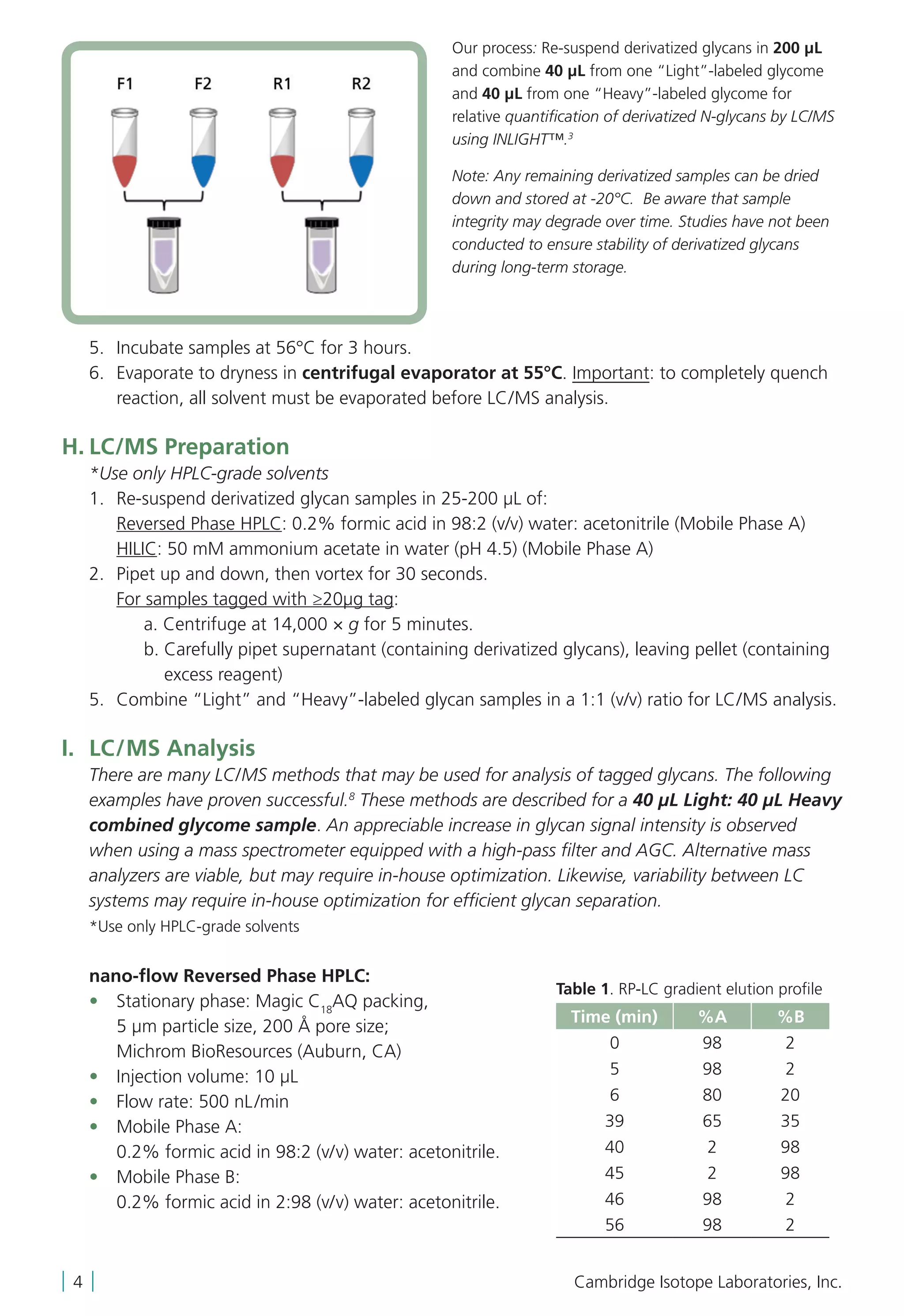
This document provides instructions for using the INLIGHTTM Glycan Tagging Kit for comparative quantification of N-linked glycans. The kit contains light and stable isotope-labeled hydrazide reagents for derivatizing free N-glycans isolated from glycoproteins. The protocol describes steps for denaturing glycoproteins, enzymatically cleaving glycans, purifying glycans using solid phase extraction, derivatizing glycans with the light and heavy reagents, and analyzing the derivatized glycans using liquid chromatography-mass spectrometry. Standard glycoprotein samples of fetuin and RNase B are recommended to optimize the method before analyzing complex biological samples.
![KIT CONTENTS
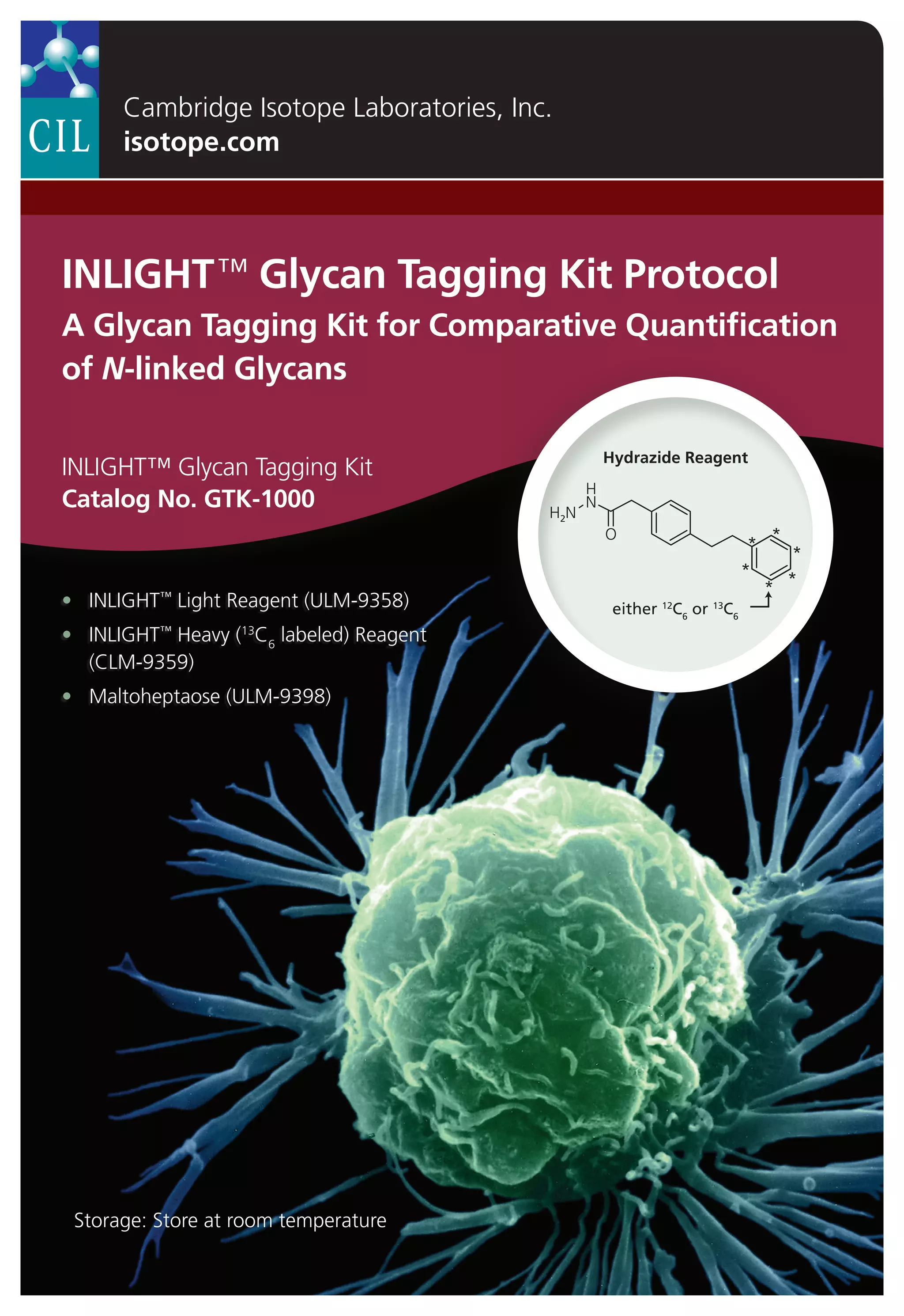
INLIGHT™ Strategy
N-Linked Glycan Cleavage and
Derivatization via Hydrazone Formation
INLIGHT Glycan Tagging Kit
GTK-1000
• INLIGHT™ Light Reagent, 5 × 0.25 mg (ULM-9358, provided)
• INLIGHT™ Heavy (13
C6
labeled) Reagent, 5 × 0.25 mg
(CLM-9359, provided)
• 1
Maltoheptaose, 5 × 10 μg (ULM-9398, provided)
REQUIRED REAGENTS
Solvents Manufacturer Part No./Cat. No.
Acetonitrile – HPLC grade (ACN) Burdick Jackson AH015-4
Methanol – HPLC grade Burdick Jackson AH230-4
Water – HPLC grade Burdick Jackson AH365-4
Formic acid Sigma-Aldrich 56302
Acetic acid (50%) solution for HPLC Sigma-Aldrich 45754
Trifluoroacetic acid, Reagent Plus®
9 (TFA) Sigma-Aldrich T6508
Ethanol (200 proof) HPLC grade Sigma-Aldrich 459828
Reagents Other Supplies Manufacturer Part No./Cat. No.
SPE graphitized carbograph columns [300 mg] Grace Discovery Sciences 210101
Ammonium bicarbonate Sigma-Aldrich A6141
Dithiothreitol 1 M aqueous solution Sigma-Aldrich 646563
2
Horseradish peroxidase (HRP) Type VI Sigma-Aldrich P8375
PNGase F – glycerol free [75,000 units] New England Biolabs P0705L
3
Fetuin New England Biolabs P6042S
4
RNase B New England Biolabs P7817S
INSTRUCTIONS FOR USE
A. Solution and Buffer Preparation
1. SPE Wash Solution (solvent #1) 1 Liter – 1 mL TFA in water (HPLC grade).
2. SPE Condition Solution (solvent #2) 1 Liter – 500 µL TFA, 800 mL ACN, 200 mL water
(HPLC grade).
3. SPE Elution Solution (solvent #3) 1 Liter – 1 mL TFA, 250 mL ACN, 750 mL water
(HPLC grade).
isotope.com | 1 |
Hydrazide Reagent
2-(4-phenethylphenyl)acetohydrazide:
N-glycan tagging reagent (see ref. 1)
(continued)](https://image.slidesharecdn.com/3ae2997d-cd0b-4fe8-88c0-81696b6dd2b5-151223220729/75/Protocol_GTK_glycan_booklet-3-2048.jpg)
![E. Glycan Purification by Solid-Phase Extraction (SPE)
Solvent #1 – SPE wash solution: 0.1% TFA in water
Solvent #2 – SPE conditioning solution: 0.05/80/20% (v/v/v) TFA/ACN/water
Solvent #3 – SPE elution solution: 0.1/25/75% (v/v/v) TFA/ACN/water
1. Condition SPE graphitized carbograph columns in the following order:
a) Two column volumes of solvent #1.
b) One column volume of solvent #2.
c) Two column volumes of solvent #1.
2. Re-suspend samples in 1 mL of solvent #1.
3. Briefly vortex to mix and spin down in a centrifuge.
4. When the last of the wash solution elutes, pour the reconstituted sample into the
SPE column.
5. Rinse sample vial with 1 mL of solvent #1, vortex, spin down in centrifuge, and pour into
the same cartridge as the initial reconstitution. Repeat rinse once more.
6. Allow meniscus to reach top of filter.
7. Wash loaded column (containing glycome solution) with 40 mL of solvent #1.
8. Elute the glycans with 1 mL of solvent #3 and collect fraction (Fraction A).
9. Repeat 3 times (Fractions B, C D).
10. Freeze collected fractions (4) at -80°C.
F. Combine SPE Fractions
1. Concentrate SPE fractions to dryness using centrifugal evaporator at 45°C (~5-6 hours).
2. Re-suspend Fractions A, C D with 75 µL HPLC water; briefly vortex to mix and spin down
in a centrifuge.
3. Combine suspended fractions into Fraction B.
4. Freeze at -80°C.
5. Concentrate to dryness at 45°C (~2-3 hours).
6. After sample is completely dry, store at -20°C until day of LC/MS analysis.
G. Glycan Derivatization
Note: Each vial contains 0.25 mg of INLIGHT™ reagent [2-(4-phenethylphenyl)acetohydrazide],
enough for up to five reactions, depending on the starting concentration of glycan. The
following quantity of tag has proven sufficient for derivatization of: 50 µg maltodextrin (linear
polysaccharide standard); 50 µg RNase B glycoprotein (1 glycosite; high mannose-type glycans);
100 µg fetuin glycoprotein (3 glycosites; complex type glycans), and 50 μL aliquots of human
plasma (approx. 30 mg/mL total protein based on Bradford assay). It is recommended that
laboratories optimize INLIGHT™ tag : glyco-substrate ratio to minimize excess tag and maximize
reaction efficiency ( 95%).
1. Make a 0.25 mg/mL solution of each: INLIGHT™ Light Reagent and INLIGHT™ Heavy
Reagent by dissolving each reagent in derivatization solution; 75/25 (v/v) methanol:
50% acetic acid.
2. Re-suspend purified glycan samples in either:
a) 200 μL Light Reagent solution (natural 2-(4-phenethylphenyl)acetohydrazide)
b) 200 μL Heavy Reagent solution (13
C6
labeled 2-(4-phenethylphenyl)acetohydrazide)
3. Pipet up and down, then vortex to further mix, and spin down by centrifuge.
4. Optional: Add 10 µg maltoheptaose (polysaccharide standard) to each reaction, prepared
fresh in derivatization buffer.
isotope.com | 3 |
(continued)](https://image.slidesharecdn.com/3ae2997d-cd0b-4fe8-88c0-81696b6dd2b5-151223220729/75/Protocol_GTK_glycan_booklet-5-2048.jpg)