
Recommended
More Related Content
What's hot
What's hot (20)
Similar to Antibody Glycan Analysis with Normal Phase PhyTip Columns
Similar to Antibody Glycan Analysis with Normal Phase PhyTip Columns (20)
More from Chris Suh
More from Chris Suh (10)
Recently uploaded
Recently uploaded (20)
Antibody Glycan Analysis with Normal Phase PhyTip Columns
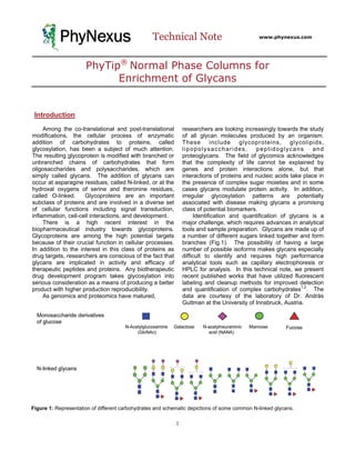
- 1. PhyNexus Technical Note www.phynexus.com PhyTip® Normal Phase Columns for Enrichment of Glycans Introduction Among the co-translational and post-translational researchers are looking increasingly towards the study modifications, the cellular process of enzymatic of all glycan molecules produced by an organism. addition of carbohydrates to proteins, called These include glycoproteins, glycolipids, glycosylation, has been a subject of much attention. lipopolysaccharides, peptidoglycans and The resulting glycoprotein is modified with branched or proteoglycans. The field of glycomics acknowledges unbranched chains of carbohydrates that form that the complexity of life cannot be explained by oligosaccharides and polysaccharides, which are genes and protein interactions alone, but that simply called glycans. The addition of glycans can interactions of proteins and nucleic acids take place in occur at asparagine residues, called N-linked, or at the the presence of complex sugar moieties and in some hydroxal oxygens of serine and theronine residues, cases glycans modulate protein activity. In addition, called O-linked. Glycoproteins are an important irregular glycosylation patterns are potentially subclass of proteins and are involved in a diverse set associated with disease making glycans a promising of cellular functions including signal transduction, class of potential biomarkers. inflammation, cell-cell interactions, and development. Identification and quantification of glycans is a There is a high recent interest in the major challenge, which requires advances in analytical biopharmaceutical industry towards glycoproteins. tools and sample preparation. Glycans are made up of Glycoproteins are among the high potential targets a number of different sugars linked together and form because of their crucial function in cellular processes. branches (Fig.1). The possibility of having a large In addition to the interest in this class of proteins as number of possible isoforms makes glycans especially drug targets, researchers are conscious of the fact that difficult to identify and requires high performance glycans are implicated in activity and efficacy of analytical tools such as capillary electrophoresis or therapeutic peptides and proteins. Any biotherapeutic HPLC for analysis. In this technical note, we present drug development program takes glycosylation into recent published works that have utilized fluorescent serious consideration as a means of producing a better labeling and cleanup methods for improved detection product with higher production reproducibility. and quantification of complex carbohydrates1,2. The As genomics and proteomics have matured, data are courtesy of the laboratory of Dr. András Guttman at the University of Innsbruck, Austria. Figure 1: Representation of different carbohydrates and schematic depictions of some common N-linked glycans. 1
- 2. PhyNexus PhyTip Normal Phase Columns Technical Note Figure 2: Flow chart of sample preparation for capillary gel electrophoresis profiling of N-glycosylation of standard glycoprotein samples. For the maltooligosaccharide ladder standard the first two steps were not applied, the sample preparation is started with the labeling process. Materials and Methods Fluorophore labeling Separation Solution (eGene). Separations were All reagents are supplied by Sigma Aldrich unless carried out at 2-8 kV. specified. Maltooligosaccharide ladder (Grain Processing Corporation) was labeled with 1 μL 0.2 M Glycoprotein analysis 8-amino-1,3,6-pyrenetrisulfonic acid (APTS) in 15% For analysis of the glycan composition of acetic acid and 1 μL 1 M NaBH3CN in tetrahydrofuran glycoproteins, the glycans are first cleaved off of the and incubated at 55º for 2 h. The reaction was protein. The proteins analyzed were IgG, RNase B, stopped by addition of 100 μL water. Unreacted, fetal bovine fetuin (FET) asialofetuin, and α-1-acid- excess APTS was removed by three different glycoprotein (AGP). 0.1 mg protein in 10 μL water methods. First, filter plates (Millipore) were loaded was denatured by addition of Endoglycosidase buffer with 100 μL Sephadex G10 gel filtration media and (New England Biolabs) and incubation at 100º for 10 conditioned. 200 μL APTS reaction mixture was minutes was followed by digestion with 2 units of loaded and centrifuged at 2500 rpm for 15 minutes. PNGaseF at 37º for 2 hours. The deglycosylated Second, the PhyNexus ME purification system was protein was removed by cold ethanol precipitation used to condition PhyTip columns containing 160 μL and centrifugation at 11,000 g for 10 minutes. The of Sephadex G10 gel filtration media with 50% glycan-containing supernatants were dried by a acetonitrile (ACN). 200 μL APTS reaction mixture centrifugal vacuum evaporator. The released glycans was loaded onto the column and 50% ACN was used were labeled and analyzed in the same manner as to chase and release the labeled the maltooligosaccharide ladder. maltooligosaccharide. Third, the PhyNexus ME purification system was used to condition PhyTip Boronic acid lectin affinity chromatography (BLAC) columns containing 10 μL of normal phase resin with 200 μL PhyTip columns containing 5 μL of BLAC 95% ACN. 200 μL of the APTS reaction mixture was captured with 8 cycles of back-and-forth flow followed resin was manufactured by mixing 2.5 μL agarose- by 24 wash cycles in 95% ACN. The labeled immobilized concanavalin A and 2.5 μL boronic acid maltooligosaccharides were eluted with 350 μL 20% agarose beads. Using the PhyNexus ME instrument, ACN. the PhyTip columns were washed with 100 μL water, 100 μL 20mM potassium phosphate pH 2.85 and Capillary electrophoresis analysis again with 100 μL water. The PhyTip columns were Maltooligosaccharides were diluted 10-fold in conditioned in 100 μL binding buffer (50 mM taurine, pure water and analyzed on a CarbCE multicapillary 1 mM CaCl2, 1mM MnCl2, 20mM MgCl2, 0.05% NaN3, electrophoresis system equipped with a blue light- pH 8.7. 100 μL human serum was diluted in 100 μL emitting diode excitation source of wavelength 460- binding buffer and the glycoproteins were captured 470 nm (eGene) using a 12-channel gel cartridge using the PhyTip columns followed by wash in (GCK-CARB). Samples were injected binding buffer and elution with 100 μL 100 mM HCl. electrokinetically (2 kV, 20 s). Emission was detected The glycoproteins were dried and prepared for by a photodiode using a 520 nm long-pass filter. PNGaseF digestions and APTS labeling and clean-up Separations using 10 cm or 30 cm capillary lengths as described. (20 μm i.d.). The capillaries were filled with CARB 2
- 3. PhyNexus PhyTip Normal Phase Columns Technical Note Figure 3: Comparison of methods for removal of unreacted derivatization reagent, APTS, from mal- tooligosaccharide ladder labeling reactions. CE electropherogram traces of unpurified, Sephadex G10 filter plate purified, Sephadex G10 PhyTip column purified, and nor- mal phase resin PhyTip column purified reaction mixtures are compared for efficiency of dye removal Normal phase PhyTip column purified and intensity of recovered G10 PhyTip column purified maltooligosacchar ides peaks. Adapted with per- G10 filter plate purified mission from Olajos et. Unpurified al. 1 2008 American Chemical Society. Results Removal of unreacted derivatization reagent Glycans are composed of monosaccharides. The similarity of the monosaccharide subunits and the ability to form branches makes identification of glycans very difficult (Fig. 1). Advanced analytical methods such as HPLC and capillary electrophoresis (CE) are required to perform the high-resolution separation. However, glycans lack chromophores making the detection of glycans a challenge. Researchers have addressed this problem by labeling glycans with fluorophore tags such as APTS, which provide fluorescence and charge that are quite amenable to a CE separation (Fig. 2). For efficient derivatization of glycans with tags, an excess of APTS is required. Unreacted APTS must be removed because co- migration of free dye with low molecular weight glycans interferes with quantification and identification. A number of strategies were tested for the ability to remove the excess dye and enrich for labeled glycans. A maltooligosaccharide ladder was labeled with APTS and split into several aliquots. The first reaction aliquot was not purified and run on a CE. The second was purified by a spin plate containing Sephadex G10 gel filtration media. The third and fourth aliquots were purified by the PhyNexus ME semi-automated purification system using pipette tip columns containing different separation media. The PhyTip columns contained either Sephadex G10 or a normal phase resin (Fig. 3). The unpurified sample and filter plate purification method shows a large dye front at that co-migrates with the low molecular weight oligosaccharides. The PhyTip columns containing G10 resin were able to remove unreacted APTS, but the enrichment of labeled Figure 4: CE electropherograms of normal phase PhyTip column purification of APTS-labeled glycans from IgG, RNaseB, FET and AGP using an effective capillary length of 10 cm (A) or 30 cm (B). Adapted with permission from Olajos et. al.1 2008 American Chemical Society. 3
- 4. PhyNexus PhyTip Normal Phase Columns Technical Note Results cont’d combination of a lectin, concanavalin A and a pseudo-lectin boronic acid3.4. This method, termed oligosaccharides was only slightly better than the filter BLAC was used to purify glycoproteins from serum plate method. The PhyTip columns containing samples of a healthy pool of individuals and from normal phase resin achieved both effective stage-four prostate cancer patients. The purification and high enrichment. glycoproteins were subject to PGNaseF digestion, APTS labeling, sample clean-up and CE analysis to Glycan composition analysis of glycoproteins compare the glycan patterns. Researchers A number of glycoproteins, immunoglobulin G discovered a number of peaks that are specific to (IgG), ribonuclease B (RNaseB), fetal bovine fetuin prostate cancer samples and set the groundwork for (FET) and α-1-acid-glycoprotein (AGP) were further study4. analyzed for their respective glycan content. The glycans were first cleaved off of the protein using Discussion peptide-N-glycosidase F (PNGaseF) followed by labeling with APTS and purification by PhyTip Advances in high-resolution separation methods columns containing normal phase resin. The purified such as HPLC and CE are a good solution for glycans were separated by CE of either 10 or 30 cm identification, quantification and profiling of glycans. capillary lengths (Fig. 4). Though these analytical tools are excellent separation methods. Detection of glycans poses an analytical Boronic acid lectin affinity chromatography challenge because of the lack of a natural (BLAC) chromophores/charged groups. Labeling strategies Glycoproteins present in human serum represent circumvent this problem by providing a simple and an attractive target for diagnostics discovery. efficient tagging procedure. Recent advances in Disease has frequently been correlated with changes sample cleanup using PhyTip columns containing in glycosylation of proteins including extra branching, normal phase resin allow users to reproducibly truncation and excessive amounts of certain process samples in a high-throughput fashion. monosaccharides. Researchers at the University of Furthermore, this purification strategy can be Innsbruck in Austria have developed a method for advanced for diagnostics discovery using the BLAC glycoproteomics work by performing an affinity method. These new tools are essential for the enrichment of glycoproteins from serum using a advance of the glycomics and glycoproteomics fields. References 1 Marcell Olajos, Peter Hajós, Guenther K. Bonn, András Guttman, “Sample Preparation for the analysis of complex carbohydrates by multicapillary gel electrophoresis with light-emitting diode induces fluorescence detection,” Analytical Chemistry (2008), 80, 4241-4246. 2 Bradley D. Prater, Kalyan R. Anumula, Jeff T. Hutchins, “Automated sample preparation facilitated by PhyNexus MEA purification system for oligosaccharide mapping of glycoproteins,” Analytical Biochemistry (2008), 392, 195-201. 3 Alex Monzo, Guenther K. Bonn, András Guttman, “Boronic acid-lectin affinity chromatography. 1. Simultaneous glycoprotein binding with selective or combined elution,” Analytical Bioanalytical Chemistry (2007), 389, 2097-2102. 4 Alex Monzo, Marcell Olajos, Lorenzo De Benedictis, Zuly Rivera, Guenther K. Bonn, András Guttman, “Boronic acid lectin affinity chromatography (BLAC). 1. Affinity micropartitioning-mediated comparative glycosylation profiling,” Analytical Bioanalytical Chemistry (2008), 392, 195-201. Ordering information For further support call PhyNexus at 408.267.7214 or e-mail support@phynexus.com For further information on PhyNexus products visit our website at www.phynexus.com PTR 92-05-09 Box of 96 PhyTip columns (200 µL volume) containing 5 µL of Normal Phase resin per column PTR 91-10-09 Box of 96 PhyTip columns (1000 µL volume) containing 10 µL of Normal Phase resin per column PTR 91-20-09 Box of 96 PhyTip columns (1000 µL volume) containing 20 µL of Normal Phase resin per column 4 Copyright © 2008, PhyNexus, Inc., All Rights Reserved