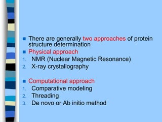
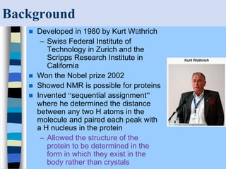
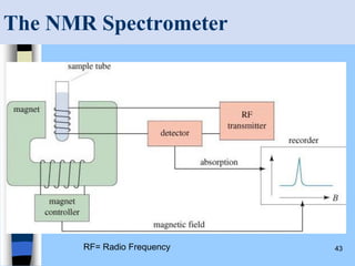
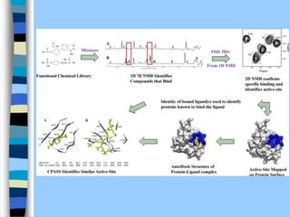
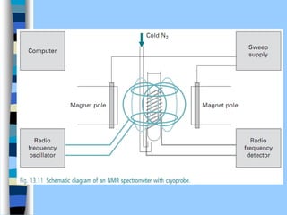
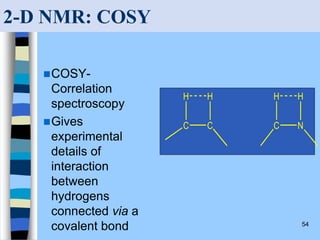
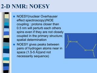
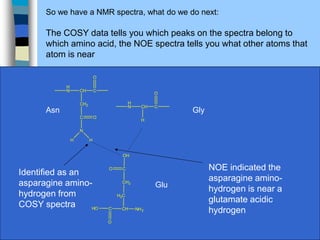
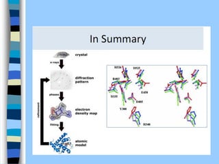
Protein Structure
determination
 There are generally two approaches of protein
structure determination
 Physical approach
1. NMR (Nuclear Magnetic Resonance)
2. X-ray crystallography
 Computational approach
1. Comparative modeling
2. Threading
3. De novo or Ab initio method
 proteins are formed by condensing the ɑ-amino group of one
amino acid or the imino group of proline with the ɑ-carboxyl
group of another, with the concomitant loss of a molecule of
water and the formation of a peptide bond
 Most proteins contain many hundreds of amino acids (ribonuclease is an
extremely small protein with only 103 amino acid residues)
 many biologically active peptides contain 20 or fewer amino acids, for example
oxytocin (9 amino acid residues), vasopressin (9), enkephalins (5), gastrin (17),
somatostatin (14) and lutenising hormone (10)
 The primary structure of a protein defines the sequence of the
amino acid residues and is dictated by the base sequence of the
corresponding gene(s)
 also defines the amino acid composition (which of the possible
20 amino acids are actually present) and content (the relative
proportions of the amino acids present)
 Secondary structure defines the localised folding of a
polypeptide chain due to hydrogen bonding
 includes structures such as the α-helix and β-pleated sheet
 Some proteins have up to 70% secondary structure but others
have none
 Tertiary structure defines the overall folding of a polypeptide
chain
 Stabilised by electrostatic attractions between oppositely
charged ionic group, by weak van der Waals forces, by
hydrogen bonding, hydrophobic interactions and, in some
proteins, by disulphide bridges
 Quaternary structure is restricted to oligomeric proteins,
(association of two or more polypeptide chains held together by
electrostatic attractions, hydrogen bonding, van der Waals
forces and occasionally disulphide bridges)
Protein Structure
NH
CH
C
CH3
NH
O
CH
C
C
H2
NH
O
CH
C
C
H2
NH
O
N
NH
CH
C
C
H2
NH
O
C
HO
O
CH
C
C
H2
C
H2
C OH
O
O
Primary structure
deals with the linear
sequence of the
amino acids in
protein
Secondary structure
deals with the special
arrangement of the
polypeptide backbone
Tertiary structure
deals with the 3D
structure of the entire
polypeptide chain
Quaternary structure:
Refers to the association of
protein subunits in their
native form
Ala
Phe
His
Asp
Glu
Nabin N. Munankarmi
 Traditionally, proteins are classified into two groups – globular
and fibrous
 Globular:
 generally water soluble and may contain a mixture of a-helix, b-
pleated sheet and random structures
 include enzymes, transport proteins and immunoglobulins.
 Fibrous:
 structural proteins, generally insoluble in water, consisting of
long cable-like structures built entirely of either helical or sheet
arrangements
 include hair keratin, silk fibroin and collagen
 We cannot see the structure of molecules however their
properties can be measured.
 Using such properties models are created to describe
structure.
Principles of Molecular Structure
Nabin N. Munankarmi
 Methods of determining properties include:
– Interaction of molecules with light or with electric, and/or
magnetic fields.
 Based on the quantitative properties, the location of atoms, groups
and subunits in 3D space (x, y, z coordinates) is predicted.
 Edman degradation still has occasional
applications in protein structure
analysis, mass spectrometry is now the
method of choice for determining amino
acid sequence data.
NMR (NUCLEAR
MAGNETIC
RESONANCE)
Magnetic phenomena
 Magnetism arises from the motion of charged
particles
 the major contribution to magnetism in molecules is
due to the spin of the charged particle
 In chemical bonds of a molecule, the negatively
charged electrons have a spin controlled by strict
quantum rules.
 A bond is constituted by two electrons with opposite
spins occupying the appropriate molecular orbital
 According to the Pauli principle, the two electrons
must have opposite spins, leading to the term paired
electrons.
 Each of the spinning electronic charges generates a
magnetic effect, but in electron pairs the effect is
almost self-cancelling
 This diamagnetism is a property of all substances
and is temperature independent
 If an electron is unpaired, there is no
counterbalancing opposing spin
 The effect of an unpaired electron exceeds the
‘background’ diamagnetism, and gives rise to
paramagnetism
 The paramagnetism of metals like iron, cobalt and
nickel is called ferromagnetism.
 Similar arguments can be made regarding atomic
nuclei
 The nucleus of an atom is constituted by protons and
neutrons, and has a net charge that is normally
compensated by the extra-nuclear electrons.
 Number of all nucleons (Z) = P + N
 P and Z determine whether a nucleus will exhibit
paramagnetism.
 Eg Carbon-12 (12 C) which consists of P=6 and N=6 and thus
Z=12. As P and Z are even 12 C nucleus possesses no nuclear
magnetism
 All other nuclei with P and Z being uneven possess residual
nuclear magnetism
 The way in which a substance behaves in an externally applied
magnetic field allows us to distinguish between dia- and
paramagnetism (attracted by external magnetic field while dia
not)
Resonance condition
 two possible energy states exist for either electronic or
nuclear magnetism in the presence of an external
magnetic field.
 In the low-energy state, the field generated by the
spinning charged particle is parallel to the external field.
 in the high-energy state, the field generated by the
spinning charged particle is antiparallel to the external
field
 When enough energy is input into the system to cause a
transition from the low- to the high-energy state, the
condition of resonance is satisfied.
 Energy must be absorbed as a discrete dose (quantum) h
 where g is a constant called spectroscopic splitting
factor, b is the magnetic moment of the electron
(termed the Bohr magneton), and B is the strength of
the applied external magnetic field
 With appropriate external magnetic fields, the
frequency of applied radiation for EPR is in the
microwave region, and for NMR in the region of radio
frequencies
NMR (Nuclear Magnetic
Resonance)
 Determination of 3D-structure of protein in aqueous solution has
become possible through the development of 2D (two
dimensional) NMR spectroscopy, now a days called Solution
NMR
 NMR structure determination of protein is very much complex
and tough
 Gives information about the no. and types of atoms in a single
molecule
 Gives information about
 Interatomic distances between specific protons that are < 0.5 A
a part in protein sequence
 The interproton distance may be
 through space, as determined by Nuclear
Overhauser Effect Spectroscopy (NOESY)
 through bonds, as determined by Correlated
Spectroscopy (COSY)
 These distances together with known geometric
constraints such as covalent bond distances and
angles, group planarity, chirality, and Van dar waals
raddii, are used to compute the proteins 3D-structure
 However, interprotonic distance measurements are
imprecise, they are insufficient to imply a unique
structure
 Therefore, an NMR structure of protein is often
represented as representative sample of
structures
 NMR method utilized to determine 3-D struc of
marcomolecules in solution. Besides, it can also
illuminate the dynamic side of protein structure,
including conformational changes, protein folding,
and interactions with other molecules
 Protein structure determined through NMR in most
favorable cases is roughly comparable to that of an
X-ray crystal structure
 Present NMR are limited to determine the structure of
macromolecules with molecular mass <40kD but
recent advancement suggest this limit soon increase
to around 100kD
Background
 Developed in 1980 by Kurt Wüthrich
– Swiss Federal Institute of
Technology in Zurich and the
Scripps Research Institute in
California
 Won the Nobel prize 2002
 Showed NMR is possible for proteins
 Invented “sequential assignment”
where he determined the distance
between any two H atoms in the
molecule and paired each peak with
a H nucleus in the protein
– Allowed the structure of the
protein to be determined in the
form in which they exist in the
body rather than crystals
Introduction
 NMR is the most powerful tool available for
structure determination.
 It is used to study a wide variety of nuclei:
– 1H (most organic chemistry) while following are
used in biochemical studies
– 13C (13C - NMR spectroscopy)
– 15N
– 19F
– 31P
How NMR works?
 Spinning charges have magnetic fields
 When brought into contact with another
magnetic field it will orient itself with
respect to it which results in +1/2 (alpha)
or –1/2 (beta) spin states
– Isotope 1H, which makes up 99.985% of all hydrogen in nature,
is one of the most sensitive nuclei available for NMR.
– 15N – only present 0.37% at natural abundance so need artificial
enrichment to make NMR experiments possible.
– 13C – present at a level of 1.1% in nature so also requires
enrichment for possible NMR experiments.
How NMR works?
 Energy gap depends on strength of the
magnetic field
 ∆E = (h/2π)/ γBo
– Bo = magnetic strength.
– Nuclei are characterized by a spin quantum number of ½
– it will then orient itself with respect to a strong magnetic
field – either parallel or anti-parallel  results in 2
quantized energy levels +1/2 and –1/2 or alpha and beta
spin states and according to Boltzmann’s law both states
are almost equally populated
How NMR works?
 The sample is exposed to radio waves of
varying frequencies, depending on the
nucleus
 Some of the radio photons have the right
energy to cause a nucleus to jump from one
energy level to the next
 When the nucleus drops back to the lower
energy level, the photon is emitted and
recorded as an NMR spectrum
Nuclear Spin
 A nucleus with an odd atomic number or an
odd mass number has a nuclear spin.
 The spinning charged nucleus generates a
magnetic field.
Chapter 13 36
External Magnetic Field
When placed in an external field, spinning
protons act like bar magnets.
=>
37
Two Energy States
 The magnetic fields
of the spinning nuclei
will align either with
the external field, or
against the field.
 A photon with the
right amount of
energy can be
absorbed and cause
the spinning proton
to flip.
38
E and Magnet Strength
 Energy difference is proportional to the
magnetic field strength.
 E = h =  h B0
2
 Gyromagnetic ratio, , is a constant for each
nucleus (26,753 s-1gauss-1 for H).
 In a 14,092 gauss field, a 60 MHz photon is
required to flip a proton.
 Low energy, radio frequency.
Magnetic Shielding
 If all protons absorbed the same amount of
energy in a given magnetic field, not much
information could be obtained.
 However protons are surrounded by electrons
that shield them from the external field.
 In such a case, the circulating electrons create
an induced magnetic field that opposes the
external magnetic field.
40
Shielded Protons
 Magnetic field strength must therefore
be increased for a shielded proton to flip
at the same frequency.
41
Protons in a Molecule
 Depending on their chemical
environment, protons in a molecule are
shielded by different amounts.
42
NMR Signals
 The number of signals shows how many
different kinds of protons are present.
 The location of the signals shows how
shielded or de-shielded the proton is.
 The intensity of the signal shows the
number of protons of that type.
 Signal splitting shows the number of
protons on adjacent atoms.
43
The NMR Spectrometer
RF= Radio Frequency
45
The NMR Graph
The number of signals
shows how many different
kinds of protons are
present.
The location of the signals
shows how shielded or de-
shielded the proton is.
The intensity of the signal
shows the number of
protons of that type.
Signal splitting shows the
number of protons on
adjacent atoms.
TMS
Signal
46
Tetramethylsilane (TMS)
 TMS is added to the
sample.
 Since silicon is less
electronegative than
carbon, TMS protons are
highly shielded. Signal
defined as zero.
 Organic protons absorb
downfield (to the left) of
the TMS signal.
Si
CH3
CH3
CH3
H3C
Protein NMR Spectroscopy
Insulin hexamer as determined by NMR spectroscopy
Magnet giving the currently
highest field in UK (18T, 800MHz,
1H resonance frequency)
COSY-
Correlation
spectroscopy
Gives
experimental
details of
interaction
between
hydrogens
connected via a
covalent bond 54
2-D NMR: COSY
C C
H H
C N
H H
2-D NMR: NOESY
 NOESY(nuclear Overhauser
effect spectroscopy)/NOE
coupling : protons closer than
0.5 nm will perturb each others
spins even if they are not closely
coupled in the primary structure;
spatial determination
 NOESY gives peaks between
pairs of hydrogen atoms near in
space (1.5-5 Å)(and not
necessarily sequence)
CH H
N
H
So we have a NMR spectra, what do we do next:
The COSY data tells you which peaks on the spectra belong to
which amino acid, the NOE spectra tells you what other atoms that
atom is near
NH2
CH
C
H2C
HO
O
CH2
C
OH
O
Asn Gly
Glu
H
N CH C
H
O
H
N CH C
CH2
O
C
N
O
H H
Identified as an
asparagine amino-
hydrogen from
COSY spectra
NOE indicated the
asparagine amino-
hydrogen is near a
glutamate acidic
hydrogen
Advantages NMR in Solving Protein
Structure
 Can be performed in aqueous solutions under in
vivo conditions
 Does not rely on specific reporter groups or
artificially attached dyes
– Essentially every single atom can be observable via its
resonance line in the spectrum.
– The entire protein can be monitored in a direct manner
without substantial intervening calculations
 Don’t need to crystallize the protein
– Many proteins can’t be crystallized
– Many detailed features of protein structure in the crystal
state can be distorted by crystal packing interactions and
the presence of high concentrations of co-solvents needed
to induce crystallization
Advantages NMR in Solving Protein
Structure
 Large array of parameters can be extracted
from the resonance lines
– ID and characterization of ligand interactions
– Mapping of ligand-binding surfaces
– Elucidation of polypeptide folding pathways
– Determination of the 3D molecular solution structure
and a description of the 3-D molecular solution
structure
– Description of its dynamic properties on time-scales
– Exploited to get the 3-D structure of proteins and the
nature of their interactions with other molecules, as
well as biological function and dynamic properties.
Application
 Molecular structure determination: Traditionally, NMR
spectroscopy is the main method of structure determination for
organic compounds.
 Solution structure of proteins and peptides: structures
of proteins up to a mass of about 50 kDa can be determined
with biomolecular NMR spectroscopy. Generally 10 mg sample
needed
 Magnetic resonance imaging (MRI): basic principles of
NMR can be applied to imaging of live samples. (MRI) can be
applied to large volumes in whole living organisms and has a
central role in routine clinical imaging of large-volume soft
tissues.
X-RAY
CRYSTALLOGRAPHY
OR DIFFRACTION
Principle
 interaction of EMR with matter causes the electrons in the
exposed sample to oscillate
 accelerated electrons, in turn, will emit radiation of the
same frequency as the incident radiation, called the
secondary waves
 The super-position of waves gives rise to the
phenomenon of interference
 Depending on the displacement (phase difference)
between two waves, their amplitudes either reinforce or
cancel each other out.
 The maximum reinforcement is called constructive
interference, the cancelling is called destructive
interference.
 interference gives rise to dark and bright rings, lines or
spots, depending on the geometry of the object causing
the diffraction
 Diffraction effects increase as the physical dimension of
the diffracting object (aperture) approaches the
wavelength of the radiation
 When the aperture has a periodic structure, for example
in a diffraction grating, repetitive layers or crystal
lattices, the features generally become sharper
 From the constructive interferences, i.e. diffraction spots
or rings, one can determine dimensions in solid
materials.
 Diffraction of atoms can be observed using
neutron and electron beam because distance
bet ions or atoms are on order of 1A (only X-
ray n neutron and electron beam coz of
similar wavelength)
X-ray Crystallography
 The Structures of proteins and nucleic acids
at the atomic level available today come from
X-ray diffraction studies.
 Is technique that directly images
molecules
 Principle: the uncertainty in locating an
object is approximately equal to the
wavelength of the radiation used to
observe it (covalent bond distance and wavelength of
x-rays used in structural studies are both -1.5A; individual
molecules cannot be seen in a light microscope becoz
visible light has min wavelength of 4000A)
 The spacing of atoms in a crystal lattice can be
determined by measuring the locations and
intensities of spots produced on photographic film by
a beam of x rays of given wavelength, after the beam
has been diffracted by the electrons of the atoms
 For eg, x-ray analysis of sodium chloride crystals
shows that Na and Cl ions are arranged in a simple
cubic lattice.
 Requirements:
– Growing a crystal
– Collecting the x-ray diffraction
pattern
– Constructing and refining a
structure model to fit the x-ray
diffraction pattern.
Takes a few days
Takes Several Months
Concepts
 X-rays - electromagnetic radiation
with short wavelengths and high
energy.
– X-rays used in structure
studies have l of ~1 Å or 0.1
nm.
 Like all other electromagnetic
radiation, X-rays are absorbed,
scattered and diffracted by
matter.
 Scattering and diffraction is
caused by interaction with
electrons (therefore electron
density map)
Concepts
 Atoms in Gases have little or no
interactions.
 Liquids and solids have significant
interactions.
 Liquid molecules “flow,” while solids
are held “rigid” because of their
restricted movements.
 The incident x-rays will simply be
scattered in all directions by the
electrons of atoms in gases and
liquids because of disorder.
 Crystals have ordered array of atoms
and scattering of x-rays is always in
certain directions only.
 The random scattering in all
directions is negligible.
Gases
Liquids
Solids
Concepts
 X-Ray Diffraction: Scattering of x-rays in a few
specific directions by crystals.
 Diffraction patterns: the positions and intensities of
scattered beams produce a diffraction pattern.
 Incidence of diffraction: Diffraction occurs only
when the l of the incident radiation is same as or
smaller than the periodicity of atoms in the crystals.
– The bond lengths in molecules is about 0.12 nm or
1.2 Å and l of x-rays is about 0.1 nm or 1 Å and
therefore diffraction can occur.
The intensities of diffraction maxima (darkness of
the spots on a film) are then used to construct
mathematically the 3D images of the crystal
structure (very complex mathematics ie Fourier
transform)
Concepts
 Characteristics of a
electromagnetic wave:
electromagnetic
radiation of any l has
two properties.
– Amplitude
– Phase
 Lenses can be used to
modulate amplitude and
phases of the waves.
– Modulation possible
for visible light.
– Modulation not
possible for x-rays
(lenses not
available)
Phase difference = 0
Phase difference = l/2
Concepts
 If the ls and phases of the diffracted x-rays can be
measured, then we can calculate the image of a molecule.
 Detectors of x-rays and other electromagnetic radiation
measure the intensity (square of the amplitude) of the
incident radiation but not phase changes.
 The phase information is measured or derived from other
methods.
 The measured diffraction or intensities is sufficient to
determine the type and size of the unit cell.
 Unit cell: the simplest repeating volume element that
produces the crystal.
 To learn about the unit cell or structure of a molecule the
phase information is needed.
 The phase information may be obtained by crystallizing the
molecules in the presence of heavy metals that do not
change the structure of the unit cell.
X-Rays
 In 1895 W. C. Roentgen accidentally
discovered x-rays; a new radiation
produced, when a beam of fast
moving electrons strike a solid
surface.
 The new radiation designated as x-
rays capable of;
– Causing fluorescence in certain
materials,
– Exposed covered photographic
plates.
– Simply passed through certain other
materials
 Later work of Roentgen showed that
x-rays are capable of piercing
materials to varying degrees.
– E.g., X-rays pass through the flesh
whereas get diffracted by the bones
(3 months after this discovery, it was
put into use in surgical wards.
 X-rays for chemical analysis are commonly obtained
by
1. rotating anode generators (in-house)
2. synchrotron facilities
 In rotating anode generators, a rotating metal target
is bombarded with high-energy (10–100 keV)
electrons that knock out core electrons.
 In synchrotrons, electrons are accelerated in a ring,
thus producing a continuous spectrum of X-rays.
Monochromators are required to select a single
wavelength
X-Rays
 Eighteen years after
the discovery of X-rays,
x-rays were used to
solve the crystal
structure of NaCl by x-
ray diffraction.
 Today x-ray diffraction
is one of the most
powerful techniques in
studying biochemical
structures.
Image Formation
 Optical image formation:
– When an object is placed in a beam of light, the light
beam is scattered in all directions.
– The wave nature of the scattered light is
characterized by its amplitude and phase.
– The pattern of scattering of light is called diffraction
pattern of the object.
– In the presence of lenses, parts of the diffraction
pattern is interpreted by the lenses and refocused to
given an image in the image plane.
Image Formation
 Image formation and x-rays: Currently available x-
ray lenses are made up of concentric rings of thin
films called Fresnel zone plates.
 These concentric rings have the ability to resolve
diffraction patterns caused by electromagnetic
radiation of ~500 Å (50 nm).
 Accordingly, images cannot be interpreted for x-ray
diffraction patterns, rather other methods are utilized
to interpret x-ray diffraction patterns.
Theory of X-ray Diffraction
 The energy of x-ray radiation of ~8,000 eV (same as
the energy of electrons in the orbitals). The energy
differences are important. In fact this is what leads
scattering of x-rays.
 The electron density in any given volume of space
(electron density) determines how strongly an atom
scatters x-rays.
 The interference of the scattered x-rays lead to the
general phenomenon of diffraction.
 The patterns of diffraction by crystals ultimately result
in crystal morphology
Theory of X-ray Diffraction
 Scattering simply refers to the ability
of an object to change the direction
of radiation wave.
 The origin of scattering can be better
explained by Huygen’s principle -
every point along a wavefront can be
considered to be the origin of a new
wavefront.
 The velocity of the new wavefront is
equal to that of the original
wavefront.
 The secondary wavefront can be
constructed by drawing circles with
r=vt at point along the starting
wavefront and joining the tangents to
each of the circles.
 The secondary wavefront is called
the scattered wave.
 If we place two objects (A
and B) in the path of the
wave, each of objects will
propagate a new
wavefront having identical
wavelengths.
 The offset in the
maximum amplitudes of
the two waves (relative
phase) depends on the
positions of A and B.
•At some point the wavefront of A will reinforce the scattered wave
from B through
–Either constructive interference if the two wave amplitudes are in phase.
–Or destructive interference if the two wave amplitudes are out of phase.
•The sum of the two waves propagated from A and B result in an
amplitude that is dependent on the relative positions A and B.
•If we make several observations of the amplitude of the new
wavefronts at different positions, we can extrapolate the information to
identify the relative positions of A and B and thus solve the structure of
molecules.
Theory of X-ray Diffraction
Bragg’s Law
 W. H. Bragg and his son W.
L. Bragg (1913) proposed
that the x-rays are
diffracted by different layers
of atoms in the crystal
leading to constructive and
destructive interferences.
Path difference (PD)
or
• The incident x-rays with a wavelength of l strike the
crystal face at an angle  and then bounce off at the
same angle.
• The rays that strike an atom in the top layer are
reflected at the angle  and rays that strike the atoms
in the second layer are also reflected at the same
angle .
• The second layer atoms are farther from the x-ray
source as compared to the first layer by a distance of
‘BC’ (see figure).
• Using trigonometry, we find that the extra distance
“BC” is equal to “d” (distance between layers) times
sin .
BC=d x sin 
2 sin 
nl
d =
Bragg’s Law
or
• Off all the variables in Bragg’s
equation, the value of the wavelength
is known, the value of sin can be
measured, the value of ‘n’ is a small
integer and assumed to be 1.
Accordingly distance between the
layers can be calculated.
BC=d x sin 
2 sin 
nl
d =
n=is an integer (1,2,3..)
X-ray
beam
Diffracted
X-ray beam
Photographic film
X-ray Diffractometer
Synchrotron Crystal mount
Detector
Applications of X-Ray Diffraction
 X-ray diffraction provides structures of molecules with
atomic resolution.
– Positions of atoms can be determined to 0.001 Å for small
molecules and 0.1 Å for macromolecules.
– Most difficult part of the project is to obtain crystals that have
enough order to diffract the x-rays.
 Structure determination is important to understand
biological function.
– Binding of oxygen to Hb
– Mutations in genes leading to diseases.
– Computational drug design.
– Cell signaling mechanism during cell division, etc.
 Structure of tRNA lead to the understanding of protein
biosynthesis and deciphering of the genetic code.
References
 https://www.frontiersin.org/articles/10.33
89/fphy.2017.00033/full?utm_source=a
d&utm_medium=fb&utm_campaign=ba
_sci_fphy&fbclid=IwAR2z8RMrW9j5nwh
nkVWHodp-
QUBsoMcBJgYYr8X_hcqqrYuMSlItGGh
vT0U

Protein Struc determination.pdf

  • 1.
  • 3.
     There aregenerally two approaches of protein structure determination  Physical approach 1. NMR (Nuclear Magnetic Resonance) 2. X-ray crystallography  Computational approach 1. Comparative modeling 2. Threading 3. De novo or Ab initio method
  • 4.
     proteins areformed by condensing the ɑ-amino group of one amino acid or the imino group of proline with the ɑ-carboxyl group of another, with the concomitant loss of a molecule of water and the formation of a peptide bond  Most proteins contain many hundreds of amino acids (ribonuclease is an extremely small protein with only 103 amino acid residues)  many biologically active peptides contain 20 or fewer amino acids, for example oxytocin (9 amino acid residues), vasopressin (9), enkephalins (5), gastrin (17), somatostatin (14) and lutenising hormone (10)
  • 6.
     The primarystructure of a protein defines the sequence of the amino acid residues and is dictated by the base sequence of the corresponding gene(s)  also defines the amino acid composition (which of the possible 20 amino acids are actually present) and content (the relative proportions of the amino acids present)  Secondary structure defines the localised folding of a polypeptide chain due to hydrogen bonding  includes structures such as the α-helix and β-pleated sheet  Some proteins have up to 70% secondary structure but others have none
  • 7.
     Tertiary structuredefines the overall folding of a polypeptide chain  Stabilised by electrostatic attractions between oppositely charged ionic group, by weak van der Waals forces, by hydrogen bonding, hydrophobic interactions and, in some proteins, by disulphide bridges  Quaternary structure is restricted to oligomeric proteins, (association of two or more polypeptide chains held together by electrostatic attractions, hydrogen bonding, van der Waals forces and occasionally disulphide bridges)
  • 10.
    Protein Structure NH CH C CH3 NH O CH C C H2 NH O CH C C H2 NH O N NH CH C C H2 NH O C HO O CH C C H2 C H2 C OH O O Primarystructure deals with the linear sequence of the amino acids in protein Secondary structure deals with the special arrangement of the polypeptide backbone Tertiary structure deals with the 3D structure of the entire polypeptide chain Quaternary structure: Refers to the association of protein subunits in their native form Ala Phe His Asp Glu Nabin N. Munankarmi
  • 11.
     Traditionally, proteinsare classified into two groups – globular and fibrous  Globular:  generally water soluble and may contain a mixture of a-helix, b- pleated sheet and random structures  include enzymes, transport proteins and immunoglobulins.  Fibrous:  structural proteins, generally insoluble in water, consisting of long cable-like structures built entirely of either helical or sheet arrangements  include hair keratin, silk fibroin and collagen
  • 12.
     We cannotsee the structure of molecules however their properties can be measured.  Using such properties models are created to describe structure. Principles of Molecular Structure
  • 13.
  • 14.
     Methods ofdetermining properties include: – Interaction of molecules with light or with electric, and/or magnetic fields.  Based on the quantitative properties, the location of atoms, groups and subunits in 3D space (x, y, z coordinates) is predicted.
  • 17.
     Edman degradationstill has occasional applications in protein structure analysis, mass spectrometry is now the method of choice for determining amino acid sequence data.
  • 18.
  • 19.
    Magnetic phenomena  Magnetismarises from the motion of charged particles  the major contribution to magnetism in molecules is due to the spin of the charged particle  In chemical bonds of a molecule, the negatively charged electrons have a spin controlled by strict quantum rules.  A bond is constituted by two electrons with opposite spins occupying the appropriate molecular orbital
  • 20.
     According tothe Pauli principle, the two electrons must have opposite spins, leading to the term paired electrons.  Each of the spinning electronic charges generates a magnetic effect, but in electron pairs the effect is almost self-cancelling  This diamagnetism is a property of all substances and is temperature independent
  • 21.
     If anelectron is unpaired, there is no counterbalancing opposing spin  The effect of an unpaired electron exceeds the ‘background’ diamagnetism, and gives rise to paramagnetism  The paramagnetism of metals like iron, cobalt and nickel is called ferromagnetism.  Similar arguments can be made regarding atomic nuclei  The nucleus of an atom is constituted by protons and neutrons, and has a net charge that is normally compensated by the extra-nuclear electrons.
  • 22.
     Number ofall nucleons (Z) = P + N  P and Z determine whether a nucleus will exhibit paramagnetism.  Eg Carbon-12 (12 C) which consists of P=6 and N=6 and thus Z=12. As P and Z are even 12 C nucleus possesses no nuclear magnetism  All other nuclei with P and Z being uneven possess residual nuclear magnetism  The way in which a substance behaves in an externally applied magnetic field allows us to distinguish between dia- and paramagnetism (attracted by external magnetic field while dia not)
  • 23.
    Resonance condition  twopossible energy states exist for either electronic or nuclear magnetism in the presence of an external magnetic field.  In the low-energy state, the field generated by the spinning charged particle is parallel to the external field.  in the high-energy state, the field generated by the spinning charged particle is antiparallel to the external field  When enough energy is input into the system to cause a transition from the low- to the high-energy state, the condition of resonance is satisfied.  Energy must be absorbed as a discrete dose (quantum) h
  • 24.
     where gis a constant called spectroscopic splitting factor, b is the magnetic moment of the electron (termed the Bohr magneton), and B is the strength of the applied external magnetic field  With appropriate external magnetic fields, the frequency of applied radiation for EPR is in the microwave region, and for NMR in the region of radio frequencies
  • 25.
    NMR (Nuclear Magnetic Resonance) Determination of 3D-structure of protein in aqueous solution has become possible through the development of 2D (two dimensional) NMR spectroscopy, now a days called Solution NMR  NMR structure determination of protein is very much complex and tough  Gives information about the no. and types of atoms in a single molecule  Gives information about  Interatomic distances between specific protons that are < 0.5 A a part in protein sequence
  • 26.
     The interprotondistance may be  through space, as determined by Nuclear Overhauser Effect Spectroscopy (NOESY)  through bonds, as determined by Correlated Spectroscopy (COSY)
  • 27.
     These distancestogether with known geometric constraints such as covalent bond distances and angles, group planarity, chirality, and Van dar waals raddii, are used to compute the proteins 3D-structure  However, interprotonic distance measurements are imprecise, they are insufficient to imply a unique structure  Therefore, an NMR structure of protein is often represented as representative sample of structures
  • 28.
     NMR methodutilized to determine 3-D struc of marcomolecules in solution. Besides, it can also illuminate the dynamic side of protein structure, including conformational changes, protein folding, and interactions with other molecules  Protein structure determined through NMR in most favorable cases is roughly comparable to that of an X-ray crystal structure  Present NMR are limited to determine the structure of macromolecules with molecular mass <40kD but recent advancement suggest this limit soon increase to around 100kD
  • 29.
    Background  Developed in1980 by Kurt Wüthrich – Swiss Federal Institute of Technology in Zurich and the Scripps Research Institute in California  Won the Nobel prize 2002  Showed NMR is possible for proteins  Invented “sequential assignment” where he determined the distance between any two H atoms in the molecule and paired each peak with a H nucleus in the protein – Allowed the structure of the protein to be determined in the form in which they exist in the body rather than crystals
  • 30.
    Introduction  NMR isthe most powerful tool available for structure determination.  It is used to study a wide variety of nuclei: – 1H (most organic chemistry) while following are used in biochemical studies – 13C (13C - NMR spectroscopy) – 15N – 19F – 31P
  • 32.
    How NMR works? Spinning charges have magnetic fields  When brought into contact with another magnetic field it will orient itself with respect to it which results in +1/2 (alpha) or –1/2 (beta) spin states – Isotope 1H, which makes up 99.985% of all hydrogen in nature, is one of the most sensitive nuclei available for NMR. – 15N – only present 0.37% at natural abundance so need artificial enrichment to make NMR experiments possible. – 13C – present at a level of 1.1% in nature so also requires enrichment for possible NMR experiments.
  • 33.
    How NMR works? Energy gap depends on strength of the magnetic field  ∆E = (h/2π)/ γBo – Bo = magnetic strength. – Nuclei are characterized by a spin quantum number of ½ – it will then orient itself with respect to a strong magnetic field – either parallel or anti-parallel  results in 2 quantized energy levels +1/2 and –1/2 or alpha and beta spin states and according to Boltzmann’s law both states are almost equally populated
  • 34.
    How NMR works? The sample is exposed to radio waves of varying frequencies, depending on the nucleus  Some of the radio photons have the right energy to cause a nucleus to jump from one energy level to the next  When the nucleus drops back to the lower energy level, the photon is emitted and recorded as an NMR spectrum
  • 35.
    Nuclear Spin  Anucleus with an odd atomic number or an odd mass number has a nuclear spin.  The spinning charged nucleus generates a magnetic field.
  • 36.
    Chapter 13 36 ExternalMagnetic Field When placed in an external field, spinning protons act like bar magnets. =>
  • 37.
    37 Two Energy States The magnetic fields of the spinning nuclei will align either with the external field, or against the field.  A photon with the right amount of energy can be absorbed and cause the spinning proton to flip.
  • 38.
    38 E and MagnetStrength  Energy difference is proportional to the magnetic field strength.  E = h =  h B0 2  Gyromagnetic ratio, , is a constant for each nucleus (26,753 s-1gauss-1 for H).  In a 14,092 gauss field, a 60 MHz photon is required to flip a proton.  Low energy, radio frequency.
  • 39.
    Magnetic Shielding  Ifall protons absorbed the same amount of energy in a given magnetic field, not much information could be obtained.  However protons are surrounded by electrons that shield them from the external field.  In such a case, the circulating electrons create an induced magnetic field that opposes the external magnetic field.
  • 40.
    40 Shielded Protons  Magneticfield strength must therefore be increased for a shielded proton to flip at the same frequency.
  • 41.
    41 Protons in aMolecule  Depending on their chemical environment, protons in a molecule are shielded by different amounts.
  • 42.
    42 NMR Signals  Thenumber of signals shows how many different kinds of protons are present.  The location of the signals shows how shielded or de-shielded the proton is.  The intensity of the signal shows the number of protons of that type.  Signal splitting shows the number of protons on adjacent atoms.
  • 43.
  • 45.
    45 The NMR Graph Thenumber of signals shows how many different kinds of protons are present. The location of the signals shows how shielded or de- shielded the proton is. The intensity of the signal shows the number of protons of that type. Signal splitting shows the number of protons on adjacent atoms. TMS Signal
  • 46.
    46 Tetramethylsilane (TMS)  TMSis added to the sample.  Since silicon is less electronegative than carbon, TMS protons are highly shielded. Signal defined as zero.  Organic protons absorb downfield (to the left) of the TMS signal. Si CH3 CH3 CH3 H3C
  • 50.
    Protein NMR Spectroscopy Insulinhexamer as determined by NMR spectroscopy
  • 51.
    Magnet giving thecurrently highest field in UK (18T, 800MHz, 1H resonance frequency)
  • 54.
  • 55.
    2-D NMR: NOESY NOESY(nuclear Overhauser effect spectroscopy)/NOE coupling : protons closer than 0.5 nm will perturb each others spins even if they are not closely coupled in the primary structure; spatial determination  NOESY gives peaks between pairs of hydrogen atoms near in space (1.5-5 Å)(and not necessarily sequence) CH H N H
  • 56.
    So we havea NMR spectra, what do we do next: The COSY data tells you which peaks on the spectra belong to which amino acid, the NOE spectra tells you what other atoms that atom is near NH2 CH C H2C HO O CH2 C OH O Asn Gly Glu H N CH C H O H N CH C CH2 O C N O H H Identified as an asparagine amino- hydrogen from COSY spectra NOE indicated the asparagine amino- hydrogen is near a glutamate acidic hydrogen
  • 57.
    Advantages NMR inSolving Protein Structure  Can be performed in aqueous solutions under in vivo conditions  Does not rely on specific reporter groups or artificially attached dyes – Essentially every single atom can be observable via its resonance line in the spectrum. – The entire protein can be monitored in a direct manner without substantial intervening calculations  Don’t need to crystallize the protein – Many proteins can’t be crystallized – Many detailed features of protein structure in the crystal state can be distorted by crystal packing interactions and the presence of high concentrations of co-solvents needed to induce crystallization
  • 58.
    Advantages NMR inSolving Protein Structure  Large array of parameters can be extracted from the resonance lines – ID and characterization of ligand interactions – Mapping of ligand-binding surfaces – Elucidation of polypeptide folding pathways – Determination of the 3D molecular solution structure and a description of the 3-D molecular solution structure – Description of its dynamic properties on time-scales – Exploited to get the 3-D structure of proteins and the nature of their interactions with other molecules, as well as biological function and dynamic properties.
  • 59.
    Application  Molecular structuredetermination: Traditionally, NMR spectroscopy is the main method of structure determination for organic compounds.  Solution structure of proteins and peptides: structures of proteins up to a mass of about 50 kDa can be determined with biomolecular NMR spectroscopy. Generally 10 mg sample needed  Magnetic resonance imaging (MRI): basic principles of NMR can be applied to imaging of live samples. (MRI) can be applied to large volumes in whole living organisms and has a central role in routine clinical imaging of large-volume soft tissues.
  • 61.
  • 62.
    Principle  interaction ofEMR with matter causes the electrons in the exposed sample to oscillate  accelerated electrons, in turn, will emit radiation of the same frequency as the incident radiation, called the secondary waves  The super-position of waves gives rise to the phenomenon of interference  Depending on the displacement (phase difference) between two waves, their amplitudes either reinforce or cancel each other out.  The maximum reinforcement is called constructive interference, the cancelling is called destructive interference.
  • 63.
     interference givesrise to dark and bright rings, lines or spots, depending on the geometry of the object causing the diffraction  Diffraction effects increase as the physical dimension of the diffracting object (aperture) approaches the wavelength of the radiation  When the aperture has a periodic structure, for example in a diffraction grating, repetitive layers or crystal lattices, the features generally become sharper  From the constructive interferences, i.e. diffraction spots or rings, one can determine dimensions in solid materials.
  • 64.
     Diffraction ofatoms can be observed using neutron and electron beam because distance bet ions or atoms are on order of 1A (only X- ray n neutron and electron beam coz of similar wavelength)
  • 65.
    X-ray Crystallography  TheStructures of proteins and nucleic acids at the atomic level available today come from X-ray diffraction studies.
  • 66.
     Is techniquethat directly images molecules  Principle: the uncertainty in locating an object is approximately equal to the wavelength of the radiation used to observe it (covalent bond distance and wavelength of x-rays used in structural studies are both -1.5A; individual molecules cannot be seen in a light microscope becoz visible light has min wavelength of 4000A)
  • 67.
     The spacingof atoms in a crystal lattice can be determined by measuring the locations and intensities of spots produced on photographic film by a beam of x rays of given wavelength, after the beam has been diffracted by the electrons of the atoms  For eg, x-ray analysis of sodium chloride crystals shows that Na and Cl ions are arranged in a simple cubic lattice.
  • 69.
     Requirements: – Growinga crystal – Collecting the x-ray diffraction pattern – Constructing and refining a structure model to fit the x-ray diffraction pattern. Takes a few days Takes Several Months
  • 70.
    Concepts  X-rays -electromagnetic radiation with short wavelengths and high energy. – X-rays used in structure studies have l of ~1 Å or 0.1 nm.  Like all other electromagnetic radiation, X-rays are absorbed, scattered and diffracted by matter.  Scattering and diffraction is caused by interaction with electrons (therefore electron density map)
  • 71.
    Concepts  Atoms inGases have little or no interactions.  Liquids and solids have significant interactions.  Liquid molecules “flow,” while solids are held “rigid” because of their restricted movements.  The incident x-rays will simply be scattered in all directions by the electrons of atoms in gases and liquids because of disorder.  Crystals have ordered array of atoms and scattering of x-rays is always in certain directions only.  The random scattering in all directions is negligible. Gases Liquids Solids
  • 72.
    Concepts  X-Ray Diffraction:Scattering of x-rays in a few specific directions by crystals.  Diffraction patterns: the positions and intensities of scattered beams produce a diffraction pattern.  Incidence of diffraction: Diffraction occurs only when the l of the incident radiation is same as or smaller than the periodicity of atoms in the crystals. – The bond lengths in molecules is about 0.12 nm or 1.2 Å and l of x-rays is about 0.1 nm or 1 Å and therefore diffraction can occur. The intensities of diffraction maxima (darkness of the spots on a film) are then used to construct mathematically the 3D images of the crystal structure (very complex mathematics ie Fourier transform)
  • 74.
    Concepts  Characteristics ofa electromagnetic wave: electromagnetic radiation of any l has two properties. – Amplitude – Phase  Lenses can be used to modulate amplitude and phases of the waves. – Modulation possible for visible light. – Modulation not possible for x-rays (lenses not available) Phase difference = 0 Phase difference = l/2
  • 75.
    Concepts  If thels and phases of the diffracted x-rays can be measured, then we can calculate the image of a molecule.  Detectors of x-rays and other electromagnetic radiation measure the intensity (square of the amplitude) of the incident radiation but not phase changes.  The phase information is measured or derived from other methods.  The measured diffraction or intensities is sufficient to determine the type and size of the unit cell.  Unit cell: the simplest repeating volume element that produces the crystal.  To learn about the unit cell or structure of a molecule the phase information is needed.  The phase information may be obtained by crystallizing the molecules in the presence of heavy metals that do not change the structure of the unit cell.
  • 78.
    X-Rays  In 1895W. C. Roentgen accidentally discovered x-rays; a new radiation produced, when a beam of fast moving electrons strike a solid surface.  The new radiation designated as x- rays capable of; – Causing fluorescence in certain materials, – Exposed covered photographic plates. – Simply passed through certain other materials  Later work of Roentgen showed that x-rays are capable of piercing materials to varying degrees. – E.g., X-rays pass through the flesh whereas get diffracted by the bones (3 months after this discovery, it was put into use in surgical wards.
  • 79.
     X-rays forchemical analysis are commonly obtained by 1. rotating anode generators (in-house) 2. synchrotron facilities  In rotating anode generators, a rotating metal target is bombarded with high-energy (10–100 keV) electrons that knock out core electrons.  In synchrotrons, electrons are accelerated in a ring, thus producing a continuous spectrum of X-rays. Monochromators are required to select a single wavelength
  • 81.
    X-Rays  Eighteen yearsafter the discovery of X-rays, x-rays were used to solve the crystal structure of NaCl by x- ray diffraction.  Today x-ray diffraction is one of the most powerful techniques in studying biochemical structures.
  • 82.
    Image Formation  Opticalimage formation: – When an object is placed in a beam of light, the light beam is scattered in all directions. – The wave nature of the scattered light is characterized by its amplitude and phase. – The pattern of scattering of light is called diffraction pattern of the object. – In the presence of lenses, parts of the diffraction pattern is interpreted by the lenses and refocused to given an image in the image plane.
  • 83.
    Image Formation  Imageformation and x-rays: Currently available x- ray lenses are made up of concentric rings of thin films called Fresnel zone plates.  These concentric rings have the ability to resolve diffraction patterns caused by electromagnetic radiation of ~500 Å (50 nm).  Accordingly, images cannot be interpreted for x-ray diffraction patterns, rather other methods are utilized to interpret x-ray diffraction patterns.
  • 84.
    Theory of X-rayDiffraction  The energy of x-ray radiation of ~8,000 eV (same as the energy of electrons in the orbitals). The energy differences are important. In fact this is what leads scattering of x-rays.  The electron density in any given volume of space (electron density) determines how strongly an atom scatters x-rays.  The interference of the scattered x-rays lead to the general phenomenon of diffraction.  The patterns of diffraction by crystals ultimately result in crystal morphology
  • 85.
    Theory of X-rayDiffraction  Scattering simply refers to the ability of an object to change the direction of radiation wave.  The origin of scattering can be better explained by Huygen’s principle - every point along a wavefront can be considered to be the origin of a new wavefront.  The velocity of the new wavefront is equal to that of the original wavefront.  The secondary wavefront can be constructed by drawing circles with r=vt at point along the starting wavefront and joining the tangents to each of the circles.  The secondary wavefront is called the scattered wave.
  • 86.
     If weplace two objects (A and B) in the path of the wave, each of objects will propagate a new wavefront having identical wavelengths.  The offset in the maximum amplitudes of the two waves (relative phase) depends on the positions of A and B. •At some point the wavefront of A will reinforce the scattered wave from B through –Either constructive interference if the two wave amplitudes are in phase. –Or destructive interference if the two wave amplitudes are out of phase. •The sum of the two waves propagated from A and B result in an amplitude that is dependent on the relative positions A and B. •If we make several observations of the amplitude of the new wavefronts at different positions, we can extrapolate the information to identify the relative positions of A and B and thus solve the structure of molecules. Theory of X-ray Diffraction
  • 87.
    Bragg’s Law  W.H. Bragg and his son W. L. Bragg (1913) proposed that the x-rays are diffracted by different layers of atoms in the crystal leading to constructive and destructive interferences. Path difference (PD) or • The incident x-rays with a wavelength of l strike the crystal face at an angle  and then bounce off at the same angle. • The rays that strike an atom in the top layer are reflected at the angle  and rays that strike the atoms in the second layer are also reflected at the same angle . • The second layer atoms are farther from the x-ray source as compared to the first layer by a distance of ‘BC’ (see figure). • Using trigonometry, we find that the extra distance “BC” is equal to “d” (distance between layers) times sin . BC=d x sin  2 sin  nl d =
  • 88.
    Bragg’s Law or • Offall the variables in Bragg’s equation, the value of the wavelength is known, the value of sin can be measured, the value of ‘n’ is a small integer and assumed to be 1. Accordingly distance between the layers can be calculated. BC=d x sin  2 sin  nl d = n=is an integer (1,2,3..)
  • 89.
  • 100.
  • 102.
    Applications of X-RayDiffraction  X-ray diffraction provides structures of molecules with atomic resolution. – Positions of atoms can be determined to 0.001 Å for small molecules and 0.1 Å for macromolecules. – Most difficult part of the project is to obtain crystals that have enough order to diffract the x-rays.  Structure determination is important to understand biological function. – Binding of oxygen to Hb – Mutations in genes leading to diseases. – Computational drug design. – Cell signaling mechanism during cell division, etc.  Structure of tRNA lead to the understanding of protein biosynthesis and deciphering of the genetic code.
  • 103.