Downloaded 230 times


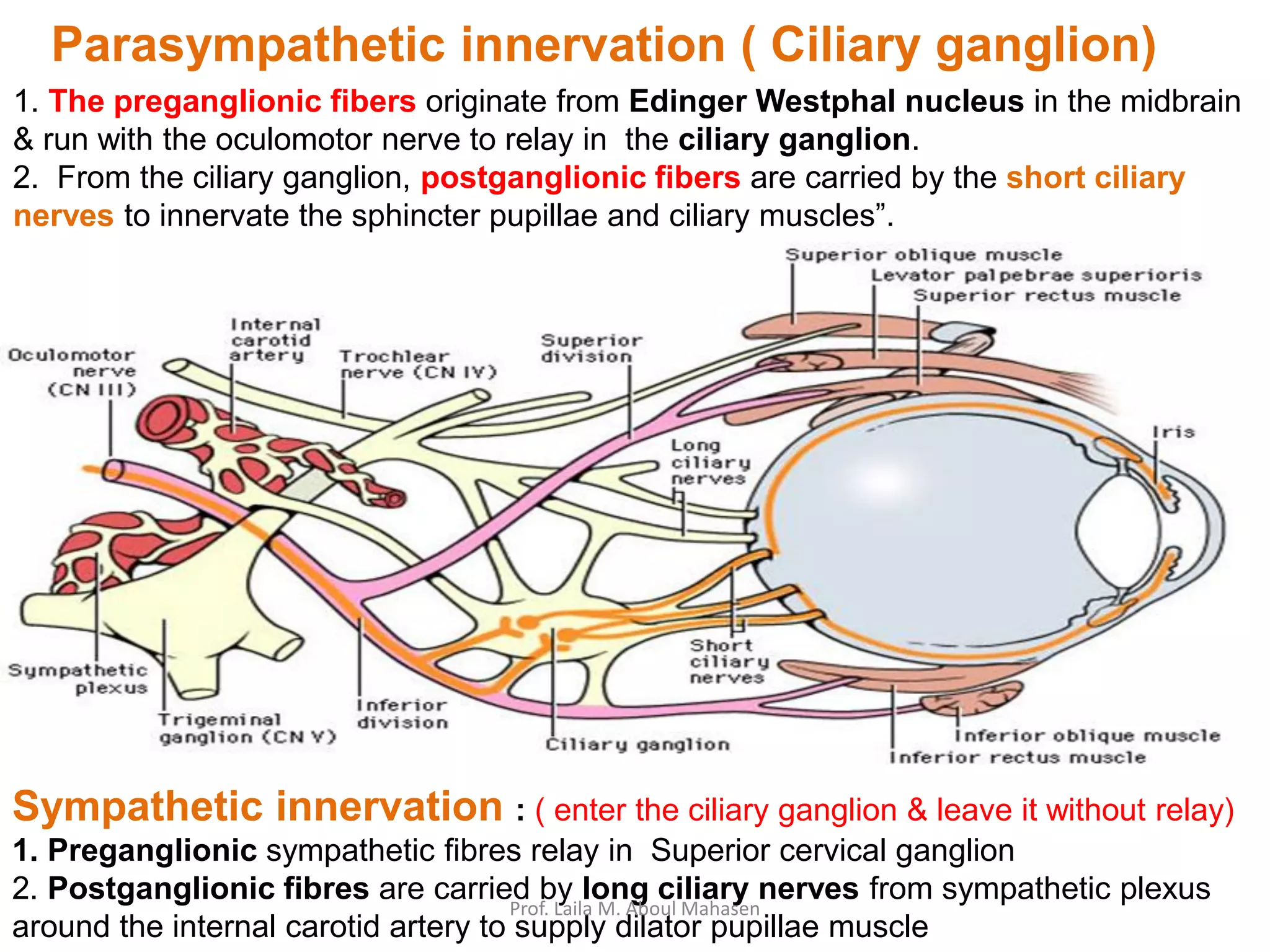


The document discusses four parasympathetic ganglia of the head: 1. The ciliary ganglion located in the orbit behind the eye supplies the sphincter pupillae and ciliary muscles. 2. The sphenopalatine ganglion in the pterygopalatine fossa supplies glands of the nasal cavity, palate and lacrimal gland. 3. The otic ganglion below the foramen ovale supplies the parotid gland. 4. The submandibular ganglion suspended from the lingual nerve supplies the submandibular and sublingual glands.


