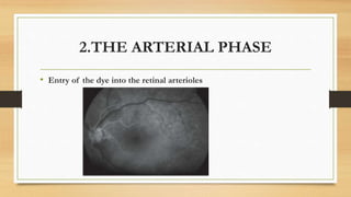
Fundus Flurescein Angiography
Punitha S.J., Boopathi Raja
INTRODUCTION
• Fluorescein angiography is an important invasive diagnostic tool for
posterior segment eye surgeons for the evaluation of the retina and choroid.
• Refers to photographing fluorescein dye in the retinal vasculature following
intravenous injection of fluorescein sodium.
• This photography can be performed with a fundus camera and either a
standard 35 mm camera back and films or video-based systems.
What is fluorescein ???
• Fluorescein (C20H12O5) refers to fluorescein sodium ( C20H10Na2O5)
• A brown or orange-red crystalline substance first synthesized in 1871 in
Germany by von Baeyer.
• It is highly water- soluble .
• Molecular weight of 376.27.
• Belongs to the group of triphenylmethane dyes.
Conti….
• It is the product of a reaction of phthalic acid anhydride and resorcinol in
which hot sulphuric acid is the catalyst.
• Available in topical solution form and as a mixture with corneal anesthetic, a
combination of benoxinate HCl.
• Also available in strip form for topical application to the cornea.
• Fluorescein solution is commercially available for injection as 10% and 25%
solution.
HISTORY OF EVOLUTION
Evaluation of fluorescein angiography has a unique and interesting aspect.
• In 1950, HaroldNovotny and Dr.David Alvis
• Novotny used the sodium fluorescein to determine the oxygen saturation into the
retinal arterioles for the first time for fluorescence observation.
• Meanwhile Dr. David Alvis used a mixed drop of it's own blood with sodium
fluorescein for fluorometric analysis.
“World’s first fluorescein angiography was done on Dr. David Alvis’s right eye”
Landmark work by Novotny and Alvis led
to evolution of fluorescein angiography….
• In April 1960, Miller and Chao, collaborators of Dr.Maumenae presented
their technique of fluorescein angiography .
• In 1969, Dr. F. Palomar presented the first article on fluorescein
angiography.
• Clinical works in Europe and USA , the better refinement of FA technique is
used for accurate and better assessment of various ocular pathologies.
In the present era of greater ophthalmological advances in the field of
diagnosis, surgical and laser management, fluorescein angiography is an
important diagnostic tool.
• There is a little difference between the Fundus Flurescein Examination
performed today and those performed when the procedure was in its infancy.
Recent advances in FA
• CUE wide field fluorescein angiography
• Digital video Angiography
• High speed fluorescein angiography
• 3D coor Doppler video angiography
PRINCIPLE
“Fluorescein dye absorbs light energy from a lower wave length and
emits at a higher wave length with fluorescence properties”.
• The absorption spectrum of fluorescein lies between 465 and 490 nm
(blue end of the visible spectrum).
• The emission spectrum lies between 520 and 530 nm ( green- yellow
area of the spectrum).
• On intravenous injection, 70 to 85 % of fluorescein molecules bind to serum
proteins,
• The remaining unbound component is responsible for fluorescence
displayed during angiography.
• The dye is excreted completely by the kidneys in 24 hours.
TECHNIQUE
A good quality angiogram requires adequate pupillary dilatation and clear
media.
Patient is explained about the procedure and an informed consent is taken.
Patient is seated in front of the camera comfortably and the sequence is as
follows:
• Color photograph of the fundus and red-free photograph.
• Securing an intravenous line.
• Fundus photograph with the exciter and barrier filters in place to look
for auto or pseudo fluorescence.
• Injection of fluorescein and switching on the timer.
• Photographs are taken 8 seconds after the beginning of the injection.
• Photographs at intervals of 11/2 to 2seconds.
• Late phase photographs at the end of 15 minutes.
SIDE EFFECTS AND
COMPLICATIONS OF FA
• Extravasations of the dye and local tissue necrosis.
• Nausea
• Vomiting
• Vasovagal attack (circulatory shock, myocardial infarction)
• Allergic reaction:
• Anaphylaxis
• Itching
• Hives
• Laryngeal edema
• Bronchospasm
Normal Fluorescein Angiogram
• Normal arm to retina time is 10 to 12 seconds.
• Fluorescein enters the eye through the ophthalmic artery, passing into the
choroidal circulation through the short posterior ciliary arteries and into the
retinal circulation through the central retinal artery.
• The inner blood retinal barrier is formed by the tight junctions of the
endothelium in the retinal vessels and the outer blood retinal barrier by the
retinal pigment epithelium; these barriers keep the fluorescein from
diffusing in to the vitreous cavity or subretinal space.
• The pigmented retinal pigment epithelium masks the choroidal fluorescence
during fluorescein angiography.
PHASES OF THE ANGIOGRAM
Choroidal
( pre- arterial)
Phase
Arterial Phase
Venous Phase
Arteriovenous
( capillary) Phase
Late Phase
1. THE CHOROIDAL PHASE
• Also called pre-arterial phase.
• One to two seconds before the dye appears in the central retinal artery.
• Patchy filling of the choroid is obscured almost immediately as the
dye diffuse out of the choriocapillaries resulting in the choroidal flush.
• A cilioretinal artery, if present will fill at this time because it is derived
from the posterior ciliary circulation.
CHOROIDAL PHASE
2.THE ARTERIAL PHASE
• Entry of the dye into the retinal arterioles
3.THE ARTERIOVENOUS PHASE
• Also called capillary phase.
• Dye enters the venules after passing through the retinal capillaries.
• Early arteriovenous phase shows laminar flow in the veins.
• In the late arteriovenous phase, the veins are filled with the dye.
Early arteriovenous
phase of the FFA
showing laminar flow in
the veins
Arteriovenous phase
showing more
pronounced laminar
flow in the veins
4. VENOUS PHASE
• Venous phase of the angiogram shows
loss of laminar flow and complete
filling of the veins.
• In the late venous phase the veins
appear brighter than the arterioles as
the concentration of the dye is more in
the venules than the arterioles.
5.THE LATE PHASE
• Due to elimination of the dye on
recirculation, fluorescence within the
retinal vessels is faint; disc staining
may appear.
• Scleral staining appears as a diffuse
background fluorescence.
Absence of blood
vessel in the foveal
zone and increased
density of xanthophyll
at the five and taller
retinal pigment
epithelial cells at the
fovea block the
choroidal fluorescence
resulting in a dark
fovea on FA.
ABNORMAL ANGIOGRAM
Abnormalities in the angiogram are classified essentially as
Hyperfluorescene
Hypofluorescence
HYPOFLUORESCENCE
Reduction or absence of normal fluorescence.
This is caused by
• Blocked fluorescence
• Hypoperfusion/vascular filling defect
Blocked
Fluorescence
• Blood, pigment,educates can
block underlying fluorescence.
• Area of blocked fluorescence
corresponds to the area of the
lesion.
Preretinal haemorrhage Corresponding FFA
shows blood blocking
underlying retinal and
choroidal fluorescence
HYPOPERFUSION/VASCULAR
FILLING DEFECT
Hypofluroscence due to non
filling of the vasculature due to
vascular occlusion or loss of
vascular bed.
Hypofluroscence due to loss of capillary bed
HYPERFLUROSCENCE
• Pre-injection fluorescence-auto/pseudofluorescence
- Autofluorescence-fluorescent characteristic of an abnormal fundus
lesion.
• Astrocytoma, optic nerve head drusen.
- Pseudofluorescence-mismatched filters
• Transmitted fluorescence.
- Due to transmission of the
choroidal fluorescence due to loss
of RPE barrier.
• Leakage.
-Leakage of the dye in the
vitreous or subretinal space due to
loss of blood retinal barrier .
• Pooling.
- Accumulation of the dye
within a space.
Hyperfluorescent spots
temporal to macular
fading away in late
phase
Hyperfluoroscene due
to leakage.Indicative of
leakage in to the vitreous
from retinal
neovascularization
FFA in CSR showing
leakage of the dye into
the subretinal space
• Staining
-Accumulation of the dye into the tissue.
WINDOW DEFECT
• The well-defined subretinal hyperfluorescent
spot appears in choroidal phase, increasing
in intensity with pro-gression of the
angiogram and fades in the late phase with
emptying of the choroidal vessels.
• Size of the lesion remains same and there
are no fuzzy margins.
BASIC PRINCIPLES OF
ANGIOGRAPHY INTERPRETATION
HYPOFLUORESCENCE
• Blocked fluorescence
1. Hemorrhages
2. Exudates
3. Glial tissue
4. Retinal pigment
• Vascular filling defects
1. Emboli
2. Arteriosclerosis
3. Vascular non perfusion
4. Vascular occlusion
HYPERFLUORESCENE
• Abnormal vessels
1. Tortuosity and dilatation
2. Neovascularization
3. Aneurysms
4. Subretinal neovascularization
• Leakage
1. Papilledema
2. Cystoid edema
3. Subretinal neovascularization
4. Retinal neovascularization
STRENGTHS
• The procedure with the fluorescein
dye is very much protective
procedure with nausea.
• Rare infection due to its side
effects.
• It allows us to assess both the anatomy and function of the retinal
and choroidal vasculature in a minimally invasive manner.
• It has become a vital component in the diagnosis, treatment, and
management of vitreoretinal disorders.
• To precisely define the location of pathologic changes, such as
those in choroidal neovascular membranes, treatment can be
initiated for the pathologic condition while the unaffected areas are
preserved.
LIMITATIONS
• The major limitation of fluorescein angiography is its inability to
precisely image the choroidal circulation.
• Another limitation of fluorescein angiography is the fact that it is a
technically involved procedure, requiring precise photographic
equipment in addition to a well-trained photographer to produce
quality results.
• Quantification of macular edema is not possible with fluorescein
angiography and to a large extent, it has been replaced with ocular
coherence tomography in the management of certain diseases
such as
• diffuse diabetic macular edema,
• uveitic macular edema, and
• the Irvine-Gass syndrome.
REFERENCES:
• PRIMARY CARE OPTOMETRY.
- Theodore Grosvenor
• CLINICAL PROCEDURES IN OPTOMETRY.
- J.Boyd Eskridge, John F. Amos, Jimmy D.Bartlett
• FUNDUS FLURESCEINANGIOGRAPHY (MINI ATLAS )
- Ashok Gorg, Josè Maria Ruiz Moreno, João J Nassaralla.
Jr, Arturo Perez Arteaga
• Jeffrey L. Olson MD, Naresh Mandava MD.,Flurescein angiography,
in retinal imaging, 2006
• Delhi Eye Centre, super speciality eyecare
• https://youtu.be/7rdHTeq4BQE
• https://youtu.be/37y1X54o_1Y
• https://youtu.be/YtzcmBNJ3ko

Presentation.pptx

  • 1.
  • 2.
    INTRODUCTION • Fluorescein angiographyis an important invasive diagnostic tool for posterior segment eye surgeons for the evaluation of the retina and choroid. • Refers to photographing fluorescein dye in the retinal vasculature following intravenous injection of fluorescein sodium. • This photography can be performed with a fundus camera and either a standard 35 mm camera back and films or video-based systems.
  • 4.
    What is fluorescein??? • Fluorescein (C20H12O5) refers to fluorescein sodium ( C20H10Na2O5) • A brown or orange-red crystalline substance first synthesized in 1871 in Germany by von Baeyer. • It is highly water- soluble . • Molecular weight of 376.27. • Belongs to the group of triphenylmethane dyes.
  • 5.
    Conti…. • It isthe product of a reaction of phthalic acid anhydride and resorcinol in which hot sulphuric acid is the catalyst. • Available in topical solution form and as a mixture with corneal anesthetic, a combination of benoxinate HCl. • Also available in strip form for topical application to the cornea. • Fluorescein solution is commercially available for injection as 10% and 25% solution.
  • 6.
    HISTORY OF EVOLUTION Evaluationof fluorescein angiography has a unique and interesting aspect. • In 1950, HaroldNovotny and Dr.David Alvis • Novotny used the sodium fluorescein to determine the oxygen saturation into the retinal arterioles for the first time for fluorescence observation. • Meanwhile Dr. David Alvis used a mixed drop of it's own blood with sodium fluorescein for fluorometric analysis. “World’s first fluorescein angiography was done on Dr. David Alvis’s right eye”
  • 7.
    Landmark work byNovotny and Alvis led to evolution of fluorescein angiography…. • In April 1960, Miller and Chao, collaborators of Dr.Maumenae presented their technique of fluorescein angiography . • In 1969, Dr. F. Palomar presented the first article on fluorescein angiography. • Clinical works in Europe and USA , the better refinement of FA technique is used for accurate and better assessment of various ocular pathologies.
  • 8.
    In the presentera of greater ophthalmological advances in the field of diagnosis, surgical and laser management, fluorescein angiography is an important diagnostic tool. • There is a little difference between the Fundus Flurescein Examination performed today and those performed when the procedure was in its infancy.
  • 9.
    Recent advances inFA • CUE wide field fluorescein angiography • Digital video Angiography • High speed fluorescein angiography • 3D coor Doppler video angiography
  • 10.
    PRINCIPLE “Fluorescein dye absorbslight energy from a lower wave length and emits at a higher wave length with fluorescence properties”. • The absorption spectrum of fluorescein lies between 465 and 490 nm (blue end of the visible spectrum). • The emission spectrum lies between 520 and 530 nm ( green- yellow area of the spectrum).
  • 12.
    • On intravenousinjection, 70 to 85 % of fluorescein molecules bind to serum proteins, • The remaining unbound component is responsible for fluorescence displayed during angiography. • The dye is excreted completely by the kidneys in 24 hours.
  • 13.
    TECHNIQUE A good qualityangiogram requires adequate pupillary dilatation and clear media. Patient is explained about the procedure and an informed consent is taken. Patient is seated in front of the camera comfortably and the sequence is as follows: • Color photograph of the fundus and red-free photograph. • Securing an intravenous line.
  • 14.
    • Fundus photographwith the exciter and barrier filters in place to look for auto or pseudo fluorescence. • Injection of fluorescein and switching on the timer. • Photographs are taken 8 seconds after the beginning of the injection. • Photographs at intervals of 11/2 to 2seconds. • Late phase photographs at the end of 15 minutes.
  • 16.
    SIDE EFFECTS AND COMPLICATIONSOF FA • Extravasations of the dye and local tissue necrosis. • Nausea • Vomiting • Vasovagal attack (circulatory shock, myocardial infarction) • Allergic reaction: • Anaphylaxis • Itching • Hives • Laryngeal edema • Bronchospasm
  • 17.
    Normal Fluorescein Angiogram •Normal arm to retina time is 10 to 12 seconds. • Fluorescein enters the eye through the ophthalmic artery, passing into the choroidal circulation through the short posterior ciliary arteries and into the retinal circulation through the central retinal artery. • The inner blood retinal barrier is formed by the tight junctions of the endothelium in the retinal vessels and the outer blood retinal barrier by the retinal pigment epithelium; these barriers keep the fluorescein from diffusing in to the vitreous cavity or subretinal space. • The pigmented retinal pigment epithelium masks the choroidal fluorescence during fluorescein angiography.
  • 18.
    PHASES OF THEANGIOGRAM Choroidal ( pre- arterial) Phase Arterial Phase Venous Phase Arteriovenous ( capillary) Phase Late Phase
  • 19.
    1. THE CHOROIDALPHASE • Also called pre-arterial phase. • One to two seconds before the dye appears in the central retinal artery. • Patchy filling of the choroid is obscured almost immediately as the dye diffuse out of the choriocapillaries resulting in the choroidal flush. • A cilioretinal artery, if present will fill at this time because it is derived from the posterior ciliary circulation.
  • 20.
  • 21.
    2.THE ARTERIAL PHASE •Entry of the dye into the retinal arterioles
  • 22.
    3.THE ARTERIOVENOUS PHASE •Also called capillary phase. • Dye enters the venules after passing through the retinal capillaries. • Early arteriovenous phase shows laminar flow in the veins. • In the late arteriovenous phase, the veins are filled with the dye.
  • 23.
    Early arteriovenous phase ofthe FFA showing laminar flow in the veins Arteriovenous phase showing more pronounced laminar flow in the veins
  • 24.
    4. VENOUS PHASE •Venous phase of the angiogram shows loss of laminar flow and complete filling of the veins. • In the late venous phase the veins appear brighter than the arterioles as the concentration of the dye is more in the venules than the arterioles.
  • 25.
    5.THE LATE PHASE •Due to elimination of the dye on recirculation, fluorescence within the retinal vessels is faint; disc staining may appear. • Scleral staining appears as a diffuse background fluorescence.
  • 26.
    Absence of blood vesselin the foveal zone and increased density of xanthophyll at the five and taller retinal pigment epithelial cells at the fovea block the choroidal fluorescence resulting in a dark fovea on FA.
  • 28.
    ABNORMAL ANGIOGRAM Abnormalities inthe angiogram are classified essentially as Hyperfluorescene Hypofluorescence
  • 29.
    HYPOFLUORESCENCE Reduction or absenceof normal fluorescence. This is caused by • Blocked fluorescence • Hypoperfusion/vascular filling defect
  • 30.
    Blocked Fluorescence • Blood, pigment,educatescan block underlying fluorescence. • Area of blocked fluorescence corresponds to the area of the lesion. Preretinal haemorrhage Corresponding FFA shows blood blocking underlying retinal and choroidal fluorescence
  • 31.
    HYPOPERFUSION/VASCULAR FILLING DEFECT Hypofluroscence dueto non filling of the vasculature due to vascular occlusion or loss of vascular bed. Hypofluroscence due to loss of capillary bed
  • 32.
    HYPERFLUROSCENCE • Pre-injection fluorescence-auto/pseudofluorescence -Autofluorescence-fluorescent characteristic of an abnormal fundus lesion. • Astrocytoma, optic nerve head drusen. - Pseudofluorescence-mismatched filters
  • 33.
    • Transmitted fluorescence. -Due to transmission of the choroidal fluorescence due to loss of RPE barrier. • Leakage. -Leakage of the dye in the vitreous or subretinal space due to loss of blood retinal barrier . • Pooling. - Accumulation of the dye within a space. Hyperfluorescent spots temporal to macular fading away in late phase Hyperfluoroscene due to leakage.Indicative of leakage in to the vitreous from retinal neovascularization FFA in CSR showing leakage of the dye into the subretinal space
  • 34.
    • Staining -Accumulation ofthe dye into the tissue.
  • 35.
    WINDOW DEFECT • Thewell-defined subretinal hyperfluorescent spot appears in choroidal phase, increasing in intensity with pro-gression of the angiogram and fades in the late phase with emptying of the choroidal vessels. • Size of the lesion remains same and there are no fuzzy margins.
  • 36.
    BASIC PRINCIPLES OF ANGIOGRAPHYINTERPRETATION HYPOFLUORESCENCE • Blocked fluorescence 1. Hemorrhages 2. Exudates 3. Glial tissue 4. Retinal pigment • Vascular filling defects 1. Emboli 2. Arteriosclerosis 3. Vascular non perfusion 4. Vascular occlusion HYPERFLUORESCENE • Abnormal vessels 1. Tortuosity and dilatation 2. Neovascularization 3. Aneurysms 4. Subretinal neovascularization • Leakage 1. Papilledema 2. Cystoid edema 3. Subretinal neovascularization 4. Retinal neovascularization
  • 38.
    STRENGTHS • The procedurewith the fluorescein dye is very much protective procedure with nausea. • Rare infection due to its side effects.
  • 39.
    • It allowsus to assess both the anatomy and function of the retinal and choroidal vasculature in a minimally invasive manner. • It has become a vital component in the diagnosis, treatment, and management of vitreoretinal disorders. • To precisely define the location of pathologic changes, such as those in choroidal neovascular membranes, treatment can be initiated for the pathologic condition while the unaffected areas are preserved.
  • 40.
    LIMITATIONS • The majorlimitation of fluorescein angiography is its inability to precisely image the choroidal circulation. • Another limitation of fluorescein angiography is the fact that it is a technically involved procedure, requiring precise photographic equipment in addition to a well-trained photographer to produce quality results.
  • 41.
    • Quantification ofmacular edema is not possible with fluorescein angiography and to a large extent, it has been replaced with ocular coherence tomography in the management of certain diseases such as • diffuse diabetic macular edema, • uveitic macular edema, and • the Irvine-Gass syndrome.
  • 42.
    REFERENCES: • PRIMARY CAREOPTOMETRY. - Theodore Grosvenor • CLINICAL PROCEDURES IN OPTOMETRY. - J.Boyd Eskridge, John F. Amos, Jimmy D.Bartlett • FUNDUS FLURESCEINANGIOGRAPHY (MINI ATLAS ) - Ashok Gorg, Josè Maria Ruiz Moreno, João J Nassaralla. Jr, Arturo Perez Arteaga
  • 43.
    • Jeffrey L.Olson MD, Naresh Mandava MD.,Flurescein angiography, in retinal imaging, 2006 • Delhi Eye Centre, super speciality eyecare • https://youtu.be/7rdHTeq4BQE • https://youtu.be/37y1X54o_1Y • https://youtu.be/YtzcmBNJ3ko