SlideShare a Scribd company logo
1 of 180
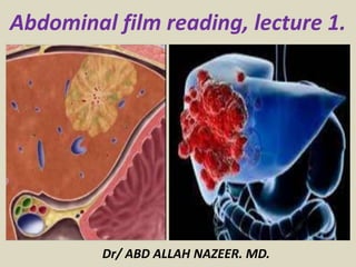
Abdominal film reading, lecture 1.
Dr/ ABD ALLAH NAZEER. MD.
Abnormal bowel gas. Too much too little gas.
Abnormal bowel gas. Too much gas.
Small bowel obstruction.
Sigmoid colon volvulus.
CT Anatomy.
Techniques for MDCT and MRI of the liver.
Autosomal dominant polycystic liver disease.
Prenatal US showing a large intra-hepatic cyst, and a normal gall bladder (*). CT
scan at births confirmed a very large hepatic cyst and the normal gall bladder (*).
Prenatal MRI confirmed hepatic cyst located in segment IV and postnatal evolution in
MRI realized preoperatively at 6 weeks of life. Postnatal US illustrated the rapid
growing of hepatic cyst between days 2 of life (D2) and the first months of life (M1).
Hepatic hemangioma lesion
at prenatal ultrasound.
Hepatic lesion at postnatal ultrasound;
marked, peripheral Doppler blood flow.
Congenital
hepatic
hemangioma.
The caudate lobe lesion (arrowheads) presents subtle hypersignal on T2-weighted sequence and signal loss on T1-weighted
out-of-phase sequence caused by the presence of intralesional fat. Such a lesion shows intense and homogeneous contrast
uptake in the arterial-phase, with decay in the portal and delayed phases, presenting greater Hepatobiliary contrast uptake
than the adjacent parenchyma, suggesting FNH as the first diagnostic hypothesis. Considering that the presence of
intralesional fat in NFH is rare, the patient will be maintained under imaging follow-up. The lesions in segments VII and VIII
(arrows) are similar, with marked hypersignal on T2-weighted, hyposignal on T1-weighted sequence, and nodular,
peripheral and discontinuous uptake in the arterial phase, a characteristic of hemangiomas.
Multiple, well-defined focal hypervascular lesions, with intermediate signal intensity on T2-
weighted sequence, with poor lesion-organ contrast-enhancement. However, the presence of
intra lesional fat was detected on out-of-phase T1-weighted sequence. The presence of intra
lesional fat is not usually found in FNH and suggests the diagnosis of adenoma – adenomatosis
Small HCC seen only in arterial phase in a patient with cirrhosis.
NECT, arterial and portal venous phase in a patient with
Hepatitis C with two lesions in the liver (arrows).
LEFT: Diffusely enhancing tumor thrombus in HCC with portal vein
invasion. RIGHT: Tumor thrombus with vessels within the thrombus.
Large HCC with mozaik pattern in a non cirrhotic patient.
Two liver nodules are seen in the segment VIII (arrows) as well as a larger nodule, in the
segment VI (arrowheads), all of them contrast-enhanced in the arterial-phase, washout in
the delayed-phase, and without uptake in the hepatobiliary-phase, characterizing HCCs.
Cholangiocarcinoma: portal venous and equilibrium phase.
Cholangiocarcinoma: Non enhanced, arterial, portal venous and equilibrium phase.
Colorectal metastasis with hyper-(rim)/hypo-/hypo- appearance. (a) Arterial phase image
shows a homogeneously enhanced hyperattenuating rim (arrows). (b) Portal phase image
shows that the lesion was homogeneously hypoattenuating. (c) Equilibrium phase image
shows that the periphery of the metastasis is hypoattenuating (arrows) relative to the
enhanced center of the lesion and the surrounding liver parenchyma.
Hepatic metastasis.
Analysis of dynamic vascular pattern(DVP) in ultrasound can be used
to distinguish benign from malignant flow patterns in focal liver lesions.
Four clinical cases show how DVP parametric images allow facilitated lesion characterization
as benign or malignant in four typical clinical examples, with malignant lesions appearing in
red, unlike benign lesions which are green or yellow-green in appearance.
Thank You.

More Related Content

What's hot

Presentation1.pptx urinary system part 11.
Presentation1.pptx urinary system part 11.Presentation1.pptx urinary system part 11.
Presentation1.pptx urinary system part 11.Abdellah Nazeer
 
Presentation1, radiological imaging of gastro intestinal stromal tumour(gist).
Presentation1, radiological imaging of gastro intestinal stromal tumour(gist).Presentation1, radiological imaging of gastro intestinal stromal tumour(gist).
Presentation1, radiological imaging of gastro intestinal stromal tumour(gist).Abdellah Nazeer
 
Imaging In Obstructive Biliopathy.Aizawl
Imaging In Obstructive Biliopathy.AizawlImaging In Obstructive Biliopathy.Aizawl
Imaging In Obstructive Biliopathy.AizawlDr. Himadri Sikhor Das
 
Diagnostic Imaging of Cholangiocarcinoma
Diagnostic Imaging of CholangiocarcinomaDiagnostic Imaging of Cholangiocarcinoma
Diagnostic Imaging of CholangiocarcinomaMohamed M.A. Zaitoun
 
Focal malignant lesions of the liver.
Focal malignant lesions of the liver.Focal malignant lesions of the liver.
Focal malignant lesions of the liver.Sanal Kumar
 
Diagnostic Imaging of Pancreatitis
Diagnostic Imaging of PancreatitisDiagnostic Imaging of Pancreatitis
Diagnostic Imaging of PancreatitisMohamed M.A. Zaitoun
 
Imaging of Malignant Liver Lesions
Imaging of Malignant Liver LesionsImaging of Malignant Liver Lesions
Imaging of Malignant Liver LesionsSahil Chaudhry
 
Pediatric liver masses
Pediatric liver massesPediatric liver masses
Pediatric liver massesSidra Afzal
 
Presentation1.pptx, radiological imaging of obstructive jaundice.
Presentation1.pptx, radiological imaging of obstructive jaundice.Presentation1.pptx, radiological imaging of obstructive jaundice.
Presentation1.pptx, radiological imaging of obstructive jaundice.Abdellah Nazeer
 
Liver imaging ct scan algorithm
Liver imaging ct scan algorithmLiver imaging ct scan algorithm
Liver imaging ct scan algorithmTabish Javed
 
Presentation1, radiological imaging of anal carcinoma.
Presentation1, radiological imaging of anal carcinoma.Presentation1, radiological imaging of anal carcinoma.
Presentation1, radiological imaging of anal carcinoma.Abdellah Nazeer
 
Imaging features of hepatobiliary and pancretic ds
Imaging features of hepatobiliary and pancretic dsImaging features of hepatobiliary and pancretic ds
Imaging features of hepatobiliary and pancretic dsradiologyoffice
 
Renal Cell Carcinoma
Renal Cell CarcinomaRenal Cell Carcinoma
Renal Cell CarcinomaSam
 
Role of imaging in urosepsis
Role of  imaging in urosepsisRole of  imaging in urosepsis
Role of imaging in urosepsisAhmed Bahnassy
 
Presentation1.pptx, ultrasound examination of the adrenal glands and kidneys.
Presentation1.pptx, ultrasound examination of the adrenal glands and kidneys.Presentation1.pptx, ultrasound examination of the adrenal glands and kidneys.
Presentation1.pptx, ultrasound examination of the adrenal glands and kidneys.Abdellah Nazeer
 

What's hot (20)

Presentation1.pptx urinary system part 11.
Presentation1.pptx urinary system part 11.Presentation1.pptx urinary system part 11.
Presentation1.pptx urinary system part 11.
 
Presentation1, radiological imaging of gastro intestinal stromal tumour(gist).
Presentation1, radiological imaging of gastro intestinal stromal tumour(gist).Presentation1, radiological imaging of gastro intestinal stromal tumour(gist).
Presentation1, radiological imaging of gastro intestinal stromal tumour(gist).
 
Malignant liver lesions
Malignant liver lesionsMalignant liver lesions
Malignant liver lesions
 
Imaging In Obstructive Biliopathy.Aizawl
Imaging In Obstructive Biliopathy.AizawlImaging In Obstructive Biliopathy.Aizawl
Imaging In Obstructive Biliopathy.Aizawl
 
Diagnostic Imaging of Cholangiocarcinoma
Diagnostic Imaging of CholangiocarcinomaDiagnostic Imaging of Cholangiocarcinoma
Diagnostic Imaging of Cholangiocarcinoma
 
Imaging of Renal Tumors
Imaging of Renal TumorsImaging of Renal Tumors
Imaging of Renal Tumors
 
Focal malignant lesions of the liver.
Focal malignant lesions of the liver.Focal malignant lesions of the liver.
Focal malignant lesions of the liver.
 
Diagnostic Imaging of Pancreatitis
Diagnostic Imaging of PancreatitisDiagnostic Imaging of Pancreatitis
Diagnostic Imaging of Pancreatitis
 
Liver lesions SYMPOSIUM RADIOLOGY
Liver lesions SYMPOSIUM RADIOLOGYLiver lesions SYMPOSIUM RADIOLOGY
Liver lesions SYMPOSIUM RADIOLOGY
 
Imaging of Malignant Liver Lesions
Imaging of Malignant Liver LesionsImaging of Malignant Liver Lesions
Imaging of Malignant Liver Lesions
 
Pediatric liver masses
Pediatric liver massesPediatric liver masses
Pediatric liver masses
 
Presentation1.pptx, radiological imaging of obstructive jaundice.
Presentation1.pptx, radiological imaging of obstructive jaundice.Presentation1.pptx, radiological imaging of obstructive jaundice.
Presentation1.pptx, radiological imaging of obstructive jaundice.
 
Liver imaging ct scan algorithm
Liver imaging ct scan algorithmLiver imaging ct scan algorithm
Liver imaging ct scan algorithm
 
Presentation1, radiological imaging of anal carcinoma.
Presentation1, radiological imaging of anal carcinoma.Presentation1, radiological imaging of anal carcinoma.
Presentation1, radiological imaging of anal carcinoma.
 
Imaging features of hepatobiliary and pancretic ds
Imaging features of hepatobiliary and pancretic dsImaging features of hepatobiliary and pancretic ds
Imaging features of hepatobiliary and pancretic ds
 
Renal Cell Carcinoma
Renal Cell CarcinomaRenal Cell Carcinoma
Renal Cell Carcinoma
 
Role of imaging in urosepsis
Role of  imaging in urosepsisRole of  imaging in urosepsis
Role of imaging in urosepsis
 
Presentation1.pptx, ultrasound examination of the adrenal glands and kidneys.
Presentation1.pptx, ultrasound examination of the adrenal glands and kidneys.Presentation1.pptx, ultrasound examination of the adrenal glands and kidneys.
Presentation1.pptx, ultrasound examination of the adrenal glands and kidneys.
 
Radiology 5th year, 3rd lecture (Dr. Nasrin Alatrushi)
Radiology 5th year, 3rd lecture (Dr. Nasrin Alatrushi)Radiology 5th year, 3rd lecture (Dr. Nasrin Alatrushi)
Radiology 5th year, 3rd lecture (Dr. Nasrin Alatrushi)
 
Renal malignancy
Renal malignancyRenal malignancy
Renal malignancy
 

Viewers also liked

Presentation1.pptx, radiological imaging of mesenteric ischemia.
Presentation1.pptx, radiological imaging of mesenteric ischemia.Presentation1.pptx, radiological imaging of mesenteric ischemia.
Presentation1.pptx, radiological imaging of mesenteric ischemia.Abdellah Nazeer
 
Presentation1.pptx, ultrasound examination of the neonatal head.
Presentation1.pptx, ultrasound examination of the neonatal head.Presentation1.pptx, ultrasound examination of the neonatal head.
Presentation1.pptx, ultrasound examination of the neonatal head.Abdellah Nazeer
 
Presentation1, radiological imaging of ear microcia.
Presentation1, radiological imaging of ear microcia.Presentation1, radiological imaging of ear microcia.
Presentation1, radiological imaging of ear microcia.Abdellah Nazeer
 
Presentation1.pptx, chest film reading. lecture 2
Presentation1.pptx, chest film reading. lecture 2Presentation1.pptx, chest film reading. lecture 2
Presentation1.pptx, chest film reading. lecture 2Abdellah Nazeer
 
Mesenteric ischemia
Mesenteric ischemiaMesenteric ischemia
Mesenteric ischemiakrisshk1989
 
Presentation1.pptx, chest film reading. lecture 1
Presentation1.pptx, chest film reading. lecture 1Presentation1.pptx, chest film reading. lecture 1
Presentation1.pptx, chest film reading. lecture 1Abdellah Nazeer
 
Ultrasound
UltrasoundUltrasound
UltrasoundRad Tech
 
Presentation1.pptx, ultrasound examination of the liver and gall bladder.
Presentation1.pptx, ultrasound examination of the liver and gall bladder.Presentation1.pptx, ultrasound examination of the liver and gall bladder.
Presentation1.pptx, ultrasound examination of the liver and gall bladder.Abdellah Nazeer
 
Presentation1, radiological imaging of placenta accreta.
Presentation1, radiological imaging of placenta accreta.Presentation1, radiological imaging of placenta accreta.
Presentation1, radiological imaging of placenta accreta.Abdellah Nazeer
 
Presentation1.pptx, ultrasound examination of the uterus and ovaries.
Presentation1.pptx, ultrasound examination of the uterus and ovaries.Presentation1.pptx, ultrasound examination of the uterus and ovaries.
Presentation1.pptx, ultrasound examination of the uterus and ovaries.Abdellah Nazeer
 
Presentation1, radiological imaging of diabetic foor and charcot joint.
Presentation1, radiological imaging of diabetic foor and charcot joint.Presentation1, radiological imaging of diabetic foor and charcot joint.
Presentation1, radiological imaging of diabetic foor and charcot joint.Abdellah Nazeer
 
Cystic liver lesions - An ultrasound perspective
Cystic liver lesions - An ultrasound perspectiveCystic liver lesions - An ultrasound perspective
Cystic liver lesions - An ultrasound perspectiveSamir Haffar
 
Presentation1.pptx, ct normal anatomy of the abdomen and pelvis.
Presentation1.pptx, ct normal anatomy of the abdomen and pelvis.Presentation1.pptx, ct normal anatomy of the abdomen and pelvis.
Presentation1.pptx, ct normal anatomy of the abdomen and pelvis.Abdellah Nazeer
 
Presentation1.pptx, radiological imaging of endometriosis.
Presentation1.pptx, radiological imaging of endometriosis.Presentation1.pptx, radiological imaging of endometriosis.
Presentation1.pptx, radiological imaging of endometriosis.Abdellah Nazeer
 
Presentation1.pptx, ultrasound study of the spleen and pancreas.
Presentation1.pptx, ultrasound study of the spleen and pancreas.Presentation1.pptx, ultrasound study of the spleen and pancreas.
Presentation1.pptx, ultrasound study of the spleen and pancreas.Abdellah Nazeer
 
Presentation1, radiological imaging of ostepopikilosis
Presentation1, radiological imaging of ostepopikilosisPresentation1, radiological imaging of ostepopikilosis
Presentation1, radiological imaging of ostepopikilosisAbdellah Nazeer
 
Presentation1.pptx, radiological imaging of osteoarthritis.
Presentation1.pptx, radiological imaging of osteoarthritis.Presentation1.pptx, radiological imaging of osteoarthritis.
Presentation1.pptx, radiological imaging of osteoarthritis.Abdellah Nazeer
 
Presentation1.pptx, ultrasound examination of the urinary bladder and prostate.
Presentation1.pptx, ultrasound examination of the urinary bladder and prostate.Presentation1.pptx, ultrasound examination of the urinary bladder and prostate.
Presentation1.pptx, ultrasound examination of the urinary bladder and prostate.Abdellah Nazeer
 
Presentation1.pptx, radiological anatomy of the knee joint.
Presentation1.pptx, radiological anatomy of the knee joint.Presentation1.pptx, radiological anatomy of the knee joint.
Presentation1.pptx, radiological anatomy of the knee joint.Abdellah Nazeer
 

Viewers also liked (20)

Presentation1.pptx, radiological imaging of mesenteric ischemia.
Presentation1.pptx, radiological imaging of mesenteric ischemia.Presentation1.pptx, radiological imaging of mesenteric ischemia.
Presentation1.pptx, radiological imaging of mesenteric ischemia.
 
Presentation1.pptx, ultrasound examination of the neonatal head.
Presentation1.pptx, ultrasound examination of the neonatal head.Presentation1.pptx, ultrasound examination of the neonatal head.
Presentation1.pptx, ultrasound examination of the neonatal head.
 
Presentation1, radiological imaging of ear microcia.
Presentation1, radiological imaging of ear microcia.Presentation1, radiological imaging of ear microcia.
Presentation1, radiological imaging of ear microcia.
 
Presentation1.pptx, chest film reading. lecture 2
Presentation1.pptx, chest film reading. lecture 2Presentation1.pptx, chest film reading. lecture 2
Presentation1.pptx, chest film reading. lecture 2
 
Mesenteric ischemia
Mesenteric ischemiaMesenteric ischemia
Mesenteric ischemia
 
Knee 2
Knee 2Knee 2
Knee 2
 
Presentation1.pptx, chest film reading. lecture 1
Presentation1.pptx, chest film reading. lecture 1Presentation1.pptx, chest film reading. lecture 1
Presentation1.pptx, chest film reading. lecture 1
 
Ultrasound
UltrasoundUltrasound
Ultrasound
 
Presentation1.pptx, ultrasound examination of the liver and gall bladder.
Presentation1.pptx, ultrasound examination of the liver and gall bladder.Presentation1.pptx, ultrasound examination of the liver and gall bladder.
Presentation1.pptx, ultrasound examination of the liver and gall bladder.
 
Presentation1, radiological imaging of placenta accreta.
Presentation1, radiological imaging of placenta accreta.Presentation1, radiological imaging of placenta accreta.
Presentation1, radiological imaging of placenta accreta.
 
Presentation1.pptx, ultrasound examination of the uterus and ovaries.
Presentation1.pptx, ultrasound examination of the uterus and ovaries.Presentation1.pptx, ultrasound examination of the uterus and ovaries.
Presentation1.pptx, ultrasound examination of the uterus and ovaries.
 
Presentation1, radiological imaging of diabetic foor and charcot joint.
Presentation1, radiological imaging of diabetic foor and charcot joint.Presentation1, radiological imaging of diabetic foor and charcot joint.
Presentation1, radiological imaging of diabetic foor and charcot joint.
 
Cystic liver lesions - An ultrasound perspective
Cystic liver lesions - An ultrasound perspectiveCystic liver lesions - An ultrasound perspective
Cystic liver lesions - An ultrasound perspective
 
Presentation1.pptx, ct normal anatomy of the abdomen and pelvis.
Presentation1.pptx, ct normal anatomy of the abdomen and pelvis.Presentation1.pptx, ct normal anatomy of the abdomen and pelvis.
Presentation1.pptx, ct normal anatomy of the abdomen and pelvis.
 
Presentation1.pptx, radiological imaging of endometriosis.
Presentation1.pptx, radiological imaging of endometriosis.Presentation1.pptx, radiological imaging of endometriosis.
Presentation1.pptx, radiological imaging of endometriosis.
 
Presentation1.pptx, ultrasound study of the spleen and pancreas.
Presentation1.pptx, ultrasound study of the spleen and pancreas.Presentation1.pptx, ultrasound study of the spleen and pancreas.
Presentation1.pptx, ultrasound study of the spleen and pancreas.
 
Presentation1, radiological imaging of ostepopikilosis
Presentation1, radiological imaging of ostepopikilosisPresentation1, radiological imaging of ostepopikilosis
Presentation1, radiological imaging of ostepopikilosis
 
Presentation1.pptx, radiological imaging of osteoarthritis.
Presentation1.pptx, radiological imaging of osteoarthritis.Presentation1.pptx, radiological imaging of osteoarthritis.
Presentation1.pptx, radiological imaging of osteoarthritis.
 
Presentation1.pptx, ultrasound examination of the urinary bladder and prostate.
Presentation1.pptx, ultrasound examination of the urinary bladder and prostate.Presentation1.pptx, ultrasound examination of the urinary bladder and prostate.
Presentation1.pptx, ultrasound examination of the urinary bladder and prostate.
 
Presentation1.pptx, radiological anatomy of the knee joint.
Presentation1.pptx, radiological anatomy of the knee joint.Presentation1.pptx, radiological anatomy of the knee joint.
Presentation1.pptx, radiological anatomy of the knee joint.
 

Similar to Abdominal Film Reading Lecture 1: Abnormal Bowel Gas, Small Bowel Obstruction, Sigmoid Colon Volvulus & More

Quick review of hepatobiliary and pancreatic lesion
Quick review of hepatobiliary and pancreatic lesionQuick review of hepatobiliary and pancreatic lesion
Quick review of hepatobiliary and pancreatic lesionVincentBatista2
 
Focal hepatic lesions
Focal hepatic lesionsFocal hepatic lesions
Focal hepatic lesionsYasser Asiri
 
LIVER SEGMENTAL ANATOMY AND IMAGINGIN IN LIVER CIRRHOSIS.pptx
LIVER SEGMENTAL ANATOMY AND IMAGINGIN IN LIVER CIRRHOSIS.pptxLIVER SEGMENTAL ANATOMY AND IMAGINGIN IN LIVER CIRRHOSIS.pptx
LIVER SEGMENTAL ANATOMY AND IMAGINGIN IN LIVER CIRRHOSIS.pptxDr vaibhavi patel
 
Carcinoma stomach seminar
Carcinoma stomach seminarCarcinoma stomach seminar
Carcinoma stomach seminarRushabh Shah
 
Hepatic inflammatory and infectious dis.
Hepatic inflammatory and infectious dis.Hepatic inflammatory and infectious dis.
Hepatic inflammatory and infectious dis.Annu Shri
 
Focal liver lesions- in the eye of a radiologist
Focal liver lesions- in the eye of a radiologistFocal liver lesions- in the eye of a radiologist
Focal liver lesions- in the eye of a radiologistDr.Santosh Atreya
 
Contouring guidelines pancreatic malignancies
Contouring guidelines  pancreatic malignancies  Contouring guidelines  pancreatic malignancies
Contouring guidelines pancreatic malignancies astha17srivastava
 
Imaging in Benign hepatic lesions
Imaging in Benign hepatic lesionsImaging in Benign hepatic lesions
Imaging in Benign hepatic lesionsGirendra Shankar
 
Presentation2, radiological imaging of gastrointestinal schwannoma.
Presentation2, radiological imaging of gastrointestinal schwannoma.Presentation2, radiological imaging of gastrointestinal schwannoma.
Presentation2, radiological imaging of gastrointestinal schwannoma.Abdellah Nazeer
 
Biliary Tract Tumours(Cholangiocarcinoma,Cystadenoma/cystadenocarcinoma,Gall ...
Biliary Tract Tumours(Cholangiocarcinoma,Cystadenoma/cystadenocarcinoma,Gall ...Biliary Tract Tumours(Cholangiocarcinoma,Cystadenoma/cystadenocarcinoma,Gall ...
Biliary Tract Tumours(Cholangiocarcinoma,Cystadenoma/cystadenocarcinoma,Gall ...Abdul Abasi
 
CT and MRI Imaging of Hepatic metastases
CT and MRI Imaging of Hepatic metastasesCT and MRI Imaging of Hepatic metastases
CT and MRI Imaging of Hepatic metastasesDr Priyanka Vishwakarma
 
Imaging of non endocrine tumour of pancreas
Imaging of non endocrine tumour of pancreasImaging of non endocrine tumour of pancreas
Imaging of non endocrine tumour of pancreasDev Lakhera
 
Ct Imaging of Abdomen Dr. Muhammad Bin Zulfiqar
Ct Imaging of Abdomen Dr. Muhammad Bin ZulfiqarCt Imaging of Abdomen Dr. Muhammad Bin Zulfiqar
Ct Imaging of Abdomen Dr. Muhammad Bin ZulfiqarDr. Muhammad Bin Zulfiqar
 
Biliary tract imaging final...........
Biliary tract imaging  final...........Biliary tract imaging  final...........
Biliary tract imaging final...........Ram Jharpula
 
Benign and Malignant Liver Disorder
Benign and Malignant Liver DisorderBenign and Malignant Liver Disorder
Benign and Malignant Liver Disorderyuyuricci
 
Liver and Gallbladder Surgical Pathology
Liver and Gallbladder Surgical PathologyLiver and Gallbladder Surgical Pathology
Liver and Gallbladder Surgical PathologyGhie Santos
 

Similar to Abdominal Film Reading Lecture 1: Abnormal Bowel Gas, Small Bowel Obstruction, Sigmoid Colon Volvulus & More (20)

Quick review of hepatobiliary and pancreatic lesion
Quick review of hepatobiliary and pancreatic lesionQuick review of hepatobiliary and pancreatic lesion
Quick review of hepatobiliary and pancreatic lesion
 
Focal hepatic lesions
Focal hepatic lesionsFocal hepatic lesions
Focal hepatic lesions
 
LIVER SEGMENTAL ANATOMY AND IMAGINGIN IN LIVER CIRRHOSIS.pptx
LIVER SEGMENTAL ANATOMY AND IMAGINGIN IN LIVER CIRRHOSIS.pptxLIVER SEGMENTAL ANATOMY AND IMAGINGIN IN LIVER CIRRHOSIS.pptx
LIVER SEGMENTAL ANATOMY AND IMAGINGIN IN LIVER CIRRHOSIS.pptx
 
Carcinoma stomach seminar
Carcinoma stomach seminarCarcinoma stomach seminar
Carcinoma stomach seminar
 
Hepatic inflammatory and infectious dis.
Hepatic inflammatory and infectious dis.Hepatic inflammatory and infectious dis.
Hepatic inflammatory and infectious dis.
 
Focal liver lesions- in the eye of a radiologist
Focal liver lesions- in the eye of a radiologistFocal liver lesions- in the eye of a radiologist
Focal liver lesions- in the eye of a radiologist
 
Contouring guidelines pancreatic malignancies
Contouring guidelines  pancreatic malignancies  Contouring guidelines  pancreatic malignancies
Contouring guidelines pancreatic malignancies
 
Gastric Carcinoma
Gastric CarcinomaGastric Carcinoma
Gastric Carcinoma
 
Imaging in Benign hepatic lesions
Imaging in Benign hepatic lesionsImaging in Benign hepatic lesions
Imaging in Benign hepatic lesions
 
Gastric cancer
Gastric cancerGastric cancer
Gastric cancer
 
Presentation2, radiological imaging of gastrointestinal schwannoma.
Presentation2, radiological imaging of gastrointestinal schwannoma.Presentation2, radiological imaging of gastrointestinal schwannoma.
Presentation2, radiological imaging of gastrointestinal schwannoma.
 
Biliary Tract Tumours(Cholangiocarcinoma,Cystadenoma/cystadenocarcinoma,Gall ...
Biliary Tract Tumours(Cholangiocarcinoma,Cystadenoma/cystadenocarcinoma,Gall ...Biliary Tract Tumours(Cholangiocarcinoma,Cystadenoma/cystadenocarcinoma,Gall ...
Biliary Tract Tumours(Cholangiocarcinoma,Cystadenoma/cystadenocarcinoma,Gall ...
 
CT and MRI Imaging of Hepatic metastases
CT and MRI Imaging of Hepatic metastasesCT and MRI Imaging of Hepatic metastases
CT and MRI Imaging of Hepatic metastases
 
Imaging of non endocrine tumour of pancreas
Imaging of non endocrine tumour of pancreasImaging of non endocrine tumour of pancreas
Imaging of non endocrine tumour of pancreas
 
gastric cancer
gastric cancergastric cancer
gastric cancer
 
Ct Imaging of Abdomen Dr. Muhammad Bin Zulfiqar
Ct Imaging of Abdomen Dr. Muhammad Bin ZulfiqarCt Imaging of Abdomen Dr. Muhammad Bin Zulfiqar
Ct Imaging of Abdomen Dr. Muhammad Bin Zulfiqar
 
Biliary tract imaging final...........
Biliary tract imaging  final...........Biliary tract imaging  final...........
Biliary tract imaging final...........
 
Benign and Malignant Liver Disorder
Benign and Malignant Liver DisorderBenign and Malignant Liver Disorder
Benign and Malignant Liver Disorder
 
Liver and Gallbladder Surgical Pathology
Liver and Gallbladder Surgical PathologyLiver and Gallbladder Surgical Pathology
Liver and Gallbladder Surgical Pathology
 
Pediatric GIT Imaging
Pediatric GIT ImagingPediatric GIT Imaging
Pediatric GIT Imaging
 

More from Abdellah Nazeer

Muculoskeletal Pediatic Imaging..pptx
Muculoskeletal Pediatic Imaging..pptxMuculoskeletal Pediatic Imaging..pptx
Muculoskeletal Pediatic Imaging..pptxAbdellah Nazeer
 
Presentation1, Ultrasound of the bowel loops and the lymph nodes..pptx
Presentation1, Ultrasound of the bowel loops and the lymph nodes..pptxPresentation1, Ultrasound of the bowel loops and the lymph nodes..pptx
Presentation1, Ultrasound of the bowel loops and the lymph nodes..pptxAbdellah Nazeer
 
Presentation1 Short cases MD..pptx
Presentation1 Short cases MD..pptxPresentation1 Short cases MD..pptx
Presentation1 Short cases MD..pptxAbdellah Nazeer
 
Presentation1, MD MCQ Cases..pptx
Presentation1, MD MCQ Cases..pptxPresentation1, MD MCQ Cases..pptx
Presentation1, MD MCQ Cases..pptxAbdellah Nazeer
 
Presentation1, Short Cases Quiz..pptx
Presentation1, Short Cases Quiz..pptxPresentation1, Short Cases Quiz..pptx
Presentation1, Short Cases Quiz..pptxAbdellah Nazeer
 
Presentation1, radiological imaging of lateral hindfoot impingement.
Presentation1, radiological imaging of lateral hindfoot impingement.Presentation1, radiological imaging of lateral hindfoot impingement.
Presentation1, radiological imaging of lateral hindfoot impingement.Abdellah Nazeer
 
Presentation2, radiological anatomy of the liver and spleen.
Presentation2, radiological anatomy of the liver and spleen.Presentation2, radiological anatomy of the liver and spleen.
Presentation2, radiological anatomy of the liver and spleen.Abdellah Nazeer
 
Presentation1, artifacts and pitfalls of the wrist and elbow joints.
Presentation1, artifacts and pitfalls of the wrist and elbow joints.Presentation1, artifacts and pitfalls of the wrist and elbow joints.
Presentation1, artifacts and pitfalls of the wrist and elbow joints.Abdellah Nazeer
 
Presentation1, artifact and pitfalls of the knee, hip and ankle joints.
Presentation1, artifact and pitfalls of the knee, hip and ankle joints.Presentation1, artifact and pitfalls of the knee, hip and ankle joints.
Presentation1, artifact and pitfalls of the knee, hip and ankle joints.Abdellah Nazeer
 
Presentation1, radiological imaging of artifact and pitfalls in shoulder join...
Presentation1, radiological imaging of artifact and pitfalls in shoulder join...Presentation1, radiological imaging of artifact and pitfalls in shoulder join...
Presentation1, radiological imaging of artifact and pitfalls in shoulder join...Abdellah Nazeer
 
Presentation1, radiological imaging of internal abdominal hernia.
Presentation1, radiological imaging of internal abdominal hernia.Presentation1, radiological imaging of internal abdominal hernia.
Presentation1, radiological imaging of internal abdominal hernia.Abdellah Nazeer
 
Presentation11, radiological imaging of ovarian torsion.
Presentation11, radiological imaging of ovarian torsion.Presentation11, radiological imaging of ovarian torsion.
Presentation11, radiological imaging of ovarian torsion.Abdellah Nazeer
 
Presentation1, musculoskeletal anatomy.
Presentation1, musculoskeletal anatomy.Presentation1, musculoskeletal anatomy.
Presentation1, musculoskeletal anatomy.Abdellah Nazeer
 
Presentation1, new mri techniques in the diagnosis and monitoring of multiple...
Presentation1, new mri techniques in the diagnosis and monitoring of multiple...Presentation1, new mri techniques in the diagnosis and monitoring of multiple...
Presentation1, new mri techniques in the diagnosis and monitoring of multiple...Abdellah Nazeer
 
Presentation1, radiological application of diffusion weighted mri in neck mas...
Presentation1, radiological application of diffusion weighted mri in neck mas...Presentation1, radiological application of diffusion weighted mri in neck mas...
Presentation1, radiological application of diffusion weighted mri in neck mas...Abdellah Nazeer
 
Presentation1, radiological application of diffusion weighted images in breas...
Presentation1, radiological application of diffusion weighted images in breas...Presentation1, radiological application of diffusion weighted images in breas...
Presentation1, radiological application of diffusion weighted images in breas...Abdellah Nazeer
 
Presentation1, radiological application of diffusion weighted images in abdom...
Presentation1, radiological application of diffusion weighted images in abdom...Presentation1, radiological application of diffusion weighted images in abdom...
Presentation1, radiological application of diffusion weighted images in abdom...Abdellah Nazeer
 
Presentation1, radiological application of diffusion weighted imges in neuror...
Presentation1, radiological application of diffusion weighted imges in neuror...Presentation1, radiological application of diffusion weighted imges in neuror...
Presentation1, radiological application of diffusion weighted imges in neuror...Abdellah Nazeer
 
Presentation1, mr physics.
Presentation1, mr physics.Presentation1, mr physics.
Presentation1, mr physics.Abdellah Nazeer
 
Presentation1. ct physics.
Presentation1. ct physics.Presentation1. ct physics.
Presentation1. ct physics.Abdellah Nazeer
 

More from Abdellah Nazeer (20)

Muculoskeletal Pediatic Imaging..pptx
Muculoskeletal Pediatic Imaging..pptxMuculoskeletal Pediatic Imaging..pptx
Muculoskeletal Pediatic Imaging..pptx
 
Presentation1, Ultrasound of the bowel loops and the lymph nodes..pptx
Presentation1, Ultrasound of the bowel loops and the lymph nodes..pptxPresentation1, Ultrasound of the bowel loops and the lymph nodes..pptx
Presentation1, Ultrasound of the bowel loops and the lymph nodes..pptx
 
Presentation1 Short cases MD..pptx
Presentation1 Short cases MD..pptxPresentation1 Short cases MD..pptx
Presentation1 Short cases MD..pptx
 
Presentation1, MD MCQ Cases..pptx
Presentation1, MD MCQ Cases..pptxPresentation1, MD MCQ Cases..pptx
Presentation1, MD MCQ Cases..pptx
 
Presentation1, Short Cases Quiz..pptx
Presentation1, Short Cases Quiz..pptxPresentation1, Short Cases Quiz..pptx
Presentation1, Short Cases Quiz..pptx
 
Presentation1, radiological imaging of lateral hindfoot impingement.
Presentation1, radiological imaging of lateral hindfoot impingement.Presentation1, radiological imaging of lateral hindfoot impingement.
Presentation1, radiological imaging of lateral hindfoot impingement.
 
Presentation2, radiological anatomy of the liver and spleen.
Presentation2, radiological anatomy of the liver and spleen.Presentation2, radiological anatomy of the liver and spleen.
Presentation2, radiological anatomy of the liver and spleen.
 
Presentation1, artifacts and pitfalls of the wrist and elbow joints.
Presentation1, artifacts and pitfalls of the wrist and elbow joints.Presentation1, artifacts and pitfalls of the wrist and elbow joints.
Presentation1, artifacts and pitfalls of the wrist and elbow joints.
 
Presentation1, artifact and pitfalls of the knee, hip and ankle joints.
Presentation1, artifact and pitfalls of the knee, hip and ankle joints.Presentation1, artifact and pitfalls of the knee, hip and ankle joints.
Presentation1, artifact and pitfalls of the knee, hip and ankle joints.
 
Presentation1, radiological imaging of artifact and pitfalls in shoulder join...
Presentation1, radiological imaging of artifact and pitfalls in shoulder join...Presentation1, radiological imaging of artifact and pitfalls in shoulder join...
Presentation1, radiological imaging of artifact and pitfalls in shoulder join...
 
Presentation1, radiological imaging of internal abdominal hernia.
Presentation1, radiological imaging of internal abdominal hernia.Presentation1, radiological imaging of internal abdominal hernia.
Presentation1, radiological imaging of internal abdominal hernia.
 
Presentation11, radiological imaging of ovarian torsion.
Presentation11, radiological imaging of ovarian torsion.Presentation11, radiological imaging of ovarian torsion.
Presentation11, radiological imaging of ovarian torsion.
 
Presentation1, musculoskeletal anatomy.
Presentation1, musculoskeletal anatomy.Presentation1, musculoskeletal anatomy.
Presentation1, musculoskeletal anatomy.
 
Presentation1, new mri techniques in the diagnosis and monitoring of multiple...
Presentation1, new mri techniques in the diagnosis and monitoring of multiple...Presentation1, new mri techniques in the diagnosis and monitoring of multiple...
Presentation1, new mri techniques in the diagnosis and monitoring of multiple...
 
Presentation1, radiological application of diffusion weighted mri in neck mas...
Presentation1, radiological application of diffusion weighted mri in neck mas...Presentation1, radiological application of diffusion weighted mri in neck mas...
Presentation1, radiological application of diffusion weighted mri in neck mas...
 
Presentation1, radiological application of diffusion weighted images in breas...
Presentation1, radiological application of diffusion weighted images in breas...Presentation1, radiological application of diffusion weighted images in breas...
Presentation1, radiological application of diffusion weighted images in breas...
 
Presentation1, radiological application of diffusion weighted images in abdom...
Presentation1, radiological application of diffusion weighted images in abdom...Presentation1, radiological application of diffusion weighted images in abdom...
Presentation1, radiological application of diffusion weighted images in abdom...
 
Presentation1, radiological application of diffusion weighted imges in neuror...
Presentation1, radiological application of diffusion weighted imges in neuror...Presentation1, radiological application of diffusion weighted imges in neuror...
Presentation1, radiological application of diffusion weighted imges in neuror...
 
Presentation1, mr physics.
Presentation1, mr physics.Presentation1, mr physics.
Presentation1, mr physics.
 
Presentation1. ct physics.
Presentation1. ct physics.Presentation1. ct physics.
Presentation1. ct physics.
 

Abdominal Film Reading Lecture 1: Abnormal Bowel Gas, Small Bowel Obstruction, Sigmoid Colon Volvulus & More

  • 1. Abdominal film reading, lecture 1. Dr/ ABD ALLAH NAZEER. MD.
  • 2.
  • 3.
  • 4.
  • 5.
  • 6.
  • 7.
  • 8.
  • 9.
  • 10.
  • 11. Abnormal bowel gas. Too much too little gas.
  • 12. Abnormal bowel gas. Too much gas.
  • 13.
  • 15.
  • 16.
  • 17.
  • 19.
  • 20.
  • 21.
  • 22.
  • 23.
  • 24.
  • 25.
  • 26.
  • 27.
  • 29.
  • 30.
  • 31.
  • 32.
  • 33.
  • 34.
  • 35.
  • 36.
  • 37.
  • 38.
  • 39.
  • 40.
  • 41.
  • 42.
  • 43.
  • 44. Techniques for MDCT and MRI of the liver.
  • 45.
  • 46.
  • 47.
  • 48.
  • 49.
  • 50.
  • 51.
  • 52.
  • 53.
  • 54.
  • 55.
  • 56.
  • 57.
  • 58.
  • 59.
  • 60.
  • 61.
  • 62.
  • 63.
  • 65. Prenatal US showing a large intra-hepatic cyst, and a normal gall bladder (*). CT scan at births confirmed a very large hepatic cyst and the normal gall bladder (*).
  • 66. Prenatal MRI confirmed hepatic cyst located in segment IV and postnatal evolution in MRI realized preoperatively at 6 weeks of life. Postnatal US illustrated the rapid growing of hepatic cyst between days 2 of life (D2) and the first months of life (M1).
  • 67.
  • 68.
  • 69.
  • 70.
  • 71.
  • 72.
  • 73.
  • 74.
  • 75.
  • 76.
  • 77.
  • 78.
  • 79.
  • 80.
  • 81.
  • 82.
  • 83.
  • 84.
  • 85.
  • 86. Hepatic hemangioma lesion at prenatal ultrasound. Hepatic lesion at postnatal ultrasound; marked, peripheral Doppler blood flow.
  • 88.
  • 89.
  • 90.
  • 91.
  • 92. The caudate lobe lesion (arrowheads) presents subtle hypersignal on T2-weighted sequence and signal loss on T1-weighted out-of-phase sequence caused by the presence of intralesional fat. Such a lesion shows intense and homogeneous contrast uptake in the arterial-phase, with decay in the portal and delayed phases, presenting greater Hepatobiliary contrast uptake than the adjacent parenchyma, suggesting FNH as the first diagnostic hypothesis. Considering that the presence of intralesional fat in NFH is rare, the patient will be maintained under imaging follow-up. The lesions in segments VII and VIII (arrows) are similar, with marked hypersignal on T2-weighted, hyposignal on T1-weighted sequence, and nodular, peripheral and discontinuous uptake in the arterial phase, a characteristic of hemangiomas.
  • 93.
  • 94.
  • 95.
  • 96.
  • 97.
  • 98.
  • 99.
  • 100.
  • 101.
  • 102.
  • 103.
  • 104.
  • 105.
  • 106. Multiple, well-defined focal hypervascular lesions, with intermediate signal intensity on T2- weighted sequence, with poor lesion-organ contrast-enhancement. However, the presence of intra lesional fat was detected on out-of-phase T1-weighted sequence. The presence of intra lesional fat is not usually found in FNH and suggests the diagnosis of adenoma – adenomatosis
  • 107.
  • 108.
  • 109.
  • 110.
  • 111.
  • 112.
  • 113.
  • 114.
  • 115.
  • 116.
  • 117.
  • 118.
  • 119.
  • 120.
  • 121.
  • 122.
  • 123.
  • 124.
  • 125.
  • 126.
  • 127.
  • 128.
  • 129.
  • 130.
  • 131.
  • 132.
  • 133.
  • 134.
  • 135.
  • 136.
  • 137.
  • 138.
  • 139.
  • 140.
  • 141.
  • 142. Small HCC seen only in arterial phase in a patient with cirrhosis.
  • 143. NECT, arterial and portal venous phase in a patient with Hepatitis C with two lesions in the liver (arrows).
  • 144.
  • 145. LEFT: Diffusely enhancing tumor thrombus in HCC with portal vein invasion. RIGHT: Tumor thrombus with vessels within the thrombus.
  • 146.
  • 147.
  • 148.
  • 149.
  • 150.
  • 151.
  • 152.
  • 153. Large HCC with mozaik pattern in a non cirrhotic patient.
  • 154.
  • 155. Two liver nodules are seen in the segment VIII (arrows) as well as a larger nodule, in the segment VI (arrowheads), all of them contrast-enhanced in the arterial-phase, washout in the delayed-phase, and without uptake in the hepatobiliary-phase, characterizing HCCs.
  • 156.
  • 157.
  • 158.
  • 159.
  • 160.
  • 161. Cholangiocarcinoma: portal venous and equilibrium phase.
  • 162. Cholangiocarcinoma: Non enhanced, arterial, portal venous and equilibrium phase.
  • 163.
  • 164.
  • 165.
  • 166.
  • 167.
  • 168.
  • 169. Colorectal metastasis with hyper-(rim)/hypo-/hypo- appearance. (a) Arterial phase image shows a homogeneously enhanced hyperattenuating rim (arrows). (b) Portal phase image shows that the lesion was homogeneously hypoattenuating. (c) Equilibrium phase image shows that the periphery of the metastasis is hypoattenuating (arrows) relative to the enhanced center of the lesion and the surrounding liver parenchyma.
  • 170.
  • 171.
  • 173.
  • 174.
  • 175.
  • 176.
  • 177.
  • 178. Analysis of dynamic vascular pattern(DVP) in ultrasound can be used to distinguish benign from malignant flow patterns in focal liver lesions.
  • 179. Four clinical cases show how DVP parametric images allow facilitated lesion characterization as benign or malignant in four typical clinical examples, with malignant lesions appearing in red, unlike benign lesions which are green or yellow-green in appearance.